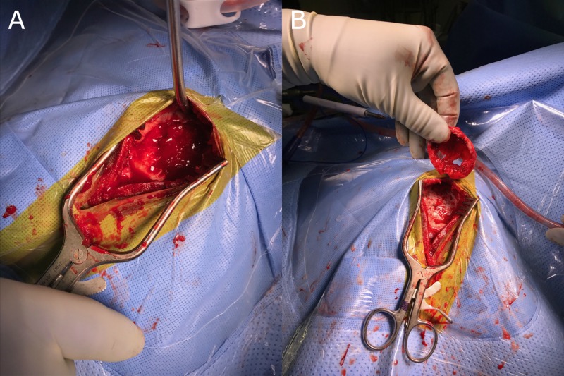
Figure 2. Intraoperative images.
A: Image revealing curettage or scraping of the lesion which kept the excision of the tumor within the walls or the capsule of the tumor, B: A wide excision involving removal of any remaining tumor, reactive zone and some normal bone tissue. All specimens are sent to pathology to be examined for microscopic cells left behind in any of the margins. Results are reported as positive or negative margins. If margins are positive, cells have been left behind and additional excision may be necessary.

