Abstract
Laparoscopy with peritoneal biopsy is a tool for rapid and accurate diagnosis of tuberculous peritonitis (TBP). However, laparoscopic procedures are not risk-free; complications include injuries to the gastrointestinal tract and major blood vessels. The purpose of the present study was to introduce a novel method for safe and straightforward laparoscopic diagnosis of TBP. A case series of 12 patients with TBP diagnosed between October 2012 and November 2013 at our hospital is presented. The patients underwent a novel method of laparoscopy involving the use of a central venous catheter (CVC). The diagnosis was confirmed by biopsy and histology. The efficacy of the method for TBP diagnosis was evaluated by the time taken for the procedure and the rate of successful completion. The safety of the method was evaluated by recording all intra- and post-operative adverse events encountered. The mean age of the patients was 41 years and 33% were male. The mean operation time was 50.6 min and the median duration of hospital stay was 7 days. In all cases, diagnostic laparoscopy was successfully performed. Targeted biopsies were taken from all of the patients and revealed caseous granulomatous inflammation. All patients tolerated the procedure without significant bleeding or digestive tract perforation. In conclusion, the present case series demonstrated a novel method of diagnostic laparoscopy with CVC that is a feasible and straightforward procedure for TBP diagnosis.
Keywords: laparoscopy, peritonitis, tuberculosis, diagnosis, catheterization, central venous, adverse effects
Introduction
Tuberculosis (TB) remains a significant health problem in developing countries and 9 million people are thought to have developed TB in 2013 (1,2). TB may occur at various anatomic locations and cases of tuberculous peritonitis (TBP) are increasing (3). TBP poses a diagnostic challenge due to the lack of specific clinical, radiological or laboratory findings (4). Due to its excellent accuracy and speed, direct observation of the entire peritoneal space and allowing for multiple targeted biopsies of the suspicious lesion, laparoscopy is the diagnostic method of choice (5–7).
However, the diagnostic failure of laparoscopy may be as high as 14% and mainly arises from interference of adhesions due to primary disease or previous surgery (5). Laparoscopic procedures are not completely risk-free. Laparoscopic entry is a blind procedure, which may pose a risk for injuries to the gastrointestinal tract and major blood vessels when attempting to gain access to the peritoneal cavity (8).
The central venous catheter (CVC) is a surgical tool frequently used during cardiac anesthesia and intensive care (9). Placement of a CVC is performed using the Seldinger technique (10), which is mastered by most surgeons. CVCs have also been used for continuous drainage of peritoneal fluid (11). As the CVC set has provided easier and safer access to the peritoneal cavity, the present study implemented it to devise a simple and rapid method for laparoscopic peritoneal biopsy. The present study presented a case series of patients subjected to laparoscopic peritoneal biopsy with CVC at our department to evaluate this novel technique for its utility and safety.
Patients and methods
Patients
A total of 12 patients diagnosed with TBP from October 2012 to November 2013 at the Department of Gastroenterology, Shenzhen People's hospital (Shenzhen, China) were enrolled in the present case series study. The inclusion criteria were suspected TBP due to exudative ascites without a clear pathological reason that was accompanied by a serum-ascites albumin gradient (SAAG) of <11 g/l. The exclusion criteria included i) ascites due to systemic illness, cirrhotic ascites, nephrotic ascites and ascites of heart failure (SAAG of >11 g/l), ii) abdominal trauma or acute abdominal pathology, iii) intraperitoneal fluid of any etiology during pregnancy, iv) defective coagulation, low platelet count (<60,000/mm3) or an international normalized ratio of >1.5, v) previous abdominal surgery and iv) TB confirmed through computed tomography (CT)-guided aspiration.
The present study was approved by the Shenzhen people's Hospital Medical Ethics Committee (Shenzhen, China). All of the recruited patients provided written informed consent.
Data collection
The clinical features of the patients, including their age, major symptoms and the results of TB tests were evaluated. Prior to laparoscopy, all patients were subjected to a purified protein derivatives (PPD) skin test, enhanced CT scan and ascitic fluid examination. All of the patients then underwent the modified technique for laparoscopic peritoneal diagnosis and biopsy with CVC. Variables including time taken to undertake the laparoscopy and duration of hospital stay were noted. All adverse events that occurred during surgery, including severe bleeding or bowel perforation or any adverse events occurring post-surgery were recorded.
Clinical assessment of patients
Prior to laparoscopy, the patients were assessed by biochemical analysis of ascites and serum tuberculosis antibodies (TB-Ab). CT was used to assess peritoneal thickness and the presence of nodules. A tuberculin skin test was performed using the PPD method according to standard techniques (12). PPD tuberculin units were diluted with sterile saline into different concentrations of ~5 tuberculin units and 0.1 ml was intradermally injected into the volar aspect of the left forearm. The injection area was then examined for a reaction after 72 hours. A rash appearance was regarded as the standard examination criterion, but local hard sections were also considered. If a slight swelling or a needle-size red dot at the needle injection site were present and the diameter of the hard section was >0.5 cm, it was defined as negative reaction. If the hard section at the injection site was between 0.5 and 1.5 cm in diameter, it was defined as a positive reaction. If the skin reactions around the injection site were strong or the hard section diameter ≥1.5 cm, it was defined as strongly positive reaction.
Laparoscopic method
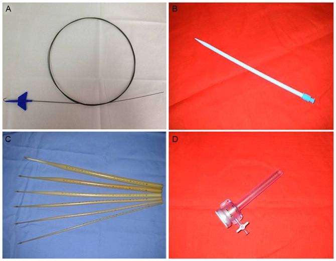
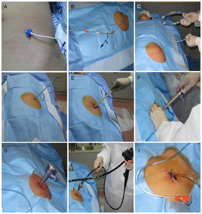
The procedure took place in an aseptic room under aseptic conditions. The equipment, including the guide-wire, dilators and trocar used during the laparoscopy procedure are shown in Figs. 1, and Fig. 2 shows the steps involved in the procedure. The patients were placed in a supine position under conscious sedation, which was achieved by intravenous administration of midazolam (0.06 mg/kg) and pethidine hydrochloride (1.2 mg/kg). An entry site was selected in the left lower quadrant of the abdomen. The abdominal wall was cleansed with iodine and the center of the cleansed area was tropical anesthetized with 2% lidocaine from the local skin to the fascia. After local anesthesia, a small incision was made in the skin and using the Seldinger technique, a central venous catheter (dual-chamber; 7 Fr; 20 cm) was inserted in the abdominal cavity (Fig. 2A). CO2 insufflation was performed through the catheter (Fig. 2B) and the intra-abdominal pressure was maintained at 8–10 mmHg. After adequate pneumoperitoneum had been obtained, a guide-wire (260 cm in length with 3-mm flexional leading end and a 0.035-inch diameter; Fig. 1A) was passed into the peritoneal cavity using the Seldinger technique (Fig. 2C). In order to obtain a suitable access tract, a 16F dilator (length, 20 cm; diameter, 4 mm; Fig. 1B) was used to dilate the tract through the guide-wire (Fig. 2D), and subsequently, shortened Savary-Gilliard dilators (diameter, 5, 7 or 9 mm; length, 40 cm; Fig. 1C) were utilized to progressively dilate the tract (Fig. 2E). A single-use 10-mm trocar (Spekath, Foshan, China; 10-mm sleeve sheath; 10-mm stem; Fig. 1D) was placed over the Savary-Gilliard dilators (Fig. 2F and G). A GIF XP260 gastroscope (Olympus Co. Ltd., Tokyo, Japan) was inserted through the trocar into the peritoneal cavity (Fig. 2H). A detailed observation of the peritoneum and intra-abdominal organs was performed from the left lower to the left upper abdomen, finally to the right lower abdomen, in an anti-clockwise direction. If there were any abnormalities such as nodules or patches, biopsy forceps were inserted through the endoscopic channel to perform a biopsy. When necessary, argon plasma coagulation was used for hemostasis. After the examination was complete, in order to monitor complications, a drainage tube was inserted and fixed to the abdominal wall, and the trocar wounds were sutured with stitches (Fig. 2I).
Figure 1.
Devices used during the laparoscopy via central venous catheter method. (A) The guide-wire; (B) 16F dilator; (C) Savary-Gilliard dilator; (D) single-use 10-mm trocar.
Figure 2.
Laparoscopy with central venous catheter procedure. (A) Insertion of the central venous catheter using the Seldinger technique. (B) Insufflation performed through the catheter. (C) Passing of the guide-wire into the peritoneal cavity. (D) Dilation of the tract with a 16F dilator. (E) Subsequent dilation of the tract with Savary-Gillard dilators. (F and G): Use of a 10-mm trocar over the Savary-Gillard dilators. (H) Insertion of the GIF XP260 gastroscope through the trocar into the peritoneal cavity. (I) Insertion of the drainage tube and suture.
Results
Patient characteristics
Table I shows the clinical features of the patients with TBP. The mean age of the patients was 41 years and 33% were male. The majority of the patients (67%) presented with ascites, including five who had an abdominal sense of flexibility, while no abdominal mass was found. CT results showed peritoneal thickening in five cases and mesenteric lymph nodes in seven cases. Ascitic fluid analysis was performed in all patients. The results are presented in Table II. Direct examination of ascitic fluids revealed an elevation of white cells. The mean concentration of lactate dehydrogenase (LDH) was 596.25 U/l and the serum-ascites albumin gradient (SAAG) was <11 g/l in all patients.
Table I.
Clinical features of the 12 patients with tuberculosis peritonitis that underwent laparoscopy through a central venous catheter.
| Case no. | Age (years) | Sex | Chief complaint | PPD | TB-Ab | History of pulmonary tuberculosis | CT showed whitish granular nodule | CT showed thickened parietal peritoneum | CT shows adhesions | Operation time (min) | Severe complications |
|---|---|---|---|---|---|---|---|---|---|---|---|
| 1 | 28 | F | Low fever, ascites | P | P | P | Yes | No | No | 46 | No |
| 2 | 63 | F | Ascites | N | N | N | Yes | No | Yes | 56 | No |
| 3 | 54 | M | Ascites | N | N | N | Yes | Yes | No | 48 | No |
| 4 | 57 | F | Ascites | N | N | N | No | Yes | Yes | 52 | No |
| 5 | 37 | M | Ascites | N | P | N | Yes | No | No | 47 | No |
| 6 | 25 | F | Hiccups, ascites | N | N | N | No | Yes | Yes | 40 | No |
| 7 | 50 | F | Ascites | N | N | N | No | Yes | Yes | 49 | No |
| 8 | 37 | M | Dyspepsia | N | N | N | Yes | No | No | 48 | No |
| 9 | 45 | F | Low fever | P | P | N | Yes | No | Yes | 72 | No |
| 10 | 38 | M | Weight-loss | N | N | N | Yes | No | No | 45 | No |
| 11 | 24 | F | Ascites | N | P | N | Yes | No | No | 43 | No |
| 12 | 34 | F | Weight loss | N | N | N | No | Yes | Yes | 61 | No |
CVC; F, female; M, male; HIV, human immunodeficiency virus; PPD, purified protein derivatives of tuberculin skin test; TB-Ab, serum tuberculosis antibody; CT, computed tomography; P, positive; N, negative.
Table II.
Ascites test results for 12 patients with tuberculosis peritonitis that underwent laparoscopy through a central venous catheter.
| Case no. | White blood cells (n/mm3) | Total protein (g/l) | Albumin (g/l) | LDH (U/l) | SAAG (g/l) |
|---|---|---|---|---|---|
| 1 | 625 | 32 | 28 | 600 | 8 |
| 2 | 700 | 32 | 27 | 201 | 7.8 |
| 3 | 350 | 29 | 23 | 369 | 6 |
| 4 | 525 | 37 | 31 | 606 | 9.2 |
| 5 | 900 | 35 | 29 | 487 | 5.9 |
| 6 | 609 | 30 | 27 | 554 | 6.3 |
| 7 | 1,001 | 31 | 26 | 587 | 7.2 |
| 8 | 789 | 28 | 25 | 625 | 7 |
| 9 | 863 | 30 | 25 | 492 | 6.9 |
| 10 | 647 | 26 | 22 | 879 | 10 |
| 11 | 539 | 39 | 32 | 791 | 8.2 |
| 12 | 896 | 28 | 23 | 964 | 6.4 |
LDH, lactate dehydrogenase; SAAG, serum-ascites albumin gradient.
Evaluation of the laparoscopic procedure
The mean operation time was 50.6 min (range, 40–72 min), the median duration of hospital stay was 7 days (range, 5–10 days). The bleeding volume ranged from 0–50 ml. The post-operative drainage time was 3 days.
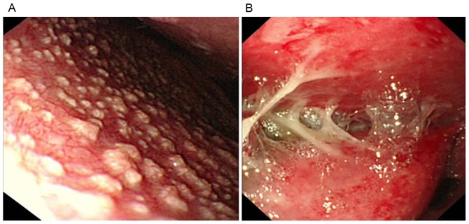
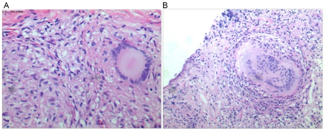
Diagnostic laparoscopy was completed in all 12 patients with TBP. Examples of images from the laparoscopy are shown in Fig. 3. Certain patients presented with thickened, hyperemic peritoneum with ascites and whitish granular nodules (<5 mm) scattered over the peritoneum (Fig. 3A). Furthermore, markedly thickened parietal peritoneum, at times with yellowish nodules and caseous material or multiple thickened adhesions were observed in certain patients (Fig. 3B). Targeted biopsies were taken from all of the patients and revealed caseous granulomatous inflammation, which is the pathological diagnostic criterion for TBP. Examples of typical histological samples for caseous necrosis and granulomas with epithelioid cells are shown in Fig. 4.
Figure 3.
Images from the diagnostic laparoscopy. (A) Whitish granular nodules scattered over the peritoneum. (B) Thickened adhesions.
Figure 4.
Histological samples stained with hematoxylin and eosin from peritoneal biopsies (magnification, ×400). (A) Caseous necrosis. (B) Granulomas with epithelioid cells
All of the patients tolerated the examination without any severe intra- or post-operative severe complications, such as significant bleeding or digestive tract perforation.
Discussion
The aim of the present case series was to evaluate a novel laparoscopic method using a CVC for patients with TBP. The procedure was successfully undertaken in all 12 patients and all of the patients tolerated the examination without any severe intra- or post-operative complications. The mean operation time was 50.6 min and the median duration of hospital stay was 7 days.
Laparoscopic diagnosis with biopsy allows for fast and accurate diagnosis of TBP, which is important for early treatment and better patient prognosis (13). Although in certain situations, laparoscopic diagnosis alone may provide false positive rates in 18%, these are avoided when supported with histopathology and ascitic fluid analysis (14). Conventionally, there are two methods of entry into the peritoneal cavity: The closed technique (Veress needle) and the open technique (Hasson technique) (15). There is no clear consensus for the best method, while Veress needle insertion is the most popular technique (16).
Meta-analysis and large multicenter studies have reported an incidence of vascular and bowel injury caused by Veress needles and subsequent trocars of 0.2 and 0.4 per 1,000, respectively (17), and a small number of complications associated with mortality occurred (18). In addition, the conventional methods have other limits: The techniques require a long time to perform, have a high cost and require endotracheal intubation and mechanical ventilation. Furthermore, they are techniques performed only by surgeons. In terms of TBP specifically, certain patients in whom adhesions occur during the disease course may require conversion to laparotomy as insertion becomes restricted (19). This problem is likely to become more evident as the disease progresses, making it safer to perform laparoscopy during the earlier stages of TBP (20).
From 2006, laparoscopy has been applied to patients with TBP by entry through the umbilicus under conscious sedation, but the rate of complications was found to be higher in patients with TBP due to Mycobacterium tuberculosis-induced peritoneal adhesions. In 2007, our group reported our experience with natural orifice transluminal endoscopic surgery (NOTES) applied to TBP (21). NOTES allows access into the abdominal cavity through a natural orifice (22). However, this method requires to be performed by an experienced endoscopist. Hence, a novel method for laparoscopic diagnosis and peritoneal biopsy with CVC was introduced in our hospital in 2012. The average time taken for the procedure was 50.6 min. All of the patients underwent an uneventful course. No complication occurred, regardless of the level of adhesions. This method allowed us to make a correct diagnosis within four days. The method clearly displays the point which allowed for direct entry into the peritoneal cavity in the majority of the cases, while the abdominal wall is distant from the underlying viscera and vessels at all times. After entering into the peritoneal cavity, the flexible laparoscope is conveniently controlled, allowing for careful observation of the peritoneum, intra-abdominal organs and mesentery. If necessary, it was possible to perform direct biopsies. This method was found to have the following potential benefits: i) Entry-associated injury, such as gastrointestinal tract perforation and bleeding rarely occur. ii) It is easy to learn and quick to perform, particularly for a physician. iii) The flexible laparoscope has a wide-angel lens with high definition and reversal, which allows for convenient observation of the peritoneum and intra-abdominal organs. iv) It may shorten the patient's hospital stay and reduce the cost, which is important in a developing country.
The present study has certain limitations: As the patient cohort was relatively small, the number of complications was expected to be low. As the study only reported on a case series and was not a randomized controlled trial, allowing comparison with other techniques, the potential benefits require to be demonstrated in further studies.
In conclusion, the novel method of laparoscopy with CVC was safe and effective in the present case series.
Acknowledgements
Not applicable.
Funding
No funding was received.
Availability of data and materials
The datasets used and/or analyzed during the current study are available from the corresponding author on reasonable request.
Authors' contributions
RZ, ZX, YZ performed the surgical procedures. RS analyzed and interpreted the patient data. LW, YM performed and analyzed ascites tests. JY, DZ, ML performed the histological examination. RZ was a major contributor in writing the manuscript. All authors read and approved the final version of the manuscript.
Ethics approval and consent to participate
The present study was approved by the Shenzhen people's Hospital Medical Ethics Committee (Shenzhen, China). All of the recruited patients provided written informed consent.
Patient consent for publication
Not applicable.
Competing interests
The authors declare that they have no competing interests.
References
- 1.Corbett EL, Watt CJ, Walker N, Maher D, Williams BG, Raviglione MC, Dye C. The growing burden of tuberculosis: Global trends and interactions with the HIV epidemic. Arch Intern Med. 2003;163:1009–1021. doi: 10.1001/archinte.163.9.1009. [DOI] [PubMed] [Google Scholar]
- 2.WHO: Global Tuberculosis Report 2014. World Health Organization, Geneva, Switzerland. 2014 [Google Scholar]
- 3.Mas MR, Comert B, Sağlamkaya U, Yamanel L, Kuzhan O, Ateşkan U, Kocabalkan F. CA-125; a new marker for diagnosis and follow-up of patients with tuberculous peritonitis. Dig Liver Dis. 2000;32:595–597. doi: 10.1016/S1590-8658(00)80841-5. [DOI] [PubMed] [Google Scholar]
- 4.Inadomi JM, Kapur S, Kinkhabwala M, Cello JP. The laparoscopic evaluation of ascites. Gastrointest Endosc Clin N Am. 2001;11:79–91. doi: 10.1016/S1052-5157(18)30088-6. [DOI] [PubMed] [Google Scholar]
- 5.Sanai FM, Bzeizi KI. Systematic review: Tuberculous peritonitis-presenting features, diagnostic strategies and treatment. Aliment Pharmacol Ther. 2005;22:685–700. doi: 10.1111/j.1365-2036.2005.02645.x. [DOI] [PubMed] [Google Scholar]
- 6.Krishnan P, Vayoth SO, Dhar P, Surendran S, Ponnambathayil S. Laparoscopy in suspected abdominal tuberculosis is useful as an early diagnostic method. ANZ J Surg. 2008;78:987–989. doi: 10.1111/j.1445-2197.2008.04717.x. [DOI] [PubMed] [Google Scholar]
- 7.Wang WN, Wallack MK, Barnhart S, Kalani AD, Storrs SL. Tuberculous peritonitis: Definitive diagnosis by laparoscopic peritoneal biopsy. Am Surg. 2008;74:1223–1224. [PubMed] [Google Scholar]
- 8.Varma R, Gupta JK. Laparoscopic entry techniques: Clinical guideline, national survey, and medicolegal ramifications. Surg Endosc. 2008;22:2686–2697. doi: 10.1007/s00464-008-9871-6. [DOI] [PubMed] [Google Scholar]
- 9.Cotogni P, Pittiruti M. Focus on peripherally inserted central catheters in critically ill patients. World J Crit Care Med. 2014;3:80–94. doi: 10.5492/wjccm.v3.i4.80. [DOI] [PMC free article] [PubMed] [Google Scholar]
- 10.Higgs ZC, Macafee DA, Braithwaite BD, Maxwell-Armstrong CA. The Seldinger technique: 50 years on. Lancet. 2005;366:1407–1409. doi: 10.1016/S0140-6736(05)66878-X. [DOI] [PubMed] [Google Scholar]
- 11.Mercadante S, Intravaia G, Ferrera P, Villari P, David F. Peritoneal catheter for continuous drainage of ascites in advanced cancer patients. Support Care Cancer. 2008;16:975–978. doi: 10.1007/s00520-008-0453-x. [DOI] [PubMed] [Google Scholar]
- 12.Nayak S, Acharjya B. Mantoux test and its interpretation. Indian Dermatol Online J. 2012;3:2–6. doi: 10.4103/2229-5178.93479. [DOI] [PMC free article] [PubMed] [Google Scholar]
- 13.Chahed J, Mekki M, Mansour A, Ben Brahim M, Maazoun K, Hidouri S, Krichene I, Sahnoun L, Jouini R, Belgith M, et al. Contribution of laparoscopy in the abdominal tuberculosis diagnosis: Retrospective study of about 11 cases. Pediatr Surg Int. 2010;26:413–418. doi: 10.1007/s00383-010-2555-z. [DOI] [PubMed] [Google Scholar]
- 14.Wani M, Mir SA, Bhat JA, Moheen HA. Ancillary tests to improve the accuracy of laparoscopy in the diagnosis of tuberculous peritonitis. Can J Surg. 2014;57:E54. doi: 10.1503/cjs.032713. [DOI] [PMC free article] [PubMed] [Google Scholar]
- 15.Vilos GA, Ternamian A, Dempster J, Laberge PY. CLINICAL PRACTICE GYNAECOLOGY COMMITTEE: Laparoscopic entry: A review of techniques, technologies, and complications. J Obstet Gynaecol Can. 2007;29:433–447. doi: 10.1016/S1701-2163(16)35497-4. (In English, French) [DOI] [PubMed] [Google Scholar]
- 16.Merlin TL, Hiller JE, Maddern GJ, Jamieson GG, Brown AR, Kolbe A. Systematic review of the safety and effectiveness of methods used to establish pneumoperitoneum in laparoscopic surgery. Br J Surg. 2003;90:668–679. doi: 10.1002/bjs.4203. [DOI] [PubMed] [Google Scholar]
- 17.Chu CM, Lin SM, Peng SM, Wu CS, Liaw YF. The role of laparoscopy in the evaluation of ascites of unknown origin. Gastrointest Endosc. 1994;40:285–289. doi: 10.1016/S0016-5107(94)70057-5. [DOI] [PubMed] [Google Scholar]
- 18.Schäfer M, Lauper M, Krähenbühl L. Trocar and veress needle injuries during laparoscopy. Surg Endosc. 2001;15:275–280. doi: 10.1007/s004640000337. [DOI] [PubMed] [Google Scholar]
- 19.Abdelaal A, Alfkey R, Abdelaziem S, Abunada M, Alfaky A, Ibrahim WH, Toro A, Di Carlo I. Role of laparoscopic peritoneal biopsy in the diagnosis of peritoneal tuberculosis. A seven-year experience. Chirurgia (Bucur) 2014;109:330–334. [PubMed] [Google Scholar]
- 20.Miyaoka H, Uesugi K, Shigematsu S, Miyake T, Furukawa E, Okita S, Okada T, Abe M, Murakami H, Matsuura B, et al. Clinical course of tuberculous peritonitis determined by laparoscopy. Intern Med. 2010;49:293–297. doi: 10.2169/internalmedicine.49.2809. [DOI] [PubMed] [Google Scholar]
- 21.Zhu H, Li Y, Wang L, Shi R, Huang X, Wang Q, Luo W. Trans-gastric peritoneoscopy with technique of natural-orifice transluminal endoscopic surgery for diagnosis of tuberculosis peritonitis: A report of 20 cases. Chin J Dig Endosc. 2011;28:252–255. (In Chinese) [Google Scholar]
- 22.Song S, Itawi EA, Saber AA. Natural orifice translumenal endoscopic surgery (NOTES) J Invest Surg. 2009;22:214–217. doi: 10.1080/08941930902866287. [DOI] [PubMed] [Google Scholar]
Associated Data
This section collects any data citations, data availability statements, or supplementary materials included in this article.
Data Availability Statement
The datasets used and/or analyzed during the current study are available from the corresponding author on reasonable request.


