Abstract
Introduction
To report 3-year treatment outcomes with intravitreal aflibercept injections for neovascular age-related macular degeneration (nAMD) in routine clinical practice.
Methods
This was a retrospective, single-centre, non-randomized interventional case series analysis. Data from treatment-naïve patients with nAMD treated between 1 October 2013 and 31 February 2014 were included in the analysis. Data including age, gender, vision acuity (VA) measured on Early Treatment of Diabetic Retinopathy Study charts (ETDRS) and injection numbers were recorded. Spectral domain optical coherence tomography (SD-OCT) data including presence or absence of macular fluid and automated central subfield macular thickness (CSMT) at year 1, 2 and 3 were also recorded.
Results
Of the 157 eyes of 148 patients treated, data from 108 eyes of 102 patients were available at 3-year follow-up. The mean (± SD) age was 80.6 ± 8.3 years with a mean of 154.5 ± 5.4 weeks follow-up. The mean VA changed from 54.4 ± 16 letters at baseline to 60.3 ± 18.1 letters (VA gain 5.9 ± 13.8 letter gain) at 1 year, to 60.8 ± 17.4 letters (VA gain 6.4 ± 14.9 letters) at 2 years and to 61.0 ± 16.6 letters (VA gain 6.6 ± 15.4 letters) at 3 years. The reduction in CSMT was 77.9 ± 101.4 µm with absence of macular fluid in 71% of eyes. The total mean number of injections was 15.9 ± 6.1 at year 3.
Conclusion
The results suggest that good long-term morphological and functional treatment outcomes can be achieved using aflibercept for nAMD in a clinical setting.
Electronic supplementary material
The online version of this article (10.1007/s40123-018-0139-5) contains supplementary material, which is available to authorized users.
Keywords: Aflibercept, AMD, Anti-VEGF, Clinical settings, Ophthalmology, Real-life outcomes
Introduction
The last 10 years have seen significant advances in the treatment of neovascular age-related macular degeneration (nAMD) [1–4]. Anti-vascular endothelial growth factor (anti-VEGF) treatment has been introduced into clinical practice following efficacy and safety outcomes based on well-conducted, robust clinical programs [5–9]. However assessing the effectiveness in real-world clinics may reveal whether treatments work in busy clinical settings [10, 11].
Real-world clinical settings differ from clinical trials in several ways: a wider range of patients are treated; patients tend to have more co-morbidities, which can impact on clinic attendance; a wider range of treatment paradigms are implemented and delays in treatment may be encountered. These factors can all impact on treatment outcomes [12–14].
The aim of this study was to report 3-year real-world visual acuity and retinal morphology outcomes of aflibercept treatment for nAMD in a clinical setting. This will allow a meaningful assessment of the robustness of the long-term efficacy response to aflibercept treatment in a real-world clinical setting.
Methods
This was a retrospective review of electronic and paper case notes supplemented with review of spectral-domain optical coherence tomography (SD-OCT) data. This study was approved and registered by Moorfields Research and Development Department (protocol number 15/059). The study conformed to the Declaration of Helsinki of 1964, as revised in 2013, concerning research in humans and animal rights. Springer’s policy concerning informed consent has been followed. This was a retrospective observational study based on previously conducted treatments and does not contain any studies with human participants or animals performed by any of the authors.
Consecutively treated eyes of patients with nAMD were included in the analysis. Any exclusions were recorded using a Consolidated Standards of Reporting Trials-like approach to minimise bias. See Fig. S1 in the electronic supplementary material for details. Treatment-naïve eyes, receiving intravitreal aflibercept injections from 1 September 2013 to 31 February 2014, were included and analysed in this study.
Aflibercept is funded in the National Health Service if criteria set by the National Institute of Health and Care Excellence (NICE) are met. See Table S1 in the electronic supplementary material for details. At Moorfields Eye Hospital, we have developed a nAMD treatment guidance protocol for aflibercept. These are similar to guidelines developed by clinicians in the UK (consensus document [15]) and are broadly based on using fixed dosing in the first year of treatment followed by a treat-and-extend regimen. See Fig. S2 in the electronic supplementary material for details.
Outcome Measures
Outcome measures included the mean change in vision acuity (VA), central subfield macular thickness (CSMT) over time and the number and frequency of injections and visits. VA measurement in clinic is carried out using an Early Treatment of Diabetic Retinopathy Study (ETDRS) chart at a starting distance of 4 m with the patient’s habitual correction where available (supplemented with pin-hole correction). In all patients with loss of 15 letters or more, an analysis of fundus photographs, SD-OCT imaging, fluorescein angiography and review of medical notes were conducted. Topcon 3DOCT-2000 (Topcon Inc) SD-OCT imaging data were used to determine the macular morphology and the CSMT. Macular morphology was assessed by three observers (M.E., M.L., M.G.)
Other outcomes included maximum gain in VA; the proportion of eyes that restarted treatment after ceasing it according to the protocol; the proportion of eyes avoiding moderate (< 15 letters) VA loss; the proportion of eyes with good VA (≥ 73 letters [20/40]) and poor VA (≤ 35 letters [20/200]), the proportion of eyes that were dry at year 1, 2 and 3. To explore whether loss to follow-up had an effect on outcomes, we compared change in mean VA in the study cohort with that of eyes that had less than 24 months of follow-up but otherwise met study inclusion criteria.
Approach to Loss to Follow-up in Data Analysis
In real-world clinical settings, patient loss to follow-up tends to be encountered more commonly than in clinical trial settings. The approach we adopted was to provide a sub-analysis by dividing the cohort into eyes of patients who presented at year 1 follow-up but who did not reach year 2 follow-up, patients who presented at year 2 follow-up but who did not reach year 3 and finally the treated eyes of patients who reached year 3 follow-up.
Statistical Analyses
Descriptive statistics were calculated including mean and standard deviation (SD) for VA and SD-OCT macular thickness data using Excel (Microsoft Excel for Mac 2011). Waterfall plots and other graphical means were used to show changes at both a per patient and cohort level to best present the data in an informative format. The chi-squared or Fisher exact test was used for categorical variables.
Results
Subject Characteristics
In total 157 consecutive treated treatment-naïve eyes from 148 patients with nAMD were included in the study. Eight eyes of eight patients were excluded from further analysis because they did not complete 1 year of follow-up. In addition, one eye of one patient was excluded because of recurrence of pre-existing intraocular inflammation. Hence, data from a total of 148 eyes (79 right eyes) of 139 patients were available for the analysis. Of these, 82 were female (58.9%) with eight patients having bilateral disease. The mean (± SD) age of patients was 80.6 (± 8.3) years (range 53–97 years) at the start of treatment, with a mean initial, pretreatment VA of 54.4 (± 16) ETDRS letters. Choroidal neovascularization (CNV) morphology included a classic component in 31 (22.5%) of cases and purely occult in 91 (65.9%) eyes. A total of 108 eyes of 102 patients completed 3-year follow-up.
Table 1 shows VA, macular morphology and SD-OCT macular thickness outcome data for patients completing 1, 2 and 3 years of follow-up. Data from 17 eyes of 16 patients were not included in the 2-year analysis as these patients were lost to follow-up in the second year of treatment and an additional 23 eyes of 21 patients were lost to follow-up during year 3. See Table S2 in the electronic supplementary material for details of the baseline demographic data of patients who completed and those who did not complete 3-year follow-up.
Table 1.
Aflibercept for neovascular age-related macular degeneration 3-year outcomes. Data regarding visual acuity, OCT macular thickness, macular morphology and number of injections for patients completing 1, 2 and 3 years of follow-up
| Analysis of the cohort | ||||
|---|---|---|---|---|
| Baseline | Year 1 | Year 2 | Year 3 | |
| 148 eyes | 148 eyes | 131 eyes | 108 eyes | |
| Eyes which lost 15 letters or more (%) | N/A | 6.8% | 9.2% | 11.1% |
| Eyes which improved by 15 letters or more (%) | N/A | 25% | 28.2% | 30.5% |
| Eyes with VA ≥ 20/40, 73 ETDRS letters (%) | 10.8% | 30.4% | 38.9% | 28.7% |
| Baseline mean BCVA (ETDRS letters) | 54.4 ± 16 | 60.3 ± 18.1 | 60.8 ± 17.4 | 61 ± 16.6 |
| Mean gain in VA (ETDRS letters) | N/A | 5.9 ± 13.8 | 6.4 ± 14.9 | 6.6 ± 15.4 |
| Change in OCT (microns) | N/A | − 71.2 ± 99.7 | − 74.7 ± 94.5 | − 77.9 ± 101.4 |
| % of eyes with no macular fluid | N/A | 66.7% | 61.3% | 66.7% |
| Number of injections | N/A | 7.2 ± 1.8 | 12 ± 3.8 | 15.9 ± 6.1 |
VA Data
VA data were available for 148 eyes of 139 patients who completed the first year of follow-up, 131 eyes of 123 patients who completed the second year of follow-up and for 108 eyes of 102 patients who completed 3 years of follow-up.
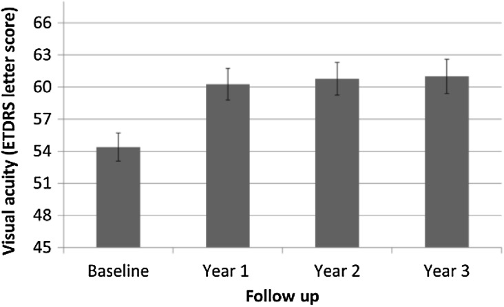
The pretreatment baseline mean initial VA increased from 54.4 (± 16) (range 17–80) ETDRS letter score at baseline to 60.3 (± 18.1) (range 22–88) at year 1, to 60.8 (± 17.4) letters (range 29–88) at year 2 and to 61.0 (± 16.6) letters (range 10–88) at year 3 (Fig. 1). The mean gain in VA was 5.9 (± 13.8) letters in year 1, 6.4 (± 14.9) gain to year 2 and 6.6 (± 15.4) gain from baseline to year 3.
Fig. 1.
Change in mean visual acuity over time: baseline, year 1, year 2 and year 3. Error bars represent the standard deviation
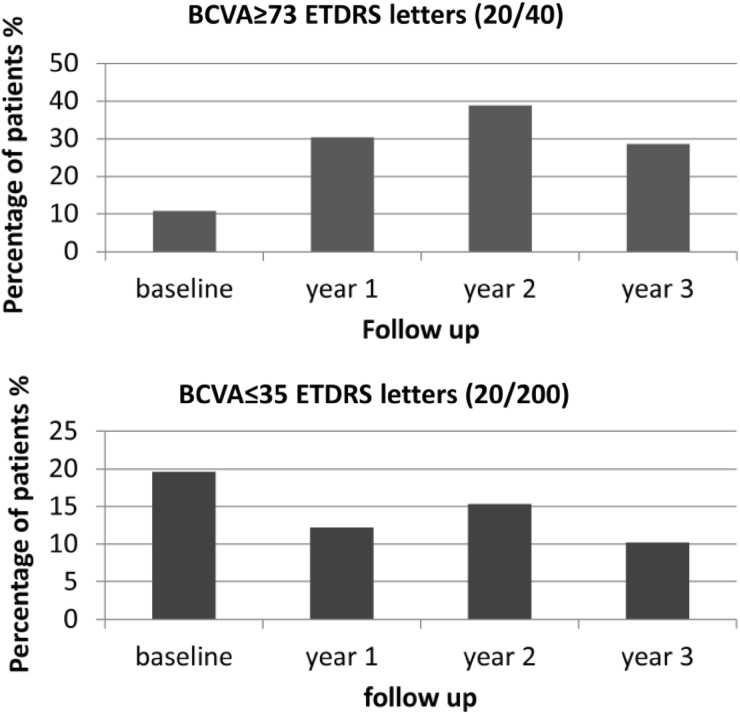
A VA of 73 letters or better (20/40) was seen in 16 eyes (10.8%) at the initial visit, 45 eyes (30.4%) eyes in year 1, 51 eyes (38.9%) in year 2 and 31 eyes (28.7%) in year 3. A VA of 35 letters or less (20/200) was seen in 29 eyes (19.6%) at the initial visit, 18 eyes (12.2%) in year 1, 20 eyes (15.3%) in year 2 and 11 eyes (10.2%) in year 3 (Fig. 2).
Fig. 2.
(Top) Percentage of patients who achieved best corrected visual acuity of 73 letters or better over time: baseline, year 1, year 2 and year 3. (Bottom) Percentage of patients who achieved best corrected visual acuity of 35 letters or worse over time: baseline, year 1, year 2 and year 3
In the 3-year analysis, 33 eyes (30.5%) achieved a VA gain of 15 letters or more from baseline with 12 eyes (11.1%) losing 15 letters or more compared to baseline. See Fig. S3 in the electronic supplementary material for details. In those eyes which lost 15 letters or more, the visual loss was attributed to the presence of foveal atrophy in seven eyes, subretinal fibrosis in four eyes and retinal pigment epithelial tear in one eye.
Subgroup Analysis of Patients who Completed 3 Years of Follow-Up
Three years of follow-up was completed in 108 eyes of 102 patients. The mean initial VA in this subgroup was 54.9 (± 15.2) ETDRS letters, with a median VA of 55 ETDRS letters which was not significantly worse compared to the 40 eyes of 37 patients with less than 36 months of follow-up. In the latter group the mean VA was 57.1 (± 16.9) letters, with a median VA of 60 letters (p > 0.05). Likewise the proportion of eyes with initial VA of 73 letters or better (20/40) was not statistically worse for eyes with 36 months of follow-up (9.7%) than for eyes lost to follow-up (15%, p > 0.05). Patients with less than 36 months of follow-up were older at initial visit (79.5 (± 2) vs 77.5 (± 8.4); p = 0.03). There was no difference between the lesion types for eyes with 24 months of follow-up and those with shorter follow-up (p > 0.05).
The mean VA letter gain in this subgroup was 7.2 ± 13.2 in year 1, 7.1 ± 15.2 in year 2 and 6.6 ± 15.4 in year 3. The total mean number of injections was 7.4 ± 1.5, 12 ± 3.8 and 15.9 ± 6.1 by the end of the first, second and third year of treatment respectively.
The median number of injections in year 1 was 8 and 4 in year 2 and 3. See Fig. S4 in the electronic supplementary material for the frequency distribution of the injections over the follow-up period. In the first year of treatment, after the first three consecutive monthly injections, 60 eyes of 58 patients (55.6%) were treated bimonthly, 23.1% of eyes were treated less frequently and 21.3% of patients more frequently. In year 2, 57 eyes (52.8% of patients) received four injections or fewer, whereas 51 eyes (47.2% of patients) had more than four injections in year 3. See Fig. S5 in the electronic supplementary material for details. Moreover, 30 eyes of 30 patients (27.8%) had not needed any injections.
SD-OCT Data Analysis
SD-OCT data were available for 138 eyes of 130 patients at baseline (93.2% of the included eyes for VA analysis). Data for year 3 were available for 100 eyes of 95 patients.
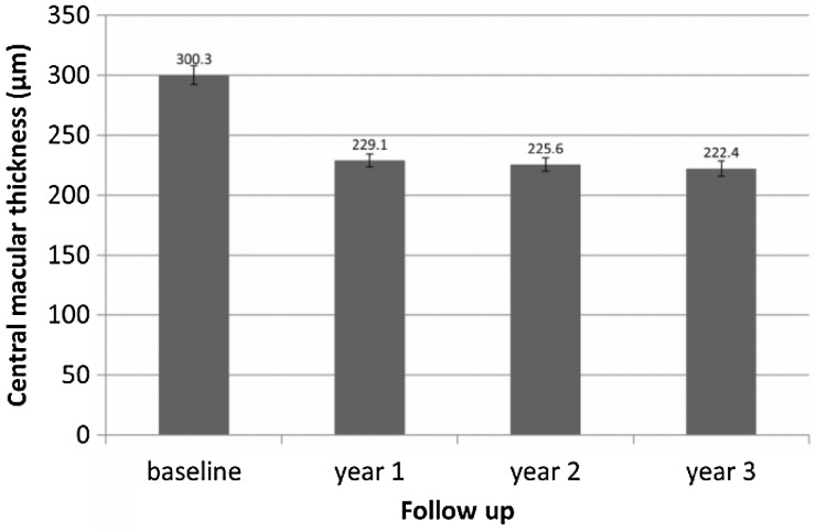
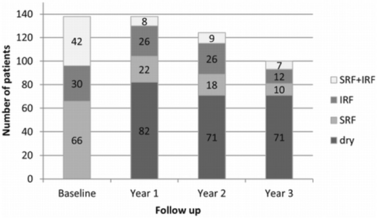
The mean (± SD) CSMT was 300.3 ± 92.6 µm at baseline, 229.1 ± 63.3 µm after 1 year, 225.6 ± 59.5 µm after 2 years and 222.4 ± 62.3 µm after 3 years (Fig. 3). Analysis of retinal morphology on SD-OCT showed that macular fluid (intraretinal fluid, subretinal fluid or both) was present in 100% of eyes at baseline, 40.5% at year 1, 42.7% at year 2 and 29% at year 3 (Fig. 4).
Fig. 3.
Change in mean central macular thickness over time: baseline, year 1, year 2 and year 3. Error bars represent the standard deviation
Fig. 4.
Bar graph showing the number of patients with macular fluid, including subretinal (SRF), intraretinal (IRF), both (IRF + SRF) and dry macula (no macula fluid) at different time points
Discussion
The results of this study show that real-world implementation of anti-VEGF therapy for the treatment of nAMD can lead to sustained improvements in VA and macular morphology. Though data from clinical trials are used to evaluate the efficacy of a therapy, it is also important to report and reflect on efficacy outcomes from real-world evidence collected from clinical settings as the latter may be more closely aligned to what patients can expect in terms of treatment outcomes outside clinical trials.
Often, we may encounter an “efficacy gap” between measures of VA gain seen in clinical trials and those reported in real-world settings. This difference in outcomes may be due to both modifiable and non-modifiable factors. The latter may include the wider range of patients who are treated in real-world settings and variable patient clinic attendance. However modifiable factors may include optimising the treatment paradigm used to achieve outcomes closer to clinical trials thereby closing the “efficacy gap”. The results of previous work from our research group [14] and others show that treat-and-extend treatment paradigms can achieve outcomes in real-world settings closer to those seen in pivotal studies. In this study we report excellent 3-year outcomes with a mean VA gain of 6.6 letters and 15.9 mean number of injections over 3 years. Moreover, 88.9% achieved maintenance of VA (loss of fewer than 15 ETDRS letters from baseline) at the end of year 3 and 30.5% eyes experienced a gain in VA of 15 ETDRS letters or more from baseline. This compares favourably to other long-term reports of anti-VEGF therapy for nAMD, though ours is the first report of outcomes using 3 years of aflibercept treatment.
Rayess et al. [16] reported 3-year treatment outcomes in a real-world setting using ranibizumab or bevacizumab delivered using a treat-and-extend paradigm in 212 eyes treated at baseline; data from 59 eyes (28%) were available at 3 years with a mean VA gain of 13.6 letters and a mean reduction in central macular thickness from 351 microns at baseline to 276 microns at 3 years. A total of 91.5% of patients lost fewer than three lines of VA and 35.6% gained three lines or more VA at 3 years. Overall 52.5% of their patients had signs of CNV activity at 3 years. Their outcomes were obtained with a median of 7.4, 5 and 5 injections in the first, second and third year of treatment respectively (derived median of 17.4 injections over 3 years)
The report from Mrejen et al. [17] followed 185 patients (210 eyes) with nAMD treated over 4 years with a mean follow-up of 3.5 years. After 3 years of follow-up, data were available from 128 eyes and the mean gain in VA was − 0.1061 logMAR with 28% of eyes gaining three lines or more in VA. The mean number of injections over 3.5 years was 28.5 and the majority of eyes received ranibizumab only (59% of eyes). In the report by Berg et al. [18] of 8 years of bevacizumab treatment for nAMD, 84 treated eyes had follow-up at 3 years and the mean gain in VA was 8.3 letters. Berg et al. reported [18] mean loss of VA of 2.9 letters for the 48 eyes compared to baseline after 7 years of follow-up in a real-world setting, highlighting the challenge for retina specialists in trying to maintain VA gains achieved in the first few years of treatment out to year 5 and beyond.
The results from our study compare favourably with these reports of long-term anti-VEGF treatment outcomes for nAMD in real-world settings with relatively high retention of treated eyes at 3 years. Of all treated eyes, 69% reached 3 years of follow-up in our study. Other strengths of our study include reporting outcomes for consecutively treated patients to help minimise bias, 3-year follow-up and reporting SD-OCT imaging outcomes including changes in macular morphology and thickness. This contrasts with other reports of outcomes of aflibercept treatment which do not report SD-OCT imaging outcomes and have shorter follow-up [19].
Limitations of our study include its retrospective nature, the limited sample size and potential variability between clinicians in use of treatment paradigms despite the use of a departmental treatment protocol. This will have led to a variable approach to treatment which may become more pronounced into the third year of treatment which means that in eyes with no signs of macular fluid in year 3, some eyes may have continued on 12 weekly treatment while others may not have received further injections if judged to be stable by the ophthalmologist after discussion with the patient. Use of habitual refractive correction-based VA measurements rather than refracted VA is also a limitation. However, this reflects the real-world setting and is also true for other real-world reports of nAMD treatment outcomes. Note that in our analysis we use an ETDRS letter score of 73 as a cut-off for a VA of 20/40. This is consistent with our previous analysis [14] of 2-year treatment outcomes but differs from the cut-off of 70 ETDRS letters used in other reports of treatment outcomes from clinical settings, making direct comparison difficult. We choose to use 73 letters as this is the cut-off applied in pivotal clinical trials when defining a VA of 20/40, allowing a direct comparison of our results with those seen in clinical trials.
Conclusions
In summary, we report 3-year treatment outcomes of aflibercept for nAMD for patients treated in a clinical setting. Our outcomes show that fixed dosing in year 1 followed by a treat-and-extend approach leads to robust functional and morphological outcomes over 3 years.
Electronic supplementary material
Below is the link to the electronic supplementary material.
Acknowledgements
We thank the participants of the study.
Funding
No funding or sponsorship was received for this study or publication of this study. All authors had full access to all of the data in this study and take complete responsibility for the integrity of the data and accuracy of the data analysis.
Authorship
All named authors meet the International Committee of Medical Journal editors (ICMJE) criteria for authorship for this article, take responsibility for the integrity of the work as a whole, and have given their approval for this version to be published.
Disclosures
Sobha Sivaprasad has received grants, speaker fees, and was an advisory board member of Novartis, Bayer, Allergan, and Roche. Philip G. Hykin has received grants, speaker fees, and was an advisory board member of Novartis, Bayer, and Allergan. Robin D. Hamilton has received grants, speaker fees, and was an advisory board member of Novartis, Bayer, Allergan, Ellex. Adnan Tufail has received grants, speaker fees, and was an advisory board member of Allergan, Bayer, Genentech, GlaxoSmithKline, Novartis, and Roche. Praveen J. Patel has received grants, speaker fees from SalutarisMD, Topcon Inc, Heidelberg Inc, Thrombogenics NV. Has served on Advisory Boards for the following companies Bayer, Novartis, Thrombogenics NV, Roche UK, Genentech Inc, Merk Inc. Maria Eleftheriadou has received speaker fees from Bayer. Maria Gemenetzi, Marko Lukic and Ranjan Rajendram have nothing to disclose.
Compliance with Ethics Guidelines
This study was approved by Moorfields Research and Development Department (protocol number 15/059). The study conformed to the Declaration of Helsinki of 1964, as revised in 2013, concerning research in humans and animal rights. Springer’s policy concerning informed consent has been followed. This was a retrospective observational study based on previously conducted treatments and does not contain any studies with human participants or animals performed by any of the authors.
Data Availability
The datasets during and/or analyzed during the current study are available from the corresponding author on reasonable request. All data generated or analyzed during this study are included in this published article/as supplementary information files.
Open Access
This article is distributed under the terms of the Creative Commons Attribution-NonCommercial 4.0 International License (http://creativecommons.org/licenses/by-nc/4.0/), which permits any noncommercial use, distribution, and reproduction in any medium, provided you give appropriate credit to the original author(s) and the source, provide a link to the Creative Commons license, and indicate if changes were made.
Footnotes
Enhanced digital features
To view enhanced digital features for this article go to 10.6084/m9.figshare.6729137.
References
- 1.Rosenfeld PJ, Brown DM, Heier JS, et al. Ranibizumab for neovascular age-related macular degeneration. N Engl J Med. 2006;355:1419–1431. doi: 10.1056/NEJMoa054481. [DOI] [PubMed] [Google Scholar]
- 2.Brown DM, Kaiser PK, Michels M, et al. Ranibizumab versus verteporfin for neovascular age-related macular degeneration. N Engl J Med. 2006;355:1432–1444. doi: 10.1056/NEJMoa062655. [DOI] [PubMed] [Google Scholar]
- 3.Arevalo JF, Lasave AF, Wu L, et al. Intravitreal bevacizumab for choroidal neovascularization in age-related macular degeneration: 5-year results of the Pan-American Collaborative Retina Study Group. Retina. 2016;36:859–867. doi: 10.1097/IAE.0000000000000827. [DOI] [PubMed] [Google Scholar]
- 4.Brown DM, Tuomi L, Shapiro H, Pier Study Group Anatomical measures as predictors of visual outcomes in ranibizumab-treated eyes with neovascular age-related macular degeneration. Retina. 2013;33:23–34. doi: 10.1097/IAE.0b013e318263cedf. [DOI] [PubMed] [Google Scholar]
- 5.Heier JS, Brown DM, Chong V, et al. Intravitreal ablibercept (VEGF trap-eye) in wet age related macular degeneration. Ophthalmlology. 2012;119:2537–2548. doi: 10.1016/j.ophtha.2012.09.006. [DOI] [PubMed] [Google Scholar]
- 6.Schmidt-Erfurth U, Kaiser PK, Korobelnik JF, et al. Intravitreal aflibercept injection for neovascualr age related macular degeneration: ninety six week results of the VIEW studies. Ophthalmology. 2014;121:193–201. doi: 10.1016/j.ophtha.2013.08.011. [DOI] [PubMed] [Google Scholar]
- 7.Singer MA, Awh CC, Sadda S, et al. HORIZON: an open-label extension trial of ranibizumab for choroidal neovascularization secondary to age-related macular degeneration. Ophthalmology. 2012;119:1175–1183. doi: 10.1016/j.ophtha.2011.12.016. [DOI] [PubMed] [Google Scholar]
- 8.Rosenfeld PJ, Shapiro H, Tuomi L, et al. Characteristics of patients losing vision after 2 years of monthly dosing in the phase III ranibizumab clinical trials. Ophthalmology. 2011;118:523–530. doi: 10.1016/j.ophtha.2010.07.011. [DOI] [PubMed] [Google Scholar]
- 9.Wolf S, Holz FG, Korobelnik JF, et al. Outcomes following three-line vision loss during treatment of neovascular age-related macular degeneration: subgroup analyses from MARINA and ANCHOR. Br J Ophthalmol. 2011;95:1713–1718. doi: 10.1136/bjophthalmol-2011-300471. [DOI] [PubMed] [Google Scholar]
- 10.Gillies MC, Campain A, Barthelmes D, et al. Long-term outcomes of treatment of neovascular age-related macular degeneration: data from an observational study. Ophthalmology. 2015;122:1837–1845. doi: 10.1016/j.ophtha.2015.05.010. [DOI] [PubMed] [Google Scholar]
- 11.Peden MC, Suñer IJ, Hammer ME, Grizzard WS. Long-term outcomes in eyes receiving fixed-interval dosing of anti-vascular endothelial growth factor agents for wet age-related macular degeneration. Ophthalmology. 2015;122:803–808. doi: 10.1016/j.ophtha.2014.11.018. [DOI] [PubMed] [Google Scholar]
- 12.Rofagha S, Bhisitkul RB, Boyer DS, Sadda SR, Zhang K, SEVEN-UP Study Group Seven-year outcomes in ranibizumab-treated patients in ANCHOR, MARINA, and HORIZON: a multicenter cohort study (SEVEN-UP) Ophthalmology. 2013;120:2292–2299. doi: 10.1016/j.ophtha.2013.03.046. [DOI] [PubMed] [Google Scholar]
- 13.Bhisitkul RB, Desai SJ, Boyer DS, Sadda SR, Zhang K. Fellow eye comparisons for 7-year outcomes in ranibizumab-treated AMD subjects from ANCHOR, MARINA, and HORIZON (SEVEN-UP Study) Ophthalmology. 2016;123:1269–1277. doi: 10.1016/j.ophtha.2016.01.033. [DOI] [PubMed] [Google Scholar]
- 14.Eleftheriadou M, Vazquez-Alfageme C, Citu CM, et al. Long-term outcomes of aflibercept treatment for neovascular age-related macular degeneration in a clinical setting. Am J Ophthalmol. 2017;174:160–168. doi: 10.1016/j.ajo.2016.09.038. [DOI] [PubMed] [Google Scholar]
- 15.McKibbin M, Devonport H, Gale R. Aflibercept in wet AMD beyond the first year of treatment: recommendations by an expert roundtable panel. Eye (Lond) 2015;29:S1–S11. doi: 10.1038/eye.2015.77. [DOI] [PMC free article] [PubMed] [Google Scholar]
- 16.Rayess N, Houston SK, 3rd, Gupta OP, et al. Treatment outcomes after 3 years in neovascular age-related macular degeneration using a treat-and-extend regimen. Am J Ophthalmol. 2015;159(3–8):e1. doi: 10.1016/j.ajo.2014.09.011. [DOI] [PubMed] [Google Scholar]
- 17.Mrejen S, Jung JJ, Chen C, et al. Long-term visual outcomes for a treat and extend anti-vascular endothelial growth factor regimen in eyes with neovascular age-related macular degeneration. J Clin Med. 2015;4:1380–1402. doi: 10.3390/jcm4071380. [DOI] [PMC free article] [PubMed] [Google Scholar]
- 18.Berg K, Roald AB, Navaratnam J, Bragadóttir R. An 8-year follow-up of anti-vascular endothelial growth factor treatment with a treat-and-extend modality for neovascular age-related macular degeneration. Acta Ophthalmol. 2017;95:796–802. doi: 10.1111/aos.13522. [DOI] [PubMed] [Google Scholar]
- 19.Almuhtaseb H, Johnston RL, Talks JS, Lotery AJ. Second-year visual acuity outcomes of nAMD patients treated with aflibercept: data analysis from the UK aflibercept users group. Eye (Lond) 2017;31:1582–1588. doi: 10.1038/eye.2017.108. [DOI] [PMC free article] [PubMed] [Google Scholar]
Associated Data
This section collects any data citations, data availability statements, or supplementary materials included in this article.
Supplementary Materials
Data Availability Statement
The datasets during and/or analyzed during the current study are available from the corresponding author on reasonable request. All data generated or analyzed during this study are included in this published article/as supplementary information files.


