Short abstract
Objective
This study was performed to compare the clinical efficacy of three internal fixation methods for distal clavicle fractures (Neer type II): clavicular hook plate (Group A), anatomical plate (Group B), and arthroscopic Endobutton (Group C).
Methods
From 2001 to 2014, 58 patients with Neer type II distal clavicle fractures were treated at our institution. The clinical results were assessed with the visual analog scale (VAS), Constant score, and Simple Shoulder Test (SST) score.
Results
All patients had anatomic reduction and bone healing at the final follow-up. Groups B and C had considerably less intraoperative blood loss than Group A. The incision was significantly shorter in Group C than in Groups A and B. The mean VAS score was significantly higher in the affected than unaffected shoulder. The Constant and SST scores were significantly higher in the unaffected than affected shoulder. The VAS, Constant, and SST scores of the affected shoulders were not significantly different among the three groups.
Conclusions
Arthroscopic Endobutton fixation has long-term clinical results similar to those of other surgical protocols for distal clavicle fractures (Neer type II). We recommend this technique because of less blood loss, shorter incision length, and less shoulder irritation than other methods.
Keywords: Clavicle, bone plates, arthroscopy, fractures, bone, Neer type II, distal, reduction, internal fixation, shoulder
Introduction
Distal clavicle fractures are common shoulder injuries. Approximately 21% to 28% of all clavicle fractures occur in the distal portion, and 10% to 52% of these are displaced.1 The Neer classification system defines five types of distal clavicular fractures. In type II fractures, the distal clavicular fragment is subjected to distal pull by the weight of the arm as well as medial pull by the strong pectoral and latissimus dorsi muscles, while the trapezius muscle pulls the proximal fragment posteriorly. These forces contribute to fracture displacement and instability in type II fractures.2 Several surgical treatments are available for distal clavicle fractures, including hook plates, anatomic locking plates, and arthroscopic treatment (flexible coracoclavicular fixation with a double Endobutton).3 We performed a retrospective study of Neer type II fractures treated by these three different fixation methods to observe their strengths and weaknesses and offer a recommendation on the most suitable treatment.
Materials and methods
Patients
The participants of this study comprised patients with Neer type II distal clavicle fractures who underwent surgical treatment at our institution from 2001 to 2014. We divided the patients into three groups based on the fixation method: patients in Group A were treated by hook plate fixation (AO Hook Plate; Synthes, Solothurn, Switzerland), those in Group B were treated with an anatomic locking plate (AO Distal Clavicle Anatomic Locking Plate; Synthes), and those in Group C were treated by arthroscopic double Endobutton fixation (15.0-mm Endobutton; Smith & Nephew Inc., London, UK) with nonabsorbable polybutylate-coated braided polyester suture (Ethibond Excel; Ethicon, Somerville, NJ, USA). Groups A and B underwent operations from 2001 to 2010, and Group C underwent operations from 2010 to 2014. All operations were performed by the same group of shoulder surgeons. Groups A and B comprised totally independent and consecutive patients, as did Group C but during different time periods. Selection of a hook plate or anatomic plate was dependent on the surgeon’s estimation of the distal clavicular fragment. If the fragment was large enough to be fixed by more than four locking screws, the surgeon chose the anatomic plate.
Surgical methods and postsurgical care
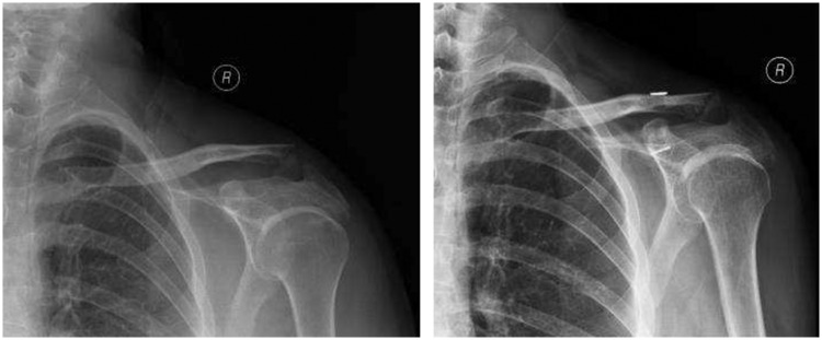
All procedures were performed with patients in the beach chair position. All patients received local cervical and/or brachial plexus anesthesia or general anesthesia. A straight incision was made along the dorsal distal clavicle to expose the acromioclavicular joint in Groups A and B. After open reduction, we inserted the hook under the acromion and fixed the fracture with screws. In Groups A and B, we routinely explored the coracoclavicular ligament and repaired it if it was totally ruptured. Arthroscopic evaluation of the shoulder structures was performed, and concomitant injuries were repaired first in Group C. We identified the lower edge of the coracoid process along the superior edge of the subscapularis muscle. A small incision was made over the clavicle, through which the director was placed under the coracoid process. After locating the distal clavicle fracture line, the fracture was reduced and the guide pin was drilled through the director under X-ray guidance. The distal clavicle and coracoid process were then connected with the united Endobuttons and nonabsorbable sutures (Figure 1). The patients wore an arm sling for 1 to 2 weeks to limit movement in the injured shoulder. They were instructed to perform passive and restricted active movements that did not cause pain. Strength training began when radiographs showed bone healing of the fracture. The rehabilitation time was lengthened in patients with concomitant injuries.
Figure 1.
Preoperative and postoperative radiographs of Neer type II distal clavicle fractures repaired with double Endobutton fixation.
Observation and evaluation indexes
All patients were followed up and radiographs were taken to evaluate fracture healing. The visual analog scale (VAS) score for shoulder pain was recorded. The Constant score and Simple Shoulder Test (SST) score were used to evaluate shoulder function.
Statistical analysis
Results were analyzed with Microsoft Excel 2007 (Microsoft Corp., Redmond, WA, USA) and SPSS 14.0 (SPSS, Inc., Chicago, IL, USA). Measurement data were tested with independent-samples t-tests and paired t-tests. The level of statistical significance was set at p ≤ 0.05.
Ethics statements
The study protocol was approved by Peking University People's Hospital ethics committee. All patients participating in the study provided verbal informed consent.
Results
In total, 58 patients (34 men, 24 women) were included in this study. Group A comprised 25 patients (type IIA fractures, n = 12; type IIB, n = 13), Group B comprised 5 patients (type IIA, n = 2; type IIB, n = 3), and Group C comprised 28 patients (type IIA, n = 12; type IIB, n = 16). The patients’ age ranged from 23 to 82 years (average, 43.5 years). The fractures resulted from sports injury in 28 patients, traffic accidents in 12, low-energy injury in 15, and other injuries in 3. The left shoulder was fractured in 33 patients and the right in 25. All injuries were closed Neer type II fractures (type IIA, 26 patients; type IIB, 32 patients). All fractures were unstable and required surgical treatment. There were no brachial plexus injuries. Concomitant injuries in the affected shoulders included a Bankart lesion in one patient, rotator cuff injury in one, glenolabral articular disruption in one, and acromioclavicular joint arthritis in one (type IIA and mild symptoms before injury). The time from injury to surgery ranged from 1 to 7 days (average, 3.5 days).
The average follow-up period was 57 months (range, 7–160 months). All patients had achieved good reduction and bone healing by the final follow-up. The average patient age, operative time, intraoperative blood loss (blood volume from the suction apparatus), length of incision (total length of the incisions including all portals), and follow-up period are shown in Table 1. The intraoperative blood loss volume was significantly lower in Groups B and C than in Group A (p < 0.05). The incision was significantly shorter in Group C than in Groups A and B (p < 0.05). The mean follow-up period in Group C was 35.6 months (range, 7–53 months), which was significantly shorter than that in Groups A and B (p < 0.05). The average VAS score for all injured shoulders was 1.2 ± 1.6, which was higher than that for the uninjured side (0.3 ± 0.8, n = 58, p < 0.05). The Constant and SST scores of the injured shoulders were 90.2 ± 12.2 and 10.2 ± 2.1, respectively; these scores on the uninjured side were 98.4 ± 5.0 and 11.7 ± 0.9 (n = 58, p < 0.05). The VAS, Constant, and SST scores were analyzed in all three groups. Up to the last follow-up visit, the VAS scores were significantly higher for the injured than uninjured shoulders in Groups A and C (p < 0.05), and the Constant and SST scores were significantly lower than those of the uninjured side (p < 0.05). However, the VAS, Constant, and SST scores were not significantly different among the three groups for either the injured or uninjured side. We repaired the coracoclavicular ligaments with sutures, which completely ruptured in eight patients in Group A and five patients in Group B. Hook plates were removed from 15 patients in Group A at an average of 18.8 months after surgery. Removal was dependent on both the healing of the fractures and the complaints of the patients. In one patient, the hook plate could not be removed because of total locking between the screw and plate. Shoulder discomfort in that patient persisted until the last follow-up. Patients in Group A complained of foreign body and/or impingement sensations from the implants; these sensations resolved in 14 patients after removal of the hook plates. The mean abduction angle was 108.6° immediately before plate removal and 171.4° at the final follow-up in these patients (n = 14, p < 0.05). Two patients treated with hook plates developed fractures proximal to the implant at 10 days and 4 weeks postoperatively, respectively. Both fractures healed after fixation with anatomic and reconstructive plates. One patient in Group C had nonunion 5 months postoperatively; healing was achieved with hook plate fixation. Three patients in Group C had concomitant injuries: Bankart injury, rotator cuff injury, and glenolabral articular disruption, respectively. These injuries were repaired arthroscopically at the time of fracture fixation. One patient with acromioclavicular joint arthritis strongly requested arthroscopic arthrectomy, and the remaining distal fragment also healed over in this patient.
Table 1.
Mean age, operation time, blood loss, incision length, and follow-up period in the three groups.
| Group | Age (years) | Operation time (hours) | Blood loss (mL) | Incision length (cm) | Follow-up time (months) |
|---|---|---|---|---|---|
| A (n = 25) | 46.5 ± 15.8 | 1.4 ± 0.7 | 78.4 ± 62.2 | 9.6 ± 1.8 | 77.4 ± 34.6 |
| B (n = 5) | 38.0 ± 14.7 | 1.7 ± 0.6 | 42.0 ± 20.5* | 10.0 ± 2.0 | 76.2 ± 72.4 |
| C (n = 28) | 41.9 ± 13.5 | 1.8 ± 0.6 | 48.9 ± 29.9* | 2.4 ± 1.4* | 35.6 ± 13.9* |
*p < 0.05 Length: Groups A and C, Groups B and C; Comparison of follow-up time: Groups A and C, Groups B and C.
Discussion
Conservative treatment of Neer type II distal clavicle fractures yields poor results and has a high rate of nonunion. Surgical treatment has shown good results.3 The use of distal clavicle hook plates provides rigid fracture fixation and yields better results than Kirschner wire techniques. We were able to clearly explore the fracture line and achieve good reduction. With extension of the incision, hook plates allow exploration and repair of ruptured coracoclavicular ligaments.2–4 Foreign body irritation from the hook beneath the acromion as well as impingement and limitation of motion are commonly seen in most patients. All of these feelings of discomfort are resolved after implant removal. Most patients in Group A complained of these sensations postoperatively and experienced relief after plate removal. The hook plate crosses the acromioclavicular joint and has minimal motion during shoulder movement. Therefore, some patients experience attrition beneath the acromion, loosening of the implant, and periprosthetic fracture.5 The hook plate method resulted in more blood loss and required a longer incision than the other methods in this study. It is more invasive and causes more procedure-associated injury than other techniques.
Anatomic plates cause less irritation than hook plates because they are located proximal to the acromioclavicular joint. However, they have some limitations. Anatomic plates can only be used in patients with a relatively intact and stable coracoclavicular ligament, and the distal fragments should be large enough to accommodate an adequate number of screws.6 The patients treated with anatomic plates in this study had no complaints of discomfort. However, the surgical procedure causes more intraoperative tissue damage than endoscopic techniques.
Although the two above-described surgeries are effective in treating distal clavicle fractures, they might result in more blood loss, a longer incision, greater intraoperative injury, and more irritation of the acromion than the Endobutton technique; internal fixation failure has also been reported.1,6 Because most distal clavicle fractures are high-energy injuries, some patients sustain soft tissue injury in addition to the fracture. It is difficult to explore and repair these injuries with these two techniques. Endobutton reconstructive fixation via arthroscopy is a minimally invasive technique developed in recent years. This flexible fixation device is usually applied in patients with acromioclavicular dislocation. Some surgeons have used Endobutton fixation for distal clavicle fractures with good results.7,8 This surgery is routinely performed via arthroscopy, which allows exploration to the bottom of the coracoid process and good reduction through an incision above the distal clavicle. Elongating the incision may be necessary in patients in whom fractures are difficult to reduce or when the soft tissue is lodged between bone fragments. The design of the Endobutton minimizes shoulder irritation from the internal fixation. Smaller incisions result in better cosmetic outcomes. It is not necessary to remove the implants postoperatively. Endobuttons are also used to repair the coracoclavicular ligament. Permanent placement of the implant potentially enhances a long-term stability of the ligament after fracture union. Routine arthroscopy with this method also allows detection and repair of related injuries (e.g., Bankart and glenolabral articular disruption), which are difficult to detect during open reduction and internal fixation surgery. In the present study, although the follow-up time was shorter in the arthroscopic group than in the other two groups, the VAS and function scores were similar because of fewer surgery-associated injuries and faster recovery.
The Endobutton technique is associated with some complications, such as suture failure and microgenesis of the coracoid process. Additionally, high staffing and technology levels are essential for performing this type of surgery. In conclusion, there is no consensus on the best method of treating distal clavicle fractures.9 Surgeons should select the optimal procedure based on their personal experience, skills, and instruments. Minimally invasive techniques such as arthroscopy are becoming more popular in traumatic orthopedics, suggesting that in addition to fracture repair, decreasing intraoperative damage is another main concern for orthopedists.
Declaration of conflicting interest
The authors declare that there is no conflict of interest.
Funding
This study was supported by the Specific Research Project of Health Pro Bono Sectors, Ministry of Health of China (201002014); the Ministry of Science and Technology 973 Project Planning (No. 2014CB542201); 863 Project (No. SS2015AA020501); and the National Natural Science Fund (Nos. 31571235, 31771322, 31671248, 31271284, 31171150, 81171146).
References
- 1.Sambandam B, Gupta R, Kumar S, et al. Fracture of distal end clavicle: a review. J Clin Orthop Trauma 2014; 5: 65–73. [DOI] [PMC free article] [PubMed] [Google Scholar]
- 2.Neer CS., 2nd. Fractures of the distal third of the clavicle. Clin Orthop Relat Res 1968; 58: 43–50. [PubMed] [Google Scholar]
- 3.Loriaut P, Moreau PE, Dallaudiere B, et al. Outcome of arthroscopic treatment for displaced lateral clavicle fractures using a double button device. Knee Surg Sports Traumatol Arthrosc 2015; 23: 1429–1433. [DOI] [PubMed] [Google Scholar]
- 4.Mechchat A, Elidrissi M, Shimi M, et al. Neer type II distal clavicle fractures: hook plate versus transacromial pin. Pan Afr Med J 2015; 20: 105. [DOI] [PMC free article] [PubMed] [Google Scholar]
- 5.Tiren D, Van Bemmel AJ, Swank DJ, et al. Hook plate fixation of acute displaced lateral clavicle fractures: mid-term results and a brief literature overview. J Orthop Surg Res 2012; 7: 2. [DOI] [PMC free article] [PubMed] [Google Scholar]
- 6.Tan HL, Zhao JK, Qian C, et al. Clinical results of treatment using a clavicular hook plate versus a T-plate in Neer type 2 distal clavicle fractures. Orthopedics 2012; 35: e1191–e1197. [DOI] [PubMed] [Google Scholar]
- 7.Soliman O, Koptan W, Zarad A. Under-corocoid-around-clavicle (UCAC) loop in type 2 distal clavicle fractures. Bone Joint J 2013; 95: 983–987. [DOI] [PubMed] [Google Scholar]
- 8.Yang SW, Lin LC, Chang SJ, et al. Treatment of acute unstable distal clavicle fractures with single coracoclavicular suture fixation. Orthopedics 2011; 34: 172. [DOI] [PubMed] [Google Scholar]
- 9.Madsen W, Yaseen Z, LaFrance R, et al. Addition of a suture anchor for coracoclavicular fixation to a superior locking plate improves stability of type 2b distal clavicle fractures. Arthroscopy 2013; 29: 998–1004. [DOI] [PubMed] [Google Scholar]


