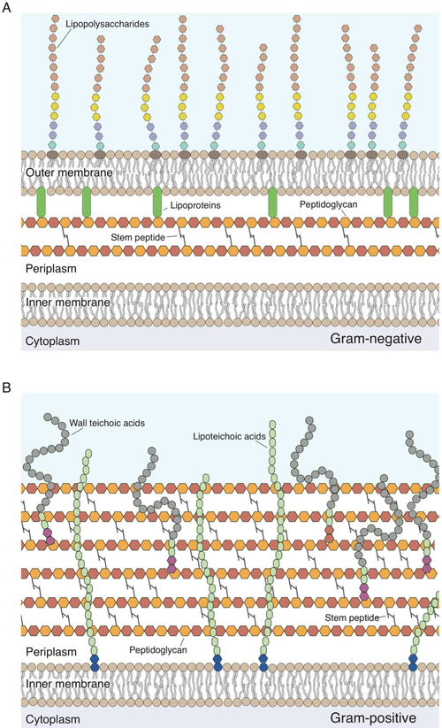
Figure 1.
Structure of the bacterial cell walls. (A) Cartoon depicting the structure of the Gram-negative cell wall. The peptidoglycan thickness is ~4 nm; monosaccharides in the peptidoglycan are represented as hexagons, and the colors demonstrate that this material consists of repeating disaccharide building blocks. Peptide cross-links in the peptidoglycan are depicted as gray lines. Monosaccharides in lipopolysaccharides are depicted as hexagons. Aqua and purple denote the inner polysaccharide core; yellow denotes the outer polysaccharide core, and brown denotes the O-antigen. Lipoproteins (green) connect the outer membrane to the peptidoglycan. (B) Cartoon depicting the Gram-positive bacterial cell wall. The peptidoglycan thickness is ~19–33 nm. Lipoteichoic acid is inserted into the membrane and consists of a glycolipid anchor (blue) and poly(glycerol phosphate) (green). The wall teichoic acid is directly cross-linked to the peptidoglycan through a linkage unit (red) and consists of glycerol phosphate (green) and poly(alditol phosphate).

