Summary
The Chromosomal Passenger Complex (CPC) localizes to centromeres in early mitosis to activate its subunit Aurora B kinase. However, it is unclear if centromeric CPC localization contributes to CPC functions beyond Aurora B activation. Here, we show that an activated CPC that cannot localize to centromeres supports functional assembly of the outer kinetochore, but is unable to correct errors in kinetochore-microtubule attachment in Xenopus egg extracts. We find that CPC has two distinct roles at centromeres: one to properly phosphorylate Ndc80 to regulate attachment, and a second, conserved kinase-independent role in the proper composition of inner kinetochore proteins. Although a fully assembled inner kinetochore is not required for outer kinetochore assembly, we find it is essential to recruit tension indicators, such as BubR1 and 3F3/2, to erroneous attachments. Thus, centromeric CPC is necessary for tension-dependent removal of erroneous attachments, and for the kinetochore composition required to detect tension loss.
Introduction
Faithful segregation of chromosomes is the key event of mitosis, and its dysregulation can lead to aneuploidy and genomic instability, both hallmarks and drivers of cancer initiation and progression (Gordon et al., 2012). Chromosome segregation requires attachment of spindle microtubules to kinetochores, large proteinaceous structures that assemble on centromeric chromatin. The kinetochore is assembled on nucleosomes at centromeres which contain the histone H3 variant, CENP-A (Westhorpe and Straight, 2016). The 16-subunit Constitutive Centromere-Associated Network (CCAN), which constitutes the inner kinetochore, resides at the centromere throughout the cell cycle and links centromeric chromatin to the microtubule-binding proteins at the outer kinetochore. The CCAN components CENP-C and the CENP-L-N, CENP-T-W-S-X and CENP-H-I-K-M complexes make numerous interactions with each other to bind centromeres and to confer structural integrity to the kinetochore (Nagpal et al., 2015). Upon entry into mitosis, interactions are altered such that the localization of all CCAN members depend on the interaction of CENP-C with CENP-A nucleosomes (McKinley et al., 2015; Nagpal et al., 2015). This suggests that CCAN remodeling is important for kinetochore function, but its cause and purpose are not known.
The outer kinetochore, comprising the Knl1-Mis12-Ndc80 (KMN) network, connects the inner kinetochore to microtubules (Cheeseman, 2014). In higher eukaryotes, the outer kinetochore assembles upon entry to M phase, through interaction of CENP-C with the Mis12 complex (Dimitrova et al., 2016; Petrovic et al., 2016; Przewloka et al., 2011; Screpanti et al., 2011) and the recruitment of the Ndc80 complex by CENP-T (Nishino et al., 2013). The KMN network attaches to microtubules, primarily through direct interaction with Ndc80. If an attachment is lost, the KMN network halts the cell cycle by activating the spindle assembly checkpoint (SAC). Mps1 kinase binds to Ndc80 in a microtubule-dependent manner to phosphorylate KNL1, which recruits the SAC proteins, including BubR1 and Mad2, to the kinetochore (Hiruma et al., 2015; Ji et al., 2015).
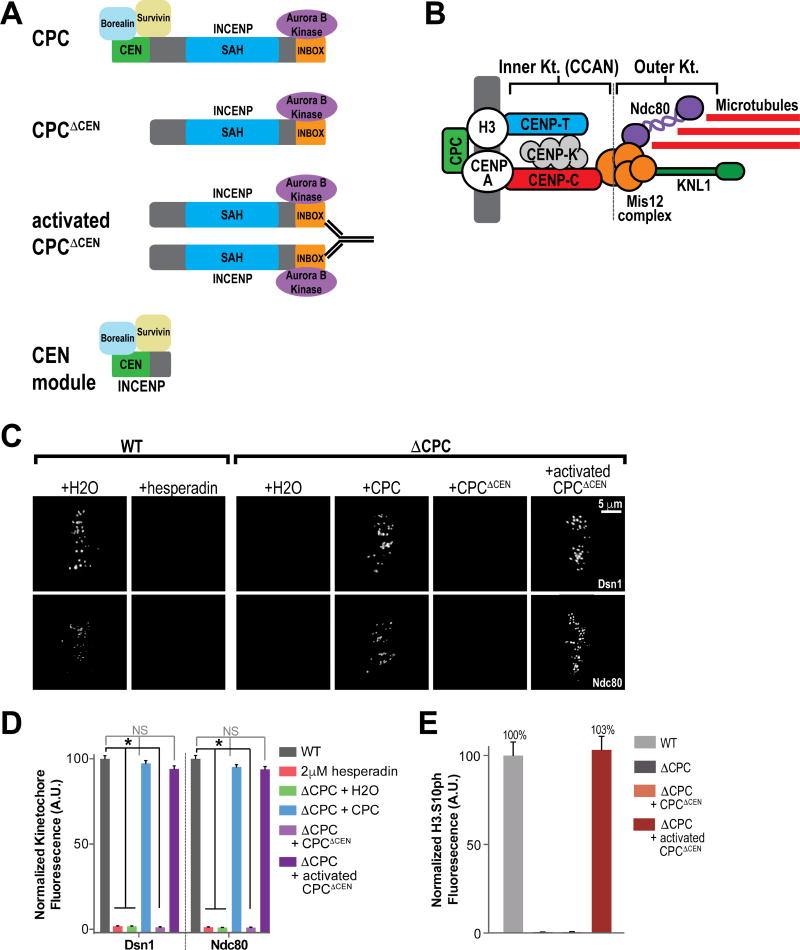
The Chromosomal Passenger Complex (CPC) is a four-protein complex which consists of the kinase Aurora B, INCENP, Survivin and Borealin. The CPC, through Aurora B phosphorylation, regulates multiple processes required for chromosome segregation (Carmena et al., 2012). INCENP is a large scaffolding protein that serves to orchestrate the localization and activity of the CPC throughout mitosis. The CPC can be separated into three functional modules based on the domain structure of INCENP (Figure 1A): a kinase module consisting of Aurora B and the C-terminus of INCENP (IN Box); a second, centromere targeting module comprising the N-terminal domain of INCENP (CEN), Survivin and Borealin; and a third module comprising the microtubule-binding SAH domain of INCENP. Full activation of Aurora B in most eukaryotes is thought to require phosphorylation of the “TSS” motif in the IN Box motif of INCENP (note: this site is absent in some species, including budding yeast Sli15) by Aurora B itself (Bishop and Schumacher, 2002; Sessa et al., 2005), which induces a conformational change in Aurora B required for full kinase activation. Multiple lines of evidence suggest this is mediated in trans by another Aurora B/INCENP complex and that kinase activation is regulated through local concentration at centromeres or microtubules (Kelly et al., 2007; Tseng et al., 2010; E. Wang et al., 2011). In early mitosis, the CPC localizes to the centromere and ensures proper kinetochore-microtubule attachments by removing improper attachments and activating the SAC. In addition, Aurora B is required for the assembly and maintenance of the outer kinetochore, through the phosphorylation of Mis12 complex subunit Dsn1 which drives the interaction of Mis12 with CENP-C (Akiyoshi et al., 2013; Dimitrova et al., 2016; Emanuele et al., 2008; Kim and Yu, 2015; Petrovic et al., 2016; Rago et al., 2015; Yang et al., 2008). CPC localization is mediated by Survivin and Borealin, which interact with Histone H3 phosphorylated at threonine 3 (H3T3ph) by Haspin (Kelly et al., 2010; F. Wang et al., 2010; Yamagishi et al., 2010), and Shugoshin, which directly interacts with histone H2AT120ph-modified nucleosomes established by Bub1 kinase (Kawashima et al., 2010; H. Liu et al., 2015). However, it remains unclear whether the conserved centromere localization of the CPC is required for its early mitotic functions (Caldas et al., 2013; Campbell and Desai, 2013; F. Wang et al., 2010; Yue et al., 2008).
Figure 1. Centromeric CPC is Not Required for Outer Kinetochore Assembly.
(A) Schematic of the Chromosomal Passenger Complex (CPC) and CPC manipulations used in this study. “CPC” indicates reconstitution of CPC depleted extracts with all four CPC subunits. “CPCΔCEN” indicates reconstitution with Aurora B kinase and a fragment of INCENP (aa 58-873; ΔCEN) of that cannot bind to centromeres (see Figure S1B). “Activated CPCΔCEN” indicates the addition of substoichiometric amounts of activating anti-INCENP antibodies. “CEN-module” indicates reconstitution with full-length Borealin (aka Dasra A), full-length Survivin, and residues 1-242 of INCENP fused to MBP.
(B) Schematic highlighting key components of the inner (or CCAN) and outer kinetochore.
(C) Representative IF images of replicated chromosomes in metaphase spindles in Xenopus laevis mock-depleted (WT) and CPC-depleted (ΔCPC) extracts with indicated CPC conditions +/− 2 µM hesperadin. Spindles were stained for kinetochore components (white). See also Figures S1A and S2A.
(D) Mean integrated fluorescence intensities of kinetochore components shown in (C) were quantified and normalized to WT. n = 96 kinetochores.
(E) Mean integrated fluorescence intensity of chromosomal H3S10ph in WT, ΔCPC, and activated CPCΔCEN extracts was quantified and normalized to WT. n = 96 kinetochores.
For all figures, error bars represent the SEM of 3 independent experiments and asterisks indicate a statistically significant difference (P<0.0001) while NS indicates no significant difference, unless otherwise noted.
Bi-orientation, where sister chromatids attach to microtubules emanating from opposing sides of the cell, is essential for chromosome segregation. Microtubule-based forces create tension across and within kinetochores in a bi-oriented state, which induces the full separation of sister kinetochores. Tension loss at kinetochores, which can be assessed experimentally through measurement of inter-kinetochore distances, is thought to signal errors in attachment which must be removed and then replaced to allow for chromosomes to bi-orient. Aurora B phosphorylates Ndc80 to weaken its interactions with microtubules to remove erroneous attachments (Cheerambathur and Desai, 2014). How Aurora B senses such errors and whether tension sensing requires centromeric CPC is currently unclear (Krenn and Musacchio, 2015).
Here we reveal that the centromere localization of the CPC is essential for error detection and correction. We demonstrate that centromere-targeting of Aurora B is required to modulate Ndc80 phosphorylation in response to tension. Importantly, we discover a kinase-independent role for the interaction of the CPC with centromeric chromatin that mediates assembly of the inner kinetochore in mitosis. We show that proper inner kinetochore composition is required for the tension responsiveness of kinetochores and the identification of erroneous attachments. Together, our data demonstrate that the CPC centromere localization controls the detection and correction of erroneous attachments by establishing proper kinetochore composition and modulating Aurora B activity.
Results
Centromeric CPC Localization is Not Required for Assembly of the Outer Kinetochore
To further understand how Aurora B and the CPC regulate kinetochore function, we first investigated the role of centromeric localization of the CPC in kinetochore assembly using Xenopus egg extracts. As previously reported (Emanuele et al., 2008; 2005), CPC immunodepletion or inhibition of Aurora B blocked the assembly of the outer kinetochore proteins Ndc80 and Dsn1 (a Mis12 Complex component, Figure 1B) onto replicated sister chromosomes in cycled mitotic Xenopus egg extracts (Figures 1C and 1D and Figure S1A). CPC depletion also prevented the phosphorylation of Ser10 on histone H3 (H3S10ph), an established Aurora B substrate, in both total extracts and on chromosomes (Figure 1E and Figure S2A). The assembly of Dsn1 and Ndc80 as well as H3S10ph were fully restored by re-expression of all four CPC subunits from pooled mRNAs in depleted extracts (Figures 1C, 1D, and 1E, Figure S1A and Figure S2A). To test whether centromeric CPC is required for kinetochore assembly, we reconstituted depleted extracts with wild-type Aurora B kinase and an INCENP deletion mutant that prevents centromere targeting (henceforth referred to as CPCΔCEN, Figure 1A). Aurora B kinase activation normally requires binding to and clustering on chromosomes or microtubules, but this requirement can be bypassed by antibody-mediated clustering of INCENP (Kelly et al., 2007; Tseng et al., 2010). Antibody-mediated clustering of INCENP increases the Aurora B-mediated phosphorylation of INCENP in trans, which is required for allosteric activation of Aurora B and its activity in egg extracts (Kelly et al., 2007). As expected, deletion of the CEN domain blocked CPC localization to chromosomes, Aurora B activation, phosphorylation of histone H3 and outer kinetochore assembly (Figures 1C and 1D, Figure S1A and Figure S2A). Remarkably, antibody-mediated clustering of CPCΔCEN (activated CPCΔCEN) fully rescued the assembly of Dsn1 and Ndc80 to kinetochores and H3S10ph on chromosomes (Figures 1C, 1D and 1E, Figure S1A and Figure S2A). We could detect no CPC subunits on centromeres or kinetochores on chromosomes in activated CPCΔCEN extracts (Figure S1B). Outer kinetochore assembly by activated CPCΔCEN was not mediated by microtubules, as addition of nocodazole had no effect on assembly (Figure S2B). Additionally, clustering of CPCΔCEN on the surface of anti-INCENP beads promoted kinetochore assembly, even though the beads were on average twelve microns away from the nearest chromosome mass (Figures S2C, S2D, and S2E) and global Aurora B kinase activity levels were very low (Figure S2F). Thus, we conclude that the CPC can promote outer kinetochore assembly without the requirement for centromere, kinetochore, or microtubule localization.
Centromeric CPC is Required to Correct Errors in Attachment
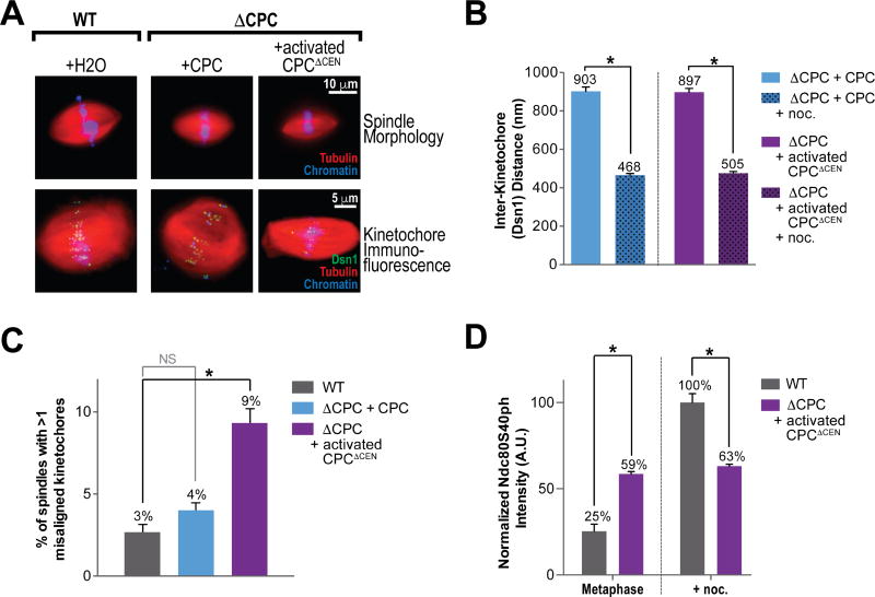
Our results thus far suggest that the localization of the CPC to centromeres is not required for assembly of the outer kinetochore. However, it is unclear whether the CPC must reside at centromeres to properly regulate kinetochore-microtubule attachment (Krenn and Musacchio, 2015). We found that activated CPCΔCEN could generate bipolar spindles with normal length and geometry as compared to control (Figure 2A). In addition, activated CPCΔCEN spindles exhibited similar levels of inter-kinetochore separation between sister chromatids (which reflects tension across kinetochores) to control and inter-kinetochore separation appropriately decreased upon treatment with nocodazole (Figure 2B). However, we observed a two- to three-fold increase in chromosome misalignment, defined as kinetochore pairs that are localized outside of the central region of the spindle (see methods), in activated CPCΔCEN spindles compared to control spindles (Figure 2C). We hypothesized that these errors could be the result of improper regulation of kinetochore-microtubule attachments. To investigate this, we measured phosphorylation of the N-terminal tail of Ndc80 at S40 (S40ph) by Aurora B (Figure 2D, Figures S3A, S3B and S3C and Figure S4D), one of several Aurora B-mediated phosphorylations that weakens the interaction of Ndc80 with microtubules to remove erroneous attachments (Cheerambathur and Desai, 2014). We observed a low level of Ndc80 phosphorylation at kinetochores in control metaphase spindles, which increased four-fold when all microtubules were depolymerized by nocodazole (Figure 2D), in agreement with findings in human cells (DeLuca et al., 2011). However, we observed elevated levels of Ndc80 phosphorylation (59% of WT nocodazole-treated) across all kinetochores in unperturbed spindles generated by activated CPCΔCEN (Figure 2D). Strikingly, these levels showed no significant change upon microtubule loss induced by nocodazole treatment. These data indicate that non-centromeric CPC results in unregulated levels of Ndc80 phosphorylation, and an increase in alignment errors.
Figure 2. Proper Chromosome Alignment and Ndc80 Phosphorylation Require Centromeric CPC.
(A) Upper panel: Representative images of spindles formed in WT and ΔCPC extracts with indicated CPC conditions. Chromatin was stained with Hoechst (blue), and rhodamine-labeled tubulin was added (red). Lower panel: Representative IF images of metaphase spindles stained for Dsn1 (green).
(B) Mean of inter-kinetochore distances observed in ΔCPC extracts with indicated CPC conditions +/− nocodazole. n = 100 kinetochores.
(C) Mean percent of bipolar spindles observed in (B) which contained multiple misaligned kinetochores. n = 75 total spindle structures per condition.
(D) Mean integrated fluorescence intensities of Ndc80S40ph in metaphase spindles in WT and activated CPCΔCEN extracts +/− nocodazole. Values are normalized to the WT + nocodazole condition, n = 96 kinetochores. See also Figure S3A–C.
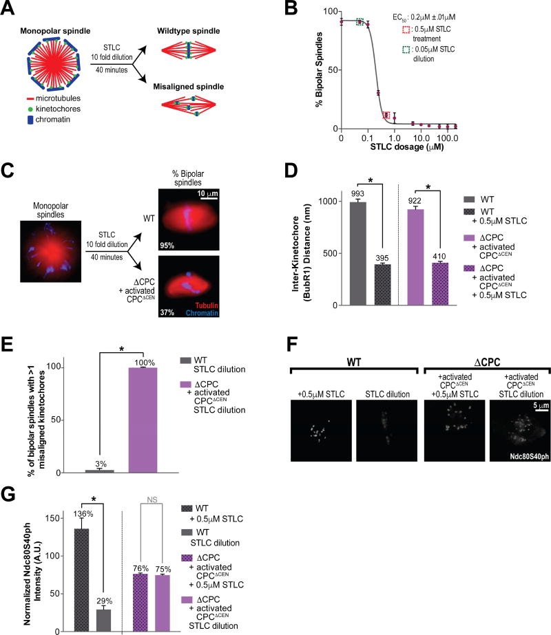
To test whether CPC must reside at centromeres to detect and correct errors in attachment, we modified an established error correction assay (Lampson et al., 2004) and adapted it to the egg extract system. Monopolar spindles are first induced by treatment with the Eg5 inhibitor, S-Trityl-L-Cysteine (STLC) (Houghtaling et al., 2009), which results in erroneous, mono-oriented (syntelic) kinetochore-microtubule attachments that do not produce tension (Figures 3A and 3B). Subsequent dilution of STLC allows for bipolar spindle formation and chromosome alignment at the metaphase plate through removal of improper kinetochore-microtubules by Aurora B kinase.
Figure 3. Centromeric CPC is Required for Error Correction.
(A) Experimental design for error correction in cell free system via STLC treatment and dilution. STLC is added to 0.5 µM upon entry into metaphase and diluted to 0.05 µM after assembly of monopolar spindles by addition of untreated CSF extract. Spindle structures are fixed 40 minutes after STLC dilution.
(B) Mean percent bipolar spindles in WT extract treated with varying doses of STLC. Error bars represent the SEM of 3 independent experiments; n = 100 spindles per dosage.
(C) Representative images of spindles with 0.5 µM STLC and after dilution of STLC to 0.05 µM, in WT and activated CPCΔCEN extracts. Monopolar spindle frequency was similar in both conditions. Percent bipolar spindle formation after dilution is indicated. Chromatin was stained with Hoechst (blue), and rhodamine-labeled tubulin was added (red). See Figure S4A for more information.
(D) Mean of inter-kinetochore distances observed in WT and activated CPCΔCEN extracts +/− 0.5 µM STLC. n = 100 kinetochores.
(E) Mean percent of bipolar spindles structures in (C) that contained multiple misaligned kinetochores after STLC dilution, n = 75 total spindle structures per condition. See Figure S4B for more information.
(F) Representative IF images of replicated chromosomes in WT and activated CPCΔCEN extracts treated with 0.5 µM STLC and after dilution of STLC to 0.05 µM. Spindles were stained for Ndc80S40ph (white).
(G) Mean integrated fluorescence intensities of Ndc80S40ph in (F). Values are normalized to the WT + nocodazole treated condition, n = 96 kinetochores per condition. See also Figure S4C.
In both control and activated CPCΔCEN extracts treated with STLC, monopolar spindles were formed and all kinetochores lost tension, as measured by the inter-kinetochore distance between sister chromatids (Figures 3C and 3D and Figure S4D). These kinetochores demonstrated no recruitment of Mad2 (Figure S5A). Since the SAC is only activated by unattached kinetochores in the egg extract system (Minshull et al., 1994), these results indicate that kinetochores remained attached to microtubules in the presence of STLC. When STLC was diluted from control extracts, 96% of spindles were bipolar and only 3% of these spindles had more than one misaligned chromosome (Figures 3C and 3E and Figure S4A). Strikingly, when STLC was diluted in activated CPCΔCEN extracts, only 37% of spindles were bipolar (Figure 3C), with the remaining structures either remaining monopolar or displaying a multipolar phenotype (Figure S4A). Of the bipolar spindles observed in activated CPCΔCEN extracts, all had multiple chromosome misalignments, a 33-fold increase compared to control (Figure 3E). The average number of misaligned kinetochores per bipolar spindle in activated CPCΔCEN extracts after STLC dilution was 4.13 (±1.5), compared to 0.14 (±0.4) in WT extracts (Figure S4B). We conclude that non-centromeric CPC has severe defects in its ability to correct errors in microtubule attachment.
Centromeric CPC is Required for Specific and Dynamic Ndc80 Phosphorylation
Since we had observed that Ndc80 phosphorylation was improperly regulated in activated CPCΔCEN metaphase spindles, we examined Ndc80 phosphorylation levels throughout the error correction process. In WT extracts, we found that levels of Ndc80 phosphorylation markedly increased upon treatment with STLC and returned to levels observed in unperturbed metaphase spindles upon STLC dilution (Figure 2D, Figures 3F and 3G and Figure S4C), in agreement with previous studies in human cells that reported an increase in Ndc80 phosphorylation in monopolar spindles (Welburn et al., 2010). In contrast, in the bipolar spindles formed in activated CPCΔCEN extracts, Ndc80 phosphorylation levels remained elevated throughout the error correction assay (Figures 3F and 3G and Figure S4C). However, the levels of phosphorylation we observed were only ~50% of those at kinetochores in WT extracts treated with STLC. Interestingly, phosphorylation levels were uniform and irrespective of chromosome alignment or spindle bipolarity (unpublished observations, J.H.; Figure 3E and Figure S4A). Thus, our results suggest that centromeric CPC localization is required for the specific and dynamic phosphorylation of Ndc80 at kinetochores in response to tension status.
Centromeric CPC is Required for BubR1 and 3F3/2 Enrichment at Tensionless Attachments
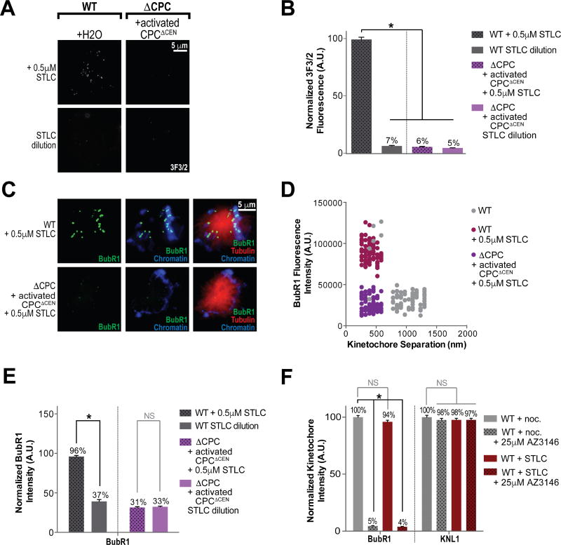
Our results thus far indicate that there may be a problem sensing a loss of tension at kinetochores in activated CPCΔCEN extracts. To test this, we performed immunofluorescence using monoclonal 3F3/2 antibodies that mark tensionless kinetochores in many species, during error correction (Cyert et al., 1988; Gorbsky and Ricketts, 1993). As expected, the 3F3/2 epitope was highly enriched at kinetochores in control extracts that were treated with STLC (Figures 4A and 4B) and 3F3/2 levels decreased by 14-fold upon STLC dilution. However, there was little detectable 3F3/2 staining at kinetochores in STLC-treated activated CPCΔCEN spindles, and remained low in bipolar spindles upon STLC dilution (Figures 4A and 4B), even though we observed many errors in alignment in this condition (Figure 3E). These results suggest that tension status is not being discerned at kinetochores assembled by activated CPCΔCEN.
Figure 4. Centromeric CPC is Necessary for the Enrichment of BubR1 and the 3F3/2 Epitope at Tensionless Kinetochore-Microtubule Attachments.
(A) Representative IF images of metaphase spindles formed in the presence of 0.5 µM STLC and after STLC dilution to 0.05 µM in WT and activated CPCΔCEN extracts. Samples were stained for 3F3/2 (white).
(B) Mean integrated fluorescence intensity of 3F3/2 in (A). Quantification was normalized to the WT STLC treated condition, n = 100 kinetochores per condition.
(C) Representative IF images of metaphase spindles formed in the presence of 0.5 µM STLC in WT and activated CPCΔCEN extracts. Samples were stained for BubR1 (green). See also Figure S5A.
(D) Scatter plot of integrated BubR1 fluorescent intensity of kinetochores, relative to sister kinetochore separation during metaphase in WT and activated CPCΔCEN extracts. STLC was added as indicated, n = 100 kinetochores per condition. See also Figure S5B.
(E) Mean integrated fluorescence intensities of BubR1 in WT and activated CPCΔCEN extracts treated with 0.5 µM STLC and after dilution to 0.05 µM STLC. Values are normalized to the nocodazole treated WT sample, n = 96 kinetochores per condition. See also Figure S5C.
(F) Mean integrated fluorescence intensities of BubR1 and KNL1 in WT extracts treated with either nocodazole or STLC. When indicated, 25 µM AZ3146 (Mps1 inhibitor) was added. Quantifications were normalized to the nocodazole treated sample without AZ3146. n = 100 kinetochores.
3F3/2 antibodies have been shown to detect the Polo-dependent phosphorylation of BubR1in Xenopus egg extracts (Wong and Fang, 2007). BubR1, which is enriched at misaligned and unattached kinetochores (Ditchfield et al., 2003; Hoffman et al., 2001; Skoufias et al., 2001), is required for the stabilization of new attachments at kinetochores that have not yet established tension, in addition to its role in SAC signaling (Elowe, 2011; Lampson and Kapoor, 2005). Indeed, during the course of our experiments, we observed a small population of highly intense BubR1 foci at kinetochores of control metaphase spindles but not in activated CPCΔCEN (Figure S5B). Furthermore, we found that while BubR1 was enriched only at kinetochores that had lost tension (inferred by the distance between sister kinetochores) in control extracts, we did not observe this enrichment at tensionless kinetochores in activated CPCΔCEN spindles (Figure S5D).
To confirm that the BubR1 enrichment we observe in control spindles is at attached but tensionless kinetochores and not at unattached kinetochores, we performed our error correction assay and measured BubR1 levels at kinetochores throughout. In control spindles treated with STLC, all kinetochores lost tension and demonstrated a robust enrichment of BubR1 (Figures 4C, 4D and 4E). Upon dilution of STLC, BubR1 levels decreased to levels found in kinetochores of aligned metaphase chromosomes (Figure 4E and Figures S5B and S5C). However, when activated CPCΔCEN extracts were treated with STLC, BubR1 levels did not increase, even though tension was lost (Figures 4C, 4D and 4E). Upon STLC dilution, BubR1 levels remained low. In contrast, BubR1 levels responded normally to loss of microtubule attachment, as induced by nocodazole, in activated CPCΔCEN extracts (Figures S5B and S5C). Checkpoint protein Mad2 also properly localized to unattached kinetochores in nocodazole-treated, activated CPCΔCEN extracts (Figures S5B and S5C). These results indicate that BubR1 enrichment at attached kinetochores that have lost tension depends on centromeric CPC, but that checkpoint-related BubR1 enrichment to unattached kinetochores does not.
The enrichment of BubR1 to unattached kinetochores depends on its interaction with the outer kinetochore proteins Bub1, Bub3, and KNL1, when KNL1 has been phosphorylated by Mps1 kinase (Krenn et al., 2014; London et al., 2012; Shepperd et al., 2012; Yamagishi et al., 2012). Mps1 kinase is targeted to kinetochores by a direct interaction with Ndc80 (Hiruma et al., 2015; Ji et al., 2015). However, it is unclear whether the tension-dependent recruitment of BubR1 to improperly attached kinetochores we observe is mediated by a similar pathway. To test this, we treated extracts with either nocodazole or STLC and added the Mps1 inhibitor AZ3146 (Hewitt et al., 2010) to each (Figure 4F and Figure S5F). As expected, BubR1 recruitment to unattached kinetochores was dependent on Mps1 activity. Surprisingly, the recruitment of BubR1 to tensionless attached kinetochores in STLC treated extracts was also Mps1 dependent. These defects in BubR1 recruitment were not due to a change in KNL1 levels (Figure 4F and Figure S5F). We then measured Mps1 kinetochore localization in control and activated CPCΔCEN extracts treated with either nocodazole or STLC. In both nocodazole or STLC, we found no difference in Mps1 levels at kinetochores assembled in either extract (Figure S5E). Altogether, these results suggest that BubR1 enrichment to attached, tensionless kinetochores and unattached kinetochores both depend on Mps1-dependent phosphorylation of KNL1 and that the BubR1 defects we observe are not due to mislocalization of Mps1.
Centromeric CPC Mediates the Complete Loading of Inner Kinetochore Proteins in a Kinase-Independent Manner
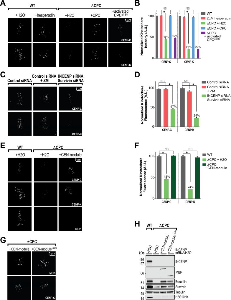
Our results thus far suggest that centromeric CPC is required for the proper response to a loss of tension at kinetochore-microtubule attachments. Proteins at the inner kinetochore not only provide a platform for outer kinetochore assembly, but have also been implicated in the response to tension (Ribeiro et al., 2010; Suzuki et al., 2014; Vargiu et al., 2017). Therefore, we hypothesized that the localization of the CPC to centromeres may play a role in the localization of inner kinetochore components (Figure 1B). Indeed, kinetochores assembled in activated CPCΔCEN extracts had reductions in certain inner kinetochore proteins at metaphase. CENP-K levels (part of the CENP-H-I-K-M complex) were decreased by 78% and CENP-C levels were decreased by 54% (Wynne and Funabiki, 2015) compared to control at centromeres of replicated sister chromatids at metaphase (Figures 5A and 5B and Figure S1A).
Figure 5. Centromeric CPC Mediates the Complete Loading of Inner Kinetochore Proteins in a Kinase-Independent Manner.
(A) Representative immunofluorescence (IF) images of replicated chromosomes in metaphase spindles in WT and ΔCPC extracts with indicated CPC conditions +/− 2 µM hesperadin. Spindles were stained for indicated kinetochore components (white). See also Figure S1A.
(B) Mean integrated fluorescence intensities of kinetochore components shown in (A) were quantified and normalized to WT. n = 96 kinetochores per condition.
(C) Representative IF images of metaphase kinetochores in human Hela cells treated with control siRNAs, control siRNA with ZM447439 (Aurora B kinase inhibitor) or INCENP + Survivin siRNAs. Samples were stained for kinetochore components (white). See also Figure S6A–D.
(D) Mean integrated fluorescence intensities of kinetochore components shown in (C) were quantified and normalized to WT. n = 96 kinetochores.
(E) Representative IF images of metaphase spindles in WT, ΔCPC, or CEN-module extracts. Spindles were stained for kinetochore components (white).
(F) Mean integrated fluorescence intensities of inner kinetochore components shown in (E) were quantified and normalized to WT. n = 96 kinetochores.
(G) Representative IF images of metaphase spindles in CEN-module and CEN-moduleΔHP1 extracts. Spindles were stained for MBP to show localization of both CEN-modules and stained for CENP-C to show kinetochore assembly (white).
(H) Western blot for the CPC components, MBP, Histone H3 phosphorylation (H3S10ph) and tubulin for samples shown in (G).
To determine whether Aurora B kinase activity or some other activity of the CPC is required for proper inner kinetochore assembly, we analyzed inner kinetochore assembly in both CPC depleted extracts and in extracts treated with the Aurora B kinase inhibitor hesperadin. We found that CPC depletion, but not Aurora B kinase inhibition with 2 µM hesperadin (Emanuele et al., 2008; Hauf et al., 2003), led to reductions in CENP-C and CENP-K kinetochore localization at metaphase (Figures 5A and 5B, and Figure S1A). These effects were specific, since defects in inner kinetochore composition in depleted extracts could be rescued by the addition of the CPC (Figures 5A and 5B and Figures S1A). These results suggest that centromeric CPC controls the assembly of the inner kinetochore in mitosis independently of Aurora B kinase activity.
To understand if control of the inner kinetochore by the CPC is conserved, we examined the localization of multiple inner kinetochore proteins in human cells arrested at metaphase. In HeLa cells, levels of the CENP-H-I-K-M complex subunits CENP-K and CENP-I were reduced by 77% and 52%, respectively, upon siRNA-meditated knockdown of the CPC but not upon Aurora B kinase inhibition (Figures 5C and 5D and Figures S6A, S6B, S6C and S6D). Similarly, CENP-C was reduced by 53% upon CPC knockdown but was not affected by Aurora B kinase inhibition. The level of CENP-A at centromeres was not changed by CPC knockdown in human cells, indicating that the observed inner kinetochore assembly defects were not due to loss of centromere identity (Figures S6A and S6B). Recent work in human cells has demonstrated that CENP-C is required for the localization of all inner kinetochore proteins in mitosis (Klare et al., 2015; McKinley et al., 2015; Nagpal et al., 2015). Indeed, the inner kinetochore protein CENP-T was reduced upon CPC knockdown by 76%, suggesting that control of inner kinetochore assembly by the CPC is upstream of CENP-C loading in mitosis (Figure S6C). These results demonstrate an evolutionarily conserved role for the CPC in inner kinetochore assembly in mitosis that is independent of Aurora B kinase activity.
The CEN-Module of the CPC Mediates Assembly of the Inner Kinetochore
To further understand the role of the CPC in inner kinetochore assembly, we tested which parts of the CPC were required to rescue the inner kinetochore defects observed upon CPC depletion from extracts. We found that Aurora B, and the SAH domain of INCENP were not required for rescue of inner kinetochore composition (Figure S6E). Strikingly, addition of only the centromere-binding module of the CPC (CEN-module, Figure 1A), in the absence of Aurora B and the remainder of INCENP, restored normal levels of the inner kinetochore components CENP-K and CENP-C at metaphase chromosomes (Figures 5E and 5F). The CEN-module localized to centromeres in extracts depleted of the CPC (Figure 5G). Accordingly, in these extracts, outer kinetochores were not formed, as determined by the absence of Dsn1 at kinetochores (Figure 5E). The rescue of inner kinetochore composition by the CEN-module was not due to HP1 binding to INCENP (Abe et al., 2016), since deletion of its interaction site did not affect CENP-C levels at kinetochores (Figures 5G and 5H). Therefore, the interaction of the CPC with centromeric chromatin is required to build a complete inner kinetochore in mitosis, independently of outer kinetochore assembly. In addition, we find that a full complement of inner kinetochore proteins is not necessary to build an outer kinetochore (Figure 1C), in agreement with previous work that demonstrates only 30% of CENP-C molecules engage outer kinetochore proteins (Suzuki et al., 2015).
A Fully Assembled Inner Kinetochore is Required for BubR1 Enrichment at Tensionless Attachments
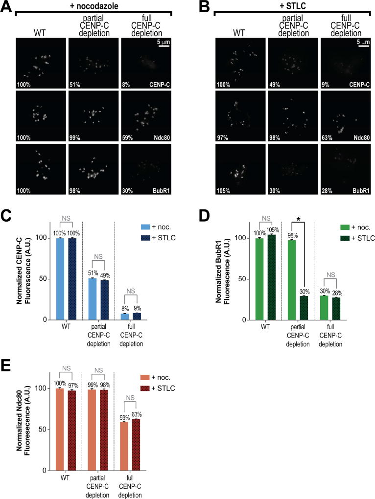
We next sought to determine if the tension-detection defect we observed in activated CPCΔCEN extracts (Figures 4A, 4B, 4C, 4D and 4E) could be due to defects in inner kinetochore composition. To do this, we sought to manipulate levels of CENP-C at kinetochores to match those observed in activated CPCΔCEN extracts (~50% of control) and measure levels of BubR1 at kinetochores in the presence of either nocodazole or STLC. Strikingly, in extracts partially depleted of CENP-C, CENP-C levels were ~50% at kinetochores (Figures 6A, 6B and 6C), and BubR1 levels remained high in nocodazole but decreased dramatically upon treatment with STLC, compared to controls (Figures 6A, 6B and 6D). Full depletion of CENP-C, which caused a significant loss of outer kinetochore Ndc80 (Figures 6A, 6B and 6E) (Krizaic et al., 2015; Milks et al., 2009), resulted in a decrease in BubR1 in both conditions (Figure 6D). We conclude that complete assembly of CENP-C at kinetochores is specifically required for the enrichment of BubR1 at tensionless attachments.
Figure 6. A Full Inner Kinetochore is Required for BubR1 Enrichment at Tensionless Attachments.
(A) Representative IF images of metaphase spindles assembled in the presence of either WT, partially CENP-C depleted, or fully CENP-C depleted extracts treated with nocodazole at metaphase. Spindles were stained for kinetochore components (white). Also indicated is the mean integrated fluorescence intensity observed at kinetochores, normalized to the WT + nocodazole condition.
(B) Representative IF images of metaphase spindles assembled in the presence of either WT, partially CENP-C depleted, or fully CENP-C depleted extracts treated with STLC at metaphase. Spindles were stained for kinetochore components (white). Also indicated is the mean integrated fluorescence intensity observed at kinetochores, normalized to the WT + nocodazole condition in (A).
(C) Mean integrated fluorescence intensities of CENP-C at kinetochores assembled in extracts as described in (A) and (B). Note that the partial depletion of CENP-C condition closely matches levels of CENP-C at kinetochores observed in CPC depleted extracts. Values are normalized to the WT CENP-C + nocodazole condition in (A). n = 104 kinetochores per condition.
(D) Mean integrated fluorescence intensities of BubR1 at kinetochores assembled in extracts as described in (A) and (B). Values are normalized to the WT + nocodazole condition in (A). n = 104 kinetochores per condition.
(E) Mean integrated fluorescence intensities of Ndc80 at kinetochores assembled in extracts as described in (A) and (B). Values are normalized to the WT + nocodazole condition in (A). n = 104 kinetochores per condition.
The Proper Response to Erroneous Attachments Requires Complete Inner Kinetochore Assembly and the Localization of Aurora B to Centromeres
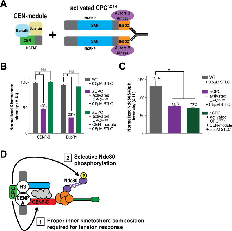
To examine whether rescuing defects inner kinetochore composition in activated CPCΔCEN extracts could also rescue tension-dependent BubR1 enrichment, we added both the CEN-module and activated CPCΔCEN in trans to CPC-depleted extracts (Figure 7A and Figure S7A). Indeed, in spindles treated with STLC, the addition of both CPC modules fully rescued the loss of BubR1 enrichment and CENP-C assembly observed in activated CPCΔCEN spindles alone (Figure 7B and Figure S7B). This demonstrates that that error sensing through the BubR1 pathway requires the CEN-module, but does not require localized Aurora B activity. We next examined whether the CEN-module could also rescue defects in Aurora B-mediated Ndc80 phosphorylation at tensionless, attached kinetochores in activated CPCΔCEN extracts. In activated CPCΔCEN extracts treated with STLC, the addition of both modules did not rescue Ndc80 phosphorylation to control levels (Figure 7C and Figure S7C). This suggests that Aurora B must be properly localized to specifically remove and correct erroneously attached microtubules, even when inner kinetochore composition and BubR1 enrichment are rescued. Together, these results indicate that an intact CPC is required at centromeres to detect and respond to errors in attachment, and suggest that local interplay between the error sensing and removal pathways controlled by the CPC is required for error correction.
Figure 7. The Proper Response to Erroneous Attachments Requires Complete Inner Kinetochore Assembly and the Localization of Aurora B to Centromeres.
(A) Schematic of experimental setup. Activating antibody was added at metaphase.
(B) Mean integrated fluorescence intensities of CENP-C and BubR1 in WT, activated CPCΔCEN, and activated CPCΔCEN + CEN-module metaphase extracts treated with STLC. Samples were treated with STLC to generate tension-less but microtubule attached kinetochores. BubR1 values are normalized to the WT + nocodazole treated condition in Figure S5C and CENP-C values are normalized to the WT condition in Figure 5A. n = 96 kinetochores per condition. See also Figures S7A and B.
(C) Mean integrated fluorescence intensity of Ndc80S40ph in WT, activated CPCΔCEN, and activated CPCΔCEN + CEN-module metaphase extracts treated with STLC. Values are normalized to the WT + nocodazole treated condition in Figure 2D. n = 96 kinetochores per condition. See also Figures S7A and C.
(D) Model for the roles of centromeric CPC in the detection and correction of attachment errors.
Discussion
Error Detection via the Inner Kinetochore
Faithful chromosome segregation requires the tension-dependent detection of errors in kinetochore-microtubule attachment. Here, we show that BubR1 enrichment and phosphorylation at kinetochores that are attached, but have lost tension (as indicated by inter-kinetochore distances), depends on a kinase-independent role of the CPC at centromeres in the formation of the inner kinetochore. However, we also show that BubR1 recruitment to unattached kinetochores does not depend on a fully formed inner kinetochore (Figure S5C). A likely explanation for this separation of function is that a fully formed inner kinetochore is required to prevent “hyper-stretching” within kinetochores by microtubule pulling forces (a defect that is not reflected in inter-kinetochore distances) (Ribeiro et al., 2010; Suzuki et al., 2014; Vargiu et al., 2017; Suzuki et al., 2011). We propose that kinetochores built in the presence of non-centromeric CPC are “hyper-stretched” and thus there is no differentiation between high and low tension states. At the molecular level, we show that BubR1 recruitment to both unattached kinetochores and attached, tensionless kinetochores depend on the kinase Mps1, which phosphorylates BubR1-docking protein KNL1 (Figure 4F) (Krenn et al., 2014; London et al., 2012; Overlack et al., 2015; Shepperd et al., 2012; Yamagishi et al., 2012). We also show that non-centromeric CPC does not affect Mps1 localization (Figure S5E) (Hiruma et al., 2015; Ji et al., 2015), which suggests that defects in BubR1 recruitment lie in control of Mps1-substrate phosphorylation. In the unattached state, Mps1 likely has full access to KNL1, regardless of the inner kinetochore state, since no microtubules are present to cause “hyper-stretching” (Aravamudhan et al., 2015; Suzuki et al., 2014). When kinetochores are attached, Mps1 may be prevented from phosphorylating KNL1 if the inner kinetochore is defective, since it has been shown that Mps1 must be in close proximity to KNL1 (Aravamudhan et al., 2015). Thus, we propose that the CPC must reside at centromeres to build a kinetochore that can withstand and differentiate tension forces in early mitosis. At anaphase, the CPC is evicted from centromeres and relocalizes to the spindle midzone (Carmena et al., 2012). An enticing possibility is that inner kinetochore assembly is a dynamic process, and that CPC relocalization blocks the tension-responsiveness of kinetochores to prevent untimely activation of error correction (Vázquez-Novelle et al., 2010).
Recent work has demonstrated that non-centromeric CPC results in weakened centromeric cohesion in human cells (Hengeveld et al., 2017), in accordance with previous reports that the CPC regulates centromeric cohesion (Marston, 2015). Interestingly, studies in budding yeast have shown that the inner kinetochore enriches centromeric cohesion by recruiting the cohesin loader complex (Fernius et al., 2013). However, multiple lines of evidence suggest that control of kinetochore composition, and not cohesion, by centromeric CPC underlies the tension-dependent enrichment of BubR1 and the 3F3/2 epitope: 1) inter-kinetochore distances are not altered in our non-centromeric CPC condition (Figure 2B), which suggests that centromeric cohesion is not drastically reduced (Rivera et al., 2012), and 2) previous reports have indicated that intra-kinetochore deformations, and not defects in cohesion between sister-kinetochores, affect tension-dependent 3F3/2 enrichment at kinetochores (Maresca and Salmon, 2009). Thus, although the CPC can regulate centromeric cohesion, which certainly contributes to bi-orientation, our results and previous findings suggest a more direct role for the CPC-dependent control of the inner kinetochore in detecting tension.
Defects in the CPC-dependent assembly of the inner kinetochore likely have deleterious effects on other processes at the kinetochore as well (Cheeseman, 2014). For example, the CENP-H-I-K-M complex, which is strongly diminished at kinetochores in the non-centromeric CPC mutant, is required to regulate microtubule dynamics (Amaro et al., 2010) as well as the checkpoint response by regulating Mad1 and Mad2 turnover (Matson and Stukenberg, 2014). Although we demonstrate Mad2 and BubR1 recruitment to unattached kinetochores does not require centromeric CPC, the halting of the cell cycle does (J.H. and M.K.B., unpublished observations), suggesting that centromeric CPC ultimately regulates both Mad2 recruitment and dynamics. Additionally, BubR1 recruits the phosphatase PP2A to kinetochores that have yet to establish tension to oppose Aurora B and to stabilize nascent attachments (Elowe et al., 2007; Foley et al., 2011; Kruse et al., 2013; Suijkerbuijk et al., 2012; Xu et al., 2013). Our results here suggest that centromeric CPC (and an intact inner kinetochore) is required for this important process, and that the CPC coordinates not just the removal but also the replacement of microtubules during error correction. Understanding how the CPC interaction with centromeric chromatin regulates inner kinetochore assembly and maintenance should be a focus of future efforts.
Regulation of Kinetochore-Microtubule Attachment by the CPC
We demonstrate that the localization of CPC to centromeres is required to properly phosphorylate Ndc80 in response to tension and attachment state. Non-centromeric CPC results in elevated yet “flat” levels of Ndc80 phosphorylation, regardless of tension status (Figure 2D and Figure 3G). These results support, but do not validate, the “spatial separation” model for tension-sensing by Aurora B, wherein tension regulates the distance between centromeric Aurora B and substrates at the kinetochore such as Ndc80 (D. Liu et al., 2009; Tanaka et al., 2002; Welburn et al., 2010). Although the in trans rescue of inner kinetochore assembly does not rescue proper Ndc80 phospho-response by CPCΔCEN (Figure 7C), “hyper-stretching” of kinetochores after inner kinetochore perturbation has been shown to dampen Ndc80 phosphorylation (Suzuki et al., 2014). Thus, the CPC may require centromeric localization to regulate substrate access as well as to titrate phosphorylation levels.
The spatial separation model has been challenged by experiments demonstrating that bi-orientation can be achieved in budding yeast when the N-terminus of INCENP homolog Sli15 is deleted (Campbell and Desai, 2013). This led to a model, in which tension controls the exposure of Aurora B kinetochore substrates for error correction, but does not require centromeric Aurora B activity. Our results here do not support a model where non-centromeric Aurora B can achieve selective error correction. However, when spindle assembly is unperturbed, we demonstrate that non-centromeric Aurora B kinase does not cause major defects in chromosome alignment (Figure 2C), most likely due to non-selective turnover of kinetochore attachments by intermediate levels of Ndc80 phosphorylation that could prevent errors in attachment (Figure 2D) (Tanaka et al., 2002). Yet, when a large number of errors are introduced (by STLC), selective removal becomes essential for the correction of erroneous attachments (Figure 3C). This suggests that initial bias towards bi-orientation (Indjeian and Murray, 2007) combined with increased microtubule dynamics can overcome a need for tension-dependent phospho-regulation of attachment (Bakhoum et al., 2009; Tanaka et al., 2002). We propose that in budding yeast, where only one microtubule interacts with each kinetochore instead of 20–25 microtubules per kinetochore in higher eukaryotes (Haase et al., 2012), intermediate, non-specific Ndc80 phosphorylation could allow for bi-orientation. Recent work in budding yeast proposed that the interaction of the CPC with microtubules, through the SAH domain of INCENP, allows for bi-orientation independently of centromere localization (Fink et al., 2017). However, although CPCΔCEN does contain the SAH domain, we find that it is not sufficient for selective error correction (Figures 3C, 3D, 3E, 3F, and 3G). Furthermore, we show that Ndc80 phosphorylation at kinetochores is not altered upon microtubule depolymerization when the CPC is non-centromeric (Figure 2D). Thus, though we cannot rule out a direct role for microtubule binding by the CPC in error correction, it is not sufficient to properly regulate kinetochore-microtubule attachment removal in egg extracts. The inner kinetochore dependent control of tension-detection may be conserved in yeast, since CPC mutants are synthetically lethal with inner kinetochore mutants (Campbell and Desai, 2013; Knockleby and Vogel, 2009). Future experiments are required to determine whether the CPC controls inner kinetochore assembly in yeast.
Our work reveals that the CPC controls assembly of the inner and outer kinetochore in mitosis (Figure 7D). We show that non-centromeric CPC cannot specifically correct errors in attachment, and we propose that this is caused by defects in Ndc80 phosphorylation and BubR1 enrichment. Our discovery of a kinase-independent role of the CPC in inner kinetochore assembly and its role in tension-responsiveness is a significant advance, since all prior models of CPC function in error correction focused mainly on the control of phosphorylation by Aurora B (Krenn and Musacchio, 2015). Furthermore, our results may explain why cancer cells are more susceptible to CPC knockdown than to Aurora B inhibition (Ditchfield et al., 2003) and why artificial targeting of CEN-deleted INCENP to centromeres does not rescue CPC function (E. Wang et al., 2011; Wheelock et al., 2017). By ultimately controlling kinetochore assembly, error detection and correction, the CPC is a master regulator of kinetochore function.
STAR METHODS
CONTACT FOR REAGENT AND RESOURCE SHARING
Please contact the corresponding author, Alexander Kelly (alexander.kelly_at_nih.gov), for reagents or further information.
EXPERIMENTAL MODEL AND SUBJECT DETAILS
Xenopus laevis
Mature Xenopus laevis were obtained from NASCO and housed in water tanks in a climate controlled environment constructed by Aquarius Aquariums. Water temperature was maintained at 16°C, and water quality (nitrites-undetectable levels, chlorine-undetectable levels, conductivity: 1200–1800 µS/cm and pH: 7.2–7.8) was tested daily. Light/dark cycles consisting of 12h/12h were followed. Frogs were fed NASCO frog brittle twice weekly.
To obtain eggs, female frogs were primed for ovulation by two injections of 50 U pregnant mare serum gonadotropin (EMD Millipore) two days apart. An injection of 500 U human chorionic gonadotropin (Prospec) followed within 21 days to induce egg laying.
To obtain sperm, mature male frogs were obtained from NASCO and housed separately from female frogs. Male frogs were euthanized with tricaine methanesulfonate (MS-222, Sigma), and testes were dissected to collect sperm.
METHOD DETAILS
Immunodepletion of Xenopus egg extracts
Xenopus laevis egg extract was prepared as previously described (Kelly et al., 2007). To prevent translation of endogenous RNAs, RNase A (Ambion) was added to a final concentration of 0.01 mg/ml to extract for 15 minutes at 12°C. RNase inhibitor (RNasin Plus RNase Inhibitor, Promega) was added at a 1:100 dilution for 5 minutes at 12°C. Yeast tRNAs (50 µg/mL) were added and extract was kept on ice. Immunodepletions of the CPC from the extract were performed as described (Kelly et al., 2007, Tseng et al., 2010) with the following modification. Complete depletion was achieved after two rounds of incubations of anti-INCENP-Protein A beads (100 µl) with RNase treated extract (100 µl) on ice for 45 minutes. Immunodepletion of CENP-C from the extract was performed using serum from rabbits raised against xCENP-C207–296 fused to GST. Depletion was performed as described above, using 10 µg serum bound to 100 µl beads for 100 µl extract.
Reconstitution of Immunodepleted Extracts
To express the CPC in immunodepleted extract, we pooled CPC mRNAs (consisting of INCENP, Aurora B, Borealin (Dasra A) and Survivin, unless otherwise indicated), and added them to extracts at the onset of interphase, using previously described methods (Kelly et al., 2007). Briefly, the CPC members were in vitro transcribed by using the mMessage Machine SP6 kit (Thermo Fisher Scientific), pooled together and then precipitated in the presence of 20 µg of glycogen. The capped mRNA pools were then resuspended in RNase-free water so that 1 µl of pooled mRNAs reconstituted 60 µl of CPC-depleted extract. To achieve partial levels of CENP-C in extract, control extract was mixed with CENP-C immunodepleted extract so that total levels of CENP-C were approximately 25% of control egg extracts (which results in 50% levels of CENP-C at kinetochores). Extract was used for spindle assembly assays as stated below.
Spindle assembly in Xenopus egg extracts
To assess kinetochore assembly and checkpoint response, sperm chromatin was replicated in extracts, and spindles were assembled according to previously published methods (Kelly et al., 2007). Briefly, 1 µl of pooled mRNAs was added to 20 µl of RNase treated control or immunodepleted extract with sperm nuclei (final concentration 500/ µl) and calcium chloride (0.3 mM). Rhodamine tubulin (Cytoskeleton, Inc.) was added at a 1:200 dilution to observe the progress of spindle assembly. Extract reactions were incubated at 20°C for 80 minutes to cycle into interphase. To drive the extract into metaphase, 40 µl of CSF extract (control or immunodepleted) was added. At the onset of metaphase, nocodazole was added as needed to a final concentration of 10 µg/ml. After 45 minutes at 20°C, metaphase spindle assembly was assayed by fixing 1 µl samples with Hoechst (10 µg/ml) (Hoechst 33258, Invitrogen) and imaging tubulin and DNA. Samples for Western blot and immunofluorescence were taken once metaphase was successfully achieved.
Error correction assay in Xenopus egg extracts
To analyze the response to defective kinetochore-microtubule attachments, an established assay for error correction in human cells was modified for extracts. The addition of an Eg5 inhibitor, S-Trityl-L-Cysteine (STLC, Sigma Aldrich), results in monopolar spindles with kinetochores in a tension-less, mono-oriented (syntelic) configuration (Houghtaling et al., 2009). Subsequent removal of Eg5 inhibition allows spindles to bipolarize and chromosomes to align at the metaphase plate through removal of improper kinetochore-microtubules by Aurora B kinase (Lampson et al., 2004). A range of concentrations of STLC was tested in extracts, and the ratio of monopolar and bipolar spindles generated were recorded in order to determine the IC50 (0.2 µM). In the modified assay for extracts, STLC was added to a final concentration of 0.5 µM at the beginning of metaphase to induce the majority of spindle structures into a monopolar state. After monopolar spindles were generated in metaphase (around 40 minutes), experiments demonstrating error correction could be performed by diluting the STLC-treated extract with ten-fold untreated CSF extract and letting the monopolar spindles re-bipolarize. At the final concentration of 0.05 µM, no monopolar spindles were observed. Samples for Western blot and immunofluorescence were taken 30 minutes after STLC was diluted.
Antibody/bead activation of Aurora B kinase
To globally activate Aurora B in experiments using INCENPΔCEN, 1 µl of anti-INCENP antibody (0.015 mg/mL) was added to 40 µl of CSF extract, for a final concentration of 0.25 ng/µl. This sub-stoichiometric amount of antibody compared to CPC in extract is sufficient to catalyze the activation of Aurora B. After incubation on ice for 20 minutes, this was added to 20 µl of experimental interphase extract to induce cycling of extract into metaphase. For bead-based activation of Aurora B using CPCΔCEN, anti-INCENP antibody was bound to protein-A Dynabeads (Invitrogen) at a concentration of 10 µg/100 µl of beads and crosslinked as above. 3 µl of antibody bound beads were added to 30ul of extract upon exit from interphase for a final antibody concentration of 10 ng/µl.
MBP-INC1-242 protein purification
The MBP-INC1-242 (CEN-module) construct was cloned into the protein expression plasmid, pET28a, and transformed into BL21(DE3) cells. Cells were induced with 1 mM IPTG for 3 hours at 37°C, and the tagged protein was purified using Ni-NTA agarose (Qiagen). The purified protein underwent buffer exchange in a PD-10 column (GE) into 150 mM sucrose, 150 mM NaCl, 20 mM HEPES, pH 7.7, 1 mM DTT. For spindle assembly experiments in extract, MBP-INC1-242 was added at the beginning of interphase at 100 nM. mRNAs for all other CPC members, including INCENPΔCEN and MBP-INC1-71 (CEN-moduleΔHP1), were also added at the beginning of interphase. For experiments using both MBP-INC1-242 and INCENPΔCEN, Aurora B was activated globally by adding anti-INCENP antibody at the onset of metaphase.
Western Blots
Primary antibodies were diluted in Licor blocking solution/PBST with a final Tween-20 concentration of 0.1% except for anti-phospho Aurora and anti-Histone3 (Ser10p), which had no Tween-20. The following antibodies and antibody dilutions were used: anti-INCENP (raised against C-terminal peptide CSNRHHLAVGYGLKY) (5.5 µg/ml), anti-Aurora B (Kelly et al., 2007) (5 µg/ml), anti-Borealin A (Dasra A) (Kelly et al., 2007) (5 µg/ml), anti-Survivin (Tseng et al., 2010) (12 µg/ml), anti-phospho Aurora (Phospho-Aurora A (Thr288)/Aurora B (Thr 232)/Aurora C (Thr198) 2914, Cell Signaling Technology) (1:200), anti-Histone3 (Ser10p) (6G3, 9706, Cell Signaling Technology) (1:500), anti-Histone3 (T3p) Phospho (Epitomics 2162-1) (1:10000), anti-MBP (New England Biolabs E8032) (1:10000), anti-alpha-tubulin (DM1, Sigma) (1:20000), Incenp (39259, Active Motif) (1:500), AIM-1 (611082, BD Transduction Laboratories) (1:500). Secondary antibodies from Licor were used (Licor goat anti-Rabbit 800 nm and Licor goat anti-mouse 680 nm) as was the Licor imaging system to scan membranes.
Microscopy and Fluorescence Quantification
To immunostain kinetochores, Xenopus egg extract was fixed for 5 min by ~20-fold dilution in BRB80 + 20% glycerol + 0.5% Triton X-100 + 3.7% formaldehyde at room temperature. Fixed reactions were layered onto a cushion of BRB80 + 40% glycerol overlaying a poly-L-lysine-coated coverslips (No. 1) placed in a 24 well plate. Nuclei were adhered onto coverslips on plate holders at 4,000 rpm for 15 min at 18°C in a centrifuge (Eppendorf 5810R). Cushions were washed with BRB80 and coverslips were postfixed in ice-cold methanol for 5 min, blocked with TBS + 0.1% Tween20 + 5% BSA over night at 4°C, incubated in primary at room temperature for 1.5 hours unless otherwise noted. All washes and antibody dilutions were done with AbDil buffer. Nuclei were stained with Hoechst 33258 prior to mounting in 80% glycerol + PBS medium. The following antibodies were used at the indicated dilutions: Dsn1 (Emanuele et al., 2008) 1:500, p-Aurora (Cell Signaling 2914S) 1:500, Knl1 (Emanuele et al., 2008) 1:200, Ndc80 (McCleland et al., 2003) 1:200, Mad2 (Chen et al., 1996) 1:200, BubR1 (Boyarchuk et al., 2007) 1:100, Borealin (Dasra A) 1:250 (Kelly et al., 2007), 3F3/2 (Gorbsky et al., 1993) 1:2500, Cenp-K (Milks et al., 2009) 1:250, Cenp-C 1:500, MBP (New England Biolabs E8032S) 1:1000, INCENP (39259, Active Motif) 1:500, Ndc80S44ph (DeLuca et al., 2011) 1:2000 and Mps1 (Zhao and Chen, 2006) 1:100.
For HeLa cells, coverslips were fixed for 10 min in chilled 100% methanol at −20°C. Dsn1 was fixed in 3% PFA in PHEM buffer + 0.05% Triton-X. Coverslips are washed in 1 X PHEM + 0.05% Triton X three times. Coverslips are incubated in 1 X PHEM + 0.5% Triton X coverslips for 10 min at room temperature. Coverslips were blocked in 2.5% BSA + 1% Boiled Donkey Serum in 1 X PHEM + 0.01% Triton-X. All washes were done in 1 X PHEM + 0.05% Triton X. The antibodies used are as follows: CENP-T (D286-3, MBL) (1:2000), CENP-C (PD030, MBL) (1:2000), CENP-A (ab13939, AbCam) (1:400), CENP-K (PD018, MBL) (1:500), CENP-I (PD032, MBL) (1:500), INCENP (39259, Active Motif) (1:1000). Secondary antibodies (goat anti-mouse cy3, donkey anti-guinea pig 647, donkey anti-rat 488, donkey anti-rabbit 488, goat-anti mouse 488) were from Jackson Immuno and used at the 1:400 in blocking buffer for 1 hr at room temperature. Cells were counterstained in Hoechst 33258 prior to mounting in Vectashield Mounting Media (H-1000, Vector Labs).
All immunofluorescence was imaged with 0.2 µm step size using an Eclipse Ti (Nikon) comprised of a Nikon Plan Apo x100/1.45, oil immersion objective, a PlanApo x40/0.95 objective, and a Hamamatsu Orca-Flash 4.0 camera. Images were captured and processed using NIS Elements AR 4.20.02 software (Nikon), and analyzed in Fiji ImageJ. The acquired Z-sections of 0.2 µm each were converted to a maximum projection using NIS Elements and Fiji. Distances between foci were measured as the distance between maximal points of fluorescence as determined by linescans. Signal intensity was measured as previously reported (Hoffman et al., 2001) using Fiji. Kinetochore intensity was measured using Fiji by centering 9 × 9 and 13 × 13 pixel regions over individual kinetochores. Total fluorescence intensity was recorded from each region. To correct for background fluorescence, the difference in intensities between the two regions was determined, and then made proportionate to the smaller region. This background value was then subtracted from the smaller region to determine kinetochore intensity with background correction as previously reported (Hoffman et al., 2001). Kinetochore alignment was determined by dividing metaphase spindles longitudinally into three equally sized zones. Kinetochore pairs, detected through immunofluorescence, were categorized as being misaligned if they were found to be outside of the central zone.
Cell culture
HeLa cells were plated in 24 well plates and maintained with DMEM (Life Technologies) supplemented with 10% FBS (Life Technologies). For immunofluorescence, cells were plated on poly-L-lysine (Sigma) coated coverslips at ~10,000 cells per well and arrested with a final concentration of 2 mM thymidine (Sigma) for 15–18 hrs at 60% confluency. Cells were released for 1–2 hrs, switched into OptiMEM media (Life Technologies) and transfected using RNAiMax (Life Technologies) according to manufacturer’s protocols with siRNA INCENP (Thermo Scientific s7423) GCUUGUACCUCAUAUCAGAtt and siRNA BIRC5 (Thermo Scientific s1458) GCAGGUUCCUUAUCUGUCAtt or non-targeting control siRNA (GE Dharmacon D0012100105) at 45 nM per siRNA (90 nM for scramble control). 2 µM final concentration of ZM 447439 (Tocris) was added at the time of transfection. After 12 hrs, MG132 (20 µM, Sigma) was added for 3 hrs. Cells were harvested protein analysis or processed for immunofluorescence.
QUANTIFICATION AND STATISTICAL ANALYSIS
All experiments were repeated at least twice and a minimum number of either 75 spindles or 96 kinetochores were analyzed for each assay. Sample size was chosen to ensure a high (>90%) theoretical statistical power in order to generate reliable P values. All graphs and statistical analysis were prepared with GraphPad Prism. Fluorescence values from experimental conditions were compared to control conditions using an ordinary one-way ANOVA with Turkey’s multiple comparison tests to determine significance. All graphs show the mean with error bars representing the s.e.m.
Supplementary Material
Highlights.
Aurora B kinase drives outer kinetochore assembly independent of spatial positioning
Error correction and selective Ndc80 phosphorylation require centromeric CPC
The CPC centromere-targeting module regulates inner kinetochore composition
Proper inner kinetochore composition is required for the detection of tension loss
Acknowledgments
We thank O. Cohen-Fix, S. Grewal, J. Cooper, M. Lichten, J. Barrowman, B. Paterson, and C. Klee for critical reading of the manuscript; and R. Chen, M. Dasso, J. DeLuca, M. Face, H. Funabiki, G. Gorbsky, A. Holland, A. Straight, P.T. Stukenberg, and A. Suzuki for kindly providing reagents and advice. This work was supported by the intramural programs of the National Cancer Institute.
Footnotes
Publisher's Disclaimer: This is a PDF file of an unedited manuscript that has been accepted for publication. As a service to our customers we are providing this early version of the manuscript. The manuscript will undergo copyediting, typesetting, and review of the resulting proof before it is published in its final citable form. Please note that during the production process errors may be discovered which could affect the content, and all legal disclaimers that apply to the journal pertain.
Author Contributions:
J.H., M.K.B., H.H., and A.K. designed the experiments. J.H., M.K.B., and H.H. performed the experiments and analyzed the data. A.K. wrote the manuscript with input from all co-authors.
The authors declare no competing financial interests.
References
- Abe Y, Sako K, Takagaki K, Hirayama Y, Uchida KSK, Herman JA, DeLuca JG, Hirota T. HP1-Assisted Aurora B Kinase Activity Prevents Chromosome Segregation Errors. Dev. Cell. 2016;36:487–497. doi: 10.1016/j.devcel.2016.02.008. [DOI] [PMC free article] [PubMed] [Google Scholar]
- Akiyoshi B, Nelson CR, Biggins S. The aurora B kinase promotes inner and outer kinetochore interactions in budding yeast. Genetics. 2013;194:785–789. doi: 10.1534/genetics.113.150839. [DOI] [PMC free article] [PubMed] [Google Scholar]
- Amaro AC, Samora CP, Holtackers R, Wang E, Kingston IJ, Alonso M, Lampson M, McAinsh AD, Meraldi P. Molecular control of kinetochore-microtubule dynamics and chromosome oscillations. Nat. Cell Biol. 2010;12:319–329. doi: 10.1038/ncb2033. [DOI] [PMC free article] [PubMed] [Google Scholar]
- Aravamudhan P, Goldfarb AA, Joglekar AP. The kinetochore encodes a mechanical switch to disrupt spindle assembly checkpoint signalling. Nat. Cell Biol. 2015;17:868–879. doi: 10.1038/ncb3179. [DOI] [PMC free article] [PubMed] [Google Scholar]
- Bakhoum SF, Thompson SL, Manning AL, Compton DA. Genome stability is ensured by temporal control of kinetochore-microtubule dynamics. Nat. Cell Biol. 2009;11:27–35. doi: 10.1038/ncb1809. [DOI] [PMC free article] [PubMed] [Google Scholar]
- Bishop JD, Schumacher JM. Phosphorylation of the carboxyl terminus of inner centromere protein (INCENP) by the Aurora B Kinase stimulates Aurora B kinase activity. J. Biol. Chem. 2002;277:27577–27580. doi: 10.1074/jbc.C200307200. [DOI] [PMC free article] [PubMed] [Google Scholar]
- Boyarchuk Y, Salic A, Dasso M, Arnaoutov A. Bub1 is essential for assembly of the functional inner centromere. 2007;176:919–928. doi: 10.1083/jcb.200609044. [DOI] [PMC free article] [PubMed] [Google Scholar]
- Caldas GV, DeLuca KF, DeLuca JG. KNL1 facilitates phosphorylation of outer kinetochore proteins by promoting Aurora B kinase activity. J. Cell Biol. 2013;203:957–969. doi: 10.1083/jcb.201306054. [DOI] [PMC free article] [PubMed] [Google Scholar]
- Campbell CS, Desai A. Tension sensing by Aurora B kinase is independent of survivin-based centromere localization. Nature. 2013;497:118–121. doi: 10.1038/nature12057. [DOI] [PMC free article] [PubMed] [Google Scholar]
- Carmena M, Wheelock M, Funabiki H, Earnshaw WC. The chromosomal passenger complex (CPC): from easy rider to the godfather of mitosis. Nat. Rev. Mol. Cell Biol. 2012;13:789–803. doi: 10.1038/nrm3474. [DOI] [PMC free article] [PubMed] [Google Scholar]
- Cheerambathur DK, Desai A. Linked in: formation and regulation of microtubule attachments during chromosome segregation. Curr. Opin. Cell Biol. 2014;26:113–122. doi: 10.1016/j.ceb.2013.12.005. [DOI] [PMC free article] [PubMed] [Google Scholar]
- Cheeseman IM. The kinetochore. Cold Spring Harb Perspect Biol. 2014;6:a015826–a015826. doi: 10.1101/cshperspect.a015826. [DOI] [PMC free article] [PubMed] [Google Scholar]
- Chen RH, Waters JC, Salmon ED, Murray AW. Association of spindle assembly checkpoint component XMAD2 with unattached kinetochores. Science. 1996;274:242–246. doi: 10.1126/science.274.5285.242. [DOI] [PubMed] [Google Scholar]
- Cyert MS, Scherson T, Kirschner MW. Monoclonal antibodies specific for thiophosphorylated proteins recognize Xenopus MPF. Dev. Biol. 1988;129:209–216. doi: 10.1016/0012-1606(88)90175-3. [DOI] [PubMed] [Google Scholar]
- DeLuca KF, Lens SMA, DeLuca JG. Temporal changes in Hec1 phosphorylation control kinetochore-microtubule attachment stability during mitosis. J. Cell. Sci. 2011;124:622–634. doi: 10.1242/jcs.072629. [DOI] [PMC free article] [PubMed] [Google Scholar]
- Dimitrova YN, Jenni S, Valverde R, Khin Y, Harrison SC. Structure of the MIND Complex Defines a Regulatory Focus for Yeast Kinetochore Assembly. Cell. 2016;167 doi: 10.1016/j.cell.2016.10.011. 1014–1027.e12. [DOI] [PMC free article] [PubMed] [Google Scholar]
- Ditchfield C, Johnson VL, Tighe A, Ellston R, Haworth C, Johnson T, Mortlock A, Keen N, Taylor SS. Aurora B couples chromosome alignment with anaphase by targeting BubR1, Mad2, and Cenp-E to kinetochores. 2003;161:267–280. doi: 10.1083/jcb.200208091. [DOI] [PMC free article] [PubMed] [Google Scholar]
- Elowe S. Bub1 and BubR1: at the interface between chromosome attachment and the spindle checkpoint. Mol. Cell. Biol. 2011;31:3085–3093. doi: 10.1128/MCB.05326-11. [DOI] [PMC free article] [PubMed] [Google Scholar]
- Elowe S, Hümmer S, Uldschmid A, Li X, Nigg EA. Tension-sensitive Plk1 phosphorylation on BubR1 regulates the stability of kinetochore microtubule interactions. Genes Dev. 2007;21:2205–2219. doi: 10.1101/gad.436007. [DOI] [PMC free article] [PubMed] [Google Scholar]
- Emanuele MJ, Lan W, Jwa M, Miller SA, Chan CSM, Stukenberg PT. Aurora B kinase and protein phosphatase 1 have opposing roles in modulating kinetochore assembly. 2008;181:241–254. doi: 10.1083/jcb.200710019. [DOI] [PMC free article] [PubMed] [Google Scholar]
- Emanuele MJ, McCleland ML, Satinover DL, Stukenberg PT. Measuring the stoichiometry and physical interactions between components elucidates the architecture of the vertebrate kinetochore. Mol. Biol. Cell. 2005;16:4882–4892. doi: 10.1091/mbc.E05-03-0239. [DOI] [PMC free article] [PubMed] [Google Scholar]
- Fernius J, Nerusheva OO, Galander S, Alves F, de L, Rappsilber J, Marston AL. Cohesin-dependent association of scc2/4 with the centromere initiates pericentromeric cohesion establishment. Curr. Biol. 2013;23:599–606. doi: 10.1016/j.cub.2013.02.022. [DOI] [PMC free article] [PubMed] [Google Scholar]
- Fink S, Turnbull K, Desai A, Campbell CS. An engineered minimal chromosomal passenger complex reveals a role for INCENP/Sli15 spindle association in chromosome biorientation. J. Cell Biol. 2017;123 doi: 10.1083/jcb.201609123. jcb.201609123. [DOI] [PMC free article] [PubMed] [Google Scholar]
- Foley EA, Maldonado M, Kapoor TM. Formation of stable attachments between kinetochores and microtubules depends on the B56-PP2A phosphatase. Nat. Cell Biol. 2011;13:1265–1271. doi: 10.1038/ncb2327. [DOI] [PMC free article] [PubMed] [Google Scholar]
- Gassmann R, Holland AJ, Varma D, Wan X, Civril F, Cleveland DW, Oegema K, Salmon ED, Desai A. Removal of Spindly from microtubule-attached kinetochores controls spindle checkpoint silencing in human cells. Genes Dev. 2010;24:957–971. doi: 10.1101/gad.1886810. [DOI] [PMC free article] [PubMed] [Google Scholar]
- Gorbsky GJ, Ricketts WA. Differential expression of a phosphoepitope at the kinetochores of moving chromosomes. 1993;122:1311–1321. doi: 10.1083/jcb.122.6.1311. [DOI] [PMC free article] [PubMed] [Google Scholar]
- Gordon DJ, Resio B, Pellman D. Causes and consequences of aneuploidy in cancer. Nat. Rev. Genet. 2012;13:189–203. doi: 10.1038/nrg3123. [DOI] [PubMed] [Google Scholar]
- Haase J, Stephens A, Verdaasdonk J, Yeh E, Bloom K. Bub1 kinase and Sgo1 modulate pericentric chromatin in response to altered microtubule dynamics. Curr. Biol. 2012;22:471–481. doi: 10.1016/j.cub.2012.02.006. [DOI] [PMC free article] [PubMed] [Google Scholar]
- Hauf S, Cole RW, LaTerra S, Zimmer C, Schnapp G, Walter R, Heckel A, van Meel J, Rieder CL, Peters J-M. The small molecule Hesperadin reveals a role for Aurora B in correcting kinetochore-microtubule attachment and in maintaining the spindle assembly checkpoint. J. Cell Biol. 2003;161:281–294. doi: 10.1083/jcb.200208092. [DOI] [PMC free article] [PubMed] [Google Scholar]
- Hengeveld RCC, Vromans MJM, Vleugel M, Hadders MA, Lens SMA. Inner centromere localization of the CPC maintains centromere cohesion and allows mitotic checkpoint silencing. Nat Commun. 2017;8:15542. doi: 10.1038/ncomms15542. [DOI] [PMC free article] [PubMed] [Google Scholar]
- Hewitt L, Tighe A, Santaguida S, White AM, Jones CD, Musacchio A, Green S, Taylor SS. Sustained Mps1 activity is required in mitosis to recruit O-Mad2 to the Mad1-C-Mad2 core complex. J. Cell Biol. 2010;190:25–34. doi: 10.1083/jcb.201002133. [DOI] [PMC free article] [PubMed] [Google Scholar]
- Hiruma Y, Sacristan C, Pachis ST, Adamopoulos A, Kuijt T, Ubbink M, Castelmur von E, Perrakis A, Kops GJPL. CELL DIVISION CYCLE. Competition between MPS1 and microtubules at kinetochores regulates spindle checkpoint signaling. Science. 2015;348:1264–1267. doi: 10.1126/science.aaa4055. [DOI] [PubMed] [Google Scholar]
- Hoffman DB, Pearson CG, Yen TJ, Howell BJ, Salmon ED. Microtubule-dependent changes in assembly of microtubule motor proteins and mitotic spindle checkpoint proteins at PtK1 kinetochores. Mol. Biol. Cell. 2001;12:1995–2009. doi: 10.1091/mbc.12.7.1995. [DOI] [PMC free article] [PubMed] [Google Scholar]
- Houghtaling BR, Yang G, Matov A, Danuser G, Kapoor TM. Op18 reveals the contribution of nonkinetochore microtubules to the dynamic organization of the vertebrate meiotic spindle. Proc. Natl. Acad. Sci. U.S.A. 2009;106:15338–15343. doi: 10.1073/pnas.0902317106. [DOI] [PMC free article] [PubMed] [Google Scholar]
- Indjeian VB, Murray AW. Budding yeast mitotic chromosomes have an intrinsic bias to biorient on the spindle. Curr. Biol. 2007;17:1837–1846. doi: 10.1016/j.cub.2007.09.056. [DOI] [PubMed] [Google Scholar]
- Ji Z, Gao H, Yu H. CELL DIVISION CYCLE. Kinetochore attachment sensed by competitive Mps1 and microtubule binding to Ndc80C. Science. 2015;348:1260–1264. doi: 10.1126/science.aaa4029. [DOI] [PubMed] [Google Scholar]
- Kawashima SA, Yamagishi Y, Honda T, Ishiguro K-I, Watanabe Y. Phosphorylation of H2A by Bub1 prevents chromosomal instability through localizing shugoshin. Science. 2010;327:172–177. doi: 10.1126/science.1180189. [DOI] [PubMed] [Google Scholar]
- Kelly AE, Ghenoiu C, Xue JZ, Zierhut C, Kimura H, Funabiki H. Survivin reads phosphorylated histone H3 threonine 3 to activate the mitotic kinase Aurora B. Science. 2010;330:235–239. doi: 10.1126/science.1189505. [DOI] [PMC free article] [PubMed] [Google Scholar]
- Kelly AE, Sampath SC, Maniar TA, Woo EM, Chait BT, Funabiki H. Chromosomal enrichment and activation of the aurora B pathway are coupled to spatially regulate spindle assembly. Dev. Cell. 2007;12:31–43. doi: 10.1016/j.devcel.2006.11.001. [DOI] [PMC free article] [PubMed] [Google Scholar]
- Kim S, Yu H. Multiple assembly mechanisms anchor the KMN spindle checkpoint platform at human mitotic kinetochores. 2015;208:181–196. doi: 10.1083/jcb.201407074. [DOI] [PMC free article] [PubMed] [Google Scholar]
- Klare K, Weir JR, Basilico F, Zimniak T, Massimiliano L, Ludwigs N, Herzog F, Musacchio A. CENP-C is a blueprint for constitutive centromere-associated network assembly within human kinetochores. 2015;210:11–22. doi: 10.1083/jcb.201412028. [DOI] [PMC free article] [PubMed] [Google Scholar]
- Knockleby J, Vogel J. The COMA complex is required for Sli15/INCENP-mediated correction of defective kinetochore attachments. Cell Cycle. 2009;8:2570–2577. doi: 10.4161/cc.8.16.9267. [DOI] [PubMed] [Google Scholar]
- Krenn V, Musacchio A. The Aurora B Kinase in Chromosome Bi-Orientation and Spindle Checkpoint Signaling. Front Oncol. 2015;5:225. doi: 10.3389/fonc.2015.00225. [DOI] [PMC free article] [PubMed] [Google Scholar]
- Krenn V, Overlack K, Primorac I, van Gerwen S, Musacchio A. KI motifs of human Knl1 enhance assembly of comprehensive spindle checkpoint complexes around MELT repeats. Curr. Biol. 2014;24:29–39. doi: 10.1016/j.cub.2013.11.046. [DOI] [PubMed] [Google Scholar]
- Krizaic I, Williams SJ, Sánchez P, Rodríguez-Corsino M, Stukenberg T, Losada A. The distinct functions of CENP-C and CENP-T/W in centromere propagation and function in Xenopus egg extracts. Nucleus. 2015;6:0–11. doi: 10.1080/19491034.2014.1003509. [DOI] [PMC free article] [PubMed] [Google Scholar]
- Kruse T, Zhang G, Larsen MSY, Lischetti T, Streicher W, Kragh Nielsen T, Bjørn SP, Nilsson J. Direct binding between BubR1 and B56-PP2A phosphatase complexes regulate mitotic progression. J. Cell. Sci. 2013;126:1086–1092. doi: 10.1242/jcs.122481. [DOI] [PubMed] [Google Scholar]
- Lampson MA, Kapoor TM. The human mitotic checkpoint protein BubR1 regulates chromosome-spindle attachments. Nat. Cell Biol. 2005;7:93–98. doi: 10.1038/ncb1208. [DOI] [PubMed] [Google Scholar]
- Lampson MA, Renduchitala K, Khodjakov A, Kapoor TM. Correcting improper chromosome-spindle attachments during cell division. Nat. Cell Biol. 2004;6:232–237. doi: 10.1038/ncb1102. [DOI] [PubMed] [Google Scholar]
- Liu D, Vader G, Vromans MJM, Lampson MA, Lens SMA. Sensing chromosome bi-orientation by spatial separation of aurora B kinase from kinetochore substrates. Science. 2009;323:1350–1353. doi: 10.1126/science.1167000. [DOI] [PMC free article] [PubMed] [Google Scholar]
- Liu H, Qu Q, Warrington R, Rice A, Cheng N, Yu H. Mitotic Transcription Installs Sgo1 at Centromeres to Coordinate Chromosome Segregation. Mol. Cell. 2015;59:426–436. doi: 10.1016/j.molcel.2015.06.018. [DOI] [PubMed] [Google Scholar]
- London N, Ceto S, Ranish JA, Biggins S. Phosphoregulation of Spc105 by Mps1 and PP1 regulates Bub1 localization to kinetochores. Curr. Biol. 2012;22:900–906. doi: 10.1016/j.cub.2012.03.052. [DOI] [PMC free article] [PubMed] [Google Scholar]
- Maresca TJ, Salmon ED. Intrakinetochore stretch is associated with changes in kinetochore phosphorylation and spindle assembly checkpoint activity. 2009;184:373–381. doi: 10.1083/jcb.200808130. [DOI] [PMC free article] [PubMed] [Google Scholar]
- Marston AL. Shugoshins: tension-sensitive pericentromeric adaptors safeguarding chromosome segregation. Mol. Cell. Biol. 2015;35:634–648. doi: 10.1128/MCB.01176-14. [DOI] [PMC free article] [PubMed] [Google Scholar]
- Matson DR, Stukenberg PT. CENP-I and Aurora B act as a molecular switch that ties RZZ/Mad1 recruitment to kinetochore attachment status. 2014;205:541–554. doi: 10.1083/jcb.201307137. [DOI] [PMC free article] [PubMed] [Google Scholar]
- McCleland ML, Gardner RD, Kallio MJ, Daum JR, Gorbsky GJ, Burke DJ, Stukenberg PT. The highly conserved Ndc80 complex is required for kinetochore assembly, chromosome congression, and spindle checkpoint activity. Genes Dev. 2003;17:101–114. doi: 10.1101/gad.1040903. [DOI] [PMC free article] [PubMed] [Google Scholar]
- McKinley KL, Sekulic N, Guo LY, Tsinman T, Black BE, Cheeseman IM. The CENP-L-N Complex Forms a Critical Node in an Integrated Meshwork of Interactions at the Centromere-Kinetochore Interface. Mol. Cell. 2015;60:886–898. doi: 10.1016/j.molcel.2015.10.027. [DOI] [PMC free article] [PubMed] [Google Scholar]
- Milks KJ, Moree B, Straight AF. Dissection of CENP-C-directed centromere and kinetochore assembly. Mol. Biol. Cell. 2009;20:4246–4255. doi: 10.1091/mbc.E09-05-0378. [DOI] [PMC free article] [PubMed] [Google Scholar]
- Minshull J, Sun H, Tonks NK, Murray AW. A MAP kinase-dependent spindle assembly checkpoint in Xenopus egg extracts. Cell. 1994;79:475–486. doi: 10.1016/0092-8674(94)90256-9. [DOI] [PubMed] [Google Scholar]
- Nagpal H, Hori T, Furukawa A, Sugase K, Osakabe A, Kurumizaka H, Fukagawa T. Dynamic changes in CCAN organization through CENP-C during cell-cycle progression. Mol. Biol. Cell. 2015;26:3768–3776. doi: 10.1091/mbc.E15-07-0531. [DOI] [PMC free article] [PubMed] [Google Scholar]
- Nishino T, Rago F, Hori T, Tomii K, Cheeseman IM, Fukagawa T. CENP-T provides a structural platform for outer kinetochore assembly. EMBO J. 2013;32:424–436. doi: 10.1038/emboj.2012.348. [DOI] [PMC free article] [PubMed] [Google Scholar]
- Overlack K, Primorac I, Vleugel M, Krenn V, Maffini S, Hoffmann I, Kops GJPL, Musacchio A. A molecular basis for the differential roles of Bub1 and BubR1 in the spindle assembly checkpoint. Elife. 2015;4:e05269. doi: 10.7554/eLife.05269. [DOI] [PMC free article] [PubMed] [Google Scholar]
- Petrovic A, Keller J, Liu Y, Overlack K, John J, Dimitrova YN, Jenni S, van Gerwen S, Stege P, Wohlgemuth S, Rombaut P, Herzog F, Harrison SC, Vetter IR, Musacchio A. Structure of the MIS12 Complex and Molecular Basis of Its Interaction with CENP-C at Human Kinetochores. Cell. 2016;167:1028–1040. doi: 10.1016/j.cell.2016.10.005. [DOI] [PMC free article] [PubMed] [Google Scholar]
- Przewloka MR, Venkei Z, Bolanos-Garcia VM, Debski J, Dadlez M, Glover DM. CENP-C is a structural platform for kinetochore assembly. Curr. Biol. 2011;21:399–405. doi: 10.1016/j.cub.2011.02.005. [DOI] [PubMed] [Google Scholar]
- Rago F, Gascoigne KE, Cheeseman IM. Distinct Organization and Regulation of the Outer Kinetochore KMN Network Downstream of CENP-C and CENP-T. Curr. Biol. 2015;25:671–677. doi: 10.1016/j.cub.2015.01.059. [DOI] [PMC free article] [PubMed] [Google Scholar]
- Ribeiro SA, Vagnarelli P, Dong Y, Hori T, McEwen BF, Fukagawa T, Flors C, Earnshaw WC. A super-resolution map of the vertebrate kinetochore. Proc. Natl. Acad. Sci. U.S.A. 2010;107:10484–10489. doi: 10.1073/pnas.1002325107. [DOI] [PMC free article] [PubMed] [Google Scholar]
- Rivera T, Ghenoiu C, Rodríguez-Corsino M, Mochida S, Funabiki H, Losada A. Xenopus Shugoshin 2 regulates the spindle assembly pathway mediated by the chromosomal passenger complex. Embo J. 2012;31:1467–1479. doi: 10.1038/emboj.2012.4. [DOI] [PMC free article] [PubMed] [Google Scholar]
- Screpanti E, De Antoni A, Alushin GM, Petrovic A, Melis T, Nogales E, Musacchio A. Direct binding of Cenp-C to the Mis12 complex joins the inner and outer kinetochore. Curr. Biol. 2011;21:391–398. doi: 10.1016/j.cub.2010.12.039. [DOI] [PMC free article] [PubMed] [Google Scholar]
- Sessa F, Mapelli M, Ciferri C, Tarricone C, Areces LB, Schneider TR, Stukenberg PT, Musacchio A. Mechanism of Aurora B activation by INCENP and inhibition by hesperadin. Mol. Cell. 2005;18:379–391. doi: 10.1016/j.molcel.2005.03.031. [DOI] [PubMed] [Google Scholar]
- Shepperd LA, Meadows JC, Sochaj AM, Lancaster TC, Zou J, Buttrick GJ, Rappsilber J, Hardwick KG, Millar JBA. Phosphodependent recruitment of Bub1 and Bub3 to Spc7/KNL1 by Mph1 kinase maintains the spindle checkpoint. Curr. Biol. 2012;22:891–899. doi: 10.1016/j.cub.2012.03.051. [DOI] [PMC free article] [PubMed] [Google Scholar]
- Skoufias DA, Andreassen PR, Lacroix FB, Wilson L, Margolis RL. Mammalian mad2 and bub1/bubR1 recognize distinct spindle-attachment and kinetochore-tension checkpoints. Proc. Natl. Acad. Sci. U.S.A. 2001;98:4492–4497. doi: 10.1073/pnas.081076898. [DOI] [PMC free article] [PubMed] [Google Scholar]
- Suijkerbuijk SJE, Vleugel M, Teixeira A, Kops GJPL. Integration of kinase and phosphatase activities by BUBR1 ensures formation of stable kinetochore-microtubule attachments. Dev. Cell. 2012;23:745–755. doi: 10.1016/j.devcel.2012.09.005. [DOI] [PubMed] [Google Scholar]
- Suzuki A, Badger BL, Salmon ED. A quantitative description of Ndc80 complex linkage to human kinetochores. Nat Commun. 2015;6:8161. doi: 10.1038/ncomms9161. [DOI] [PMC free article] [PubMed] [Google Scholar]
- Suzuki A, Badger BL, Wan X, DeLuca JG, Salmon ED. The Architecture of CCAN Proteins Creates a Structural Integrity to Resist Spindle Forces and Achieve Proper Intrakinetochore Stretch. Dev. Cell. 2014;30:717–730. doi: 10.1016/j.devcel.2014.08.003. [DOI] [PMC free article] [PubMed] [Google Scholar]
- Suzuki A, Hori T, Nishino T, Usukura J, Miyagi A, Morikawa K, Fukagawa T. Spindle microtubules generate tension-dependent changes in the distribution of inner kinetochore proteins. J. Cell Biol. 2011;193:125–140. doi: 10.1083/jcb.201012050. [DOI] [PMC free article] [PubMed] [Google Scholar]
- Tanaka TU, Rachidi N, Janke C, Pereira G, Galova M, Schiebel E, Stark MJR, Nasmyth K. Evidence that the Ipl1-Sli15 (Aurora kinase-INCENP) complex promotes chromosome bi-orientation by altering kinetochore-spindle pole connections. Cell. 2002;108:317–329. doi: 10.1016/s0092-8674(02)00633-5. [DOI] [PubMed] [Google Scholar]
- Tseng BS, Tan L, Kapoor TM, Funabiki H. Dual detection of chromosomes and microtubules by the chromosomal passenger complex drives spindle assembly. Dev. Cell. 2010;18:903–912. doi: 10.1016/j.devcel.2010.05.018. [DOI] [PMC free article] [PubMed] [Google Scholar]
- Vargiu G, Makarov AA, Allan J, Fukagawa T, Booth DG, Earnshaw WC. Stepwise unfolding supports a subunit model for vertebrate kinetochores. Proc. Natl. Acad. Sci. U.S.A. 2017;114:3133–3138. doi: 10.1073/pnas.1614145114. [DOI] [PMC free article] [PubMed] [Google Scholar]
- Vázquez-Novelle MD, Mirchenko L, Uhlmann F, Petronczki M. The “anaphase problem”: how to disable the mitotic checkpoint when sisters split. Biochem. Soc. Trans. 2010;38:1660–1666. doi: 10.1042/BST0381660. [DOI] [PubMed] [Google Scholar]
- Wang E, Ballister ER, Lampson MA. Aurora B dynamics at centromeres create a diffusion-based phosphorylation gradient. 2011;194:539–549. doi: 10.1083/jcb.201103044. [DOI] [PMC free article] [PubMed] [Google Scholar]
- Wang F, Dai J, Daum JR, Niedzialkowska E, Banerjee B, Stukenberg PT, Gorbsky GJ, Higgins JMG. Histone H3 Thr-3 phosphorylation by Haspin positions Aurora B at centromeres in mitosis. Science. 2010;330:231–235. doi: 10.1126/science.1189435. [DOI] [PMC free article] [PubMed] [Google Scholar]
- Welburn JPI, Vleugel M, Liu D, Yates JR, Lampson MA, Fukagawa T, Cheeseman IM. Aurora B phosphorylates spatially distinct targets to differentially regulate the kinetochore-microtubule interface. Mol. Cell. 2010;38:383–392. doi: 10.1016/j.molcel.2010.02.034. [DOI] [PMC free article] [PubMed] [Google Scholar]
- Westhorpe FG, Straight AF. Chromosome Segregation: Reconstituting the Kinetochore. Curr. Biol. 2016;26:R1242–R1245. doi: 10.1016/j.cub.2016.09.051. [DOI] [PubMed] [Google Scholar]
- Wheelock MS, Wynne DJ, Tseng BS, Funabiki H. Dual recognition of chromatin and microtubules by INCENP is important for mitotic progression. J. Cell Biol. 2017 doi: 10.1083/jcb.201609061. jcb.201609061. [DOI] [PMC free article] [PubMed] [Google Scholar]
- Wong OK, Fang G. Cdk1 phosphorylation of BubR1 controls spindle checkpoint arrest and Plk1-mediated formation of the 3F3/2 epitope. 2007;179:611–617. doi: 10.1083/jcb.200708044. [DOI] [PMC free article] [PubMed] [Google Scholar]
- Wynne DJ, Funabiki H. Kinetochore function is controlled by a phospho-dependent coexpansion of inner and outer components. 2015;210:899–916. doi: 10.1083/jcb.201506020. [DOI] [PMC free article] [PubMed] [Google Scholar]
- Xu P, Raetz EA, Kitagawa M, Virshup DM, Lee SH. BUBR1 recruits PP2A via the B56 family of targeting subunits to promote chromosome congression. Biol Open. 2013;2:479–486. doi: 10.1242/bio.20134051. [DOI] [PMC free article] [PubMed] [Google Scholar]
- Yamagishi Y, Honda T, Tanno Y, Watanabe Y. Two histone marks establish the inner centromere and chromosome bi-orientation. Science. 2010;330:239–243. doi: 10.1126/science.1194498. [DOI] [PubMed] [Google Scholar]
- Yamagishi Y, Yang C-H, Tanno Y, Watanabe Y. MPS1/Mph1 phosphorylates the kinetochore protein KNL1/Spc7 to recruit SAC components. Nat. Cell Biol. 2012;14:746–752. doi: 10.1038/ncb2515. [DOI] [PubMed] [Google Scholar]
- Yang Y, Wu F, Ward T, Yan F, Wu Q, Wang Z, McGlothen T, Peng W, You T, Sun M, Cui T, Hu R, Dou Z, Zhu J, Xie W, Rao Z, Ding X, Yao X. Phosphorylation of HsMis13 by Aurora B kinase is essential for assembly of functional kinetochore. J. Biol. Chem. 2008;283:26726–26736. doi: 10.1074/jbc.M804207200. [DOI] [PMC free article] [PubMed] [Google Scholar]
- Yue Z, Carvalho A, Xu Z, Yuan X, Cardinale S, Ribeiro S, Lai F, Ogawa H, Gudmundsdottir E, Gassmann R, Morrison CG, Ruchaud S, Earnshaw WC. Deconstructing Survivin: comprehensive genetic analysis of Survivin function by conditional knockout in a vertebrate cell line. 2008;183:279–296. doi: 10.1083/jcb.200806118. [DOI] [PMC free article] [PubMed] [Google Scholar]
- Zhao Y, Chen R-H. Mps1 phosphorylation by MAP kinase is required for kinetochore localization of spindle-checkpoint proteins. Curr. Biol. 2006;16:1764–1769. doi: 10.1016/j.cub.2006.07.058. [DOI] [PubMed] [Google Scholar]
Associated Data
This section collects any data citations, data availability statements, or supplementary materials included in this article.


