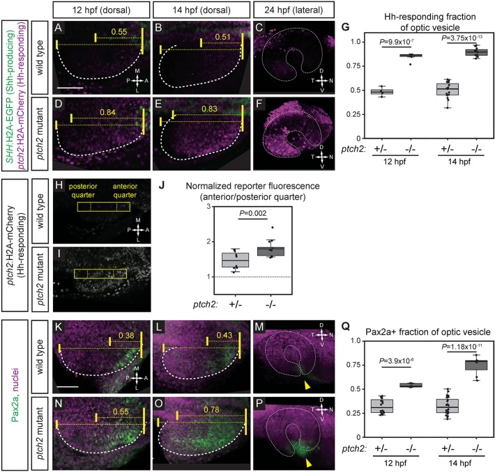
Fig. 6.
Hedgehog signaling is increased at early optic vesicle stages in the ptch2tc294z mutant. (A-G) Hh signaling reporter expression is expanded in the ptch2tc294z mutant at 12, 14 and 24 hpf. Embryos are labeled for Shh-producing cells [green; Tg(SHH:H2A-GFP)z205] and Hh-responding cells [magenta; Tg(GBS-ptch2:H2A-mCherry)z206]. (A-C) Wild-type double transgenic. (D-F) ptch2tc294z mutant double transgenic. (G) Quantification of the anterior-posterior fraction of the optic vesicle occupied by reporter-positive cells. n=4 wt, 6 mut at 12 hpf; 15 wt, 11 mut at 14 hpf. (H-J) Hh signaling reporter expression [Tg(GBS-ptch2:H2A-mCherry)z206] is upregulated in the ptch2tc294z mutant at 13 hpf. A rectangle (44 pixels wide) spanning the anterior-posterior length of the optic vesicle was drawn. Fluorescence intensity in the anterior quarter was normalized to the posterior quarter. A value of 1 (J, dotted line) represents equal fluorescence intensity in the quarters. (H) Wild-type transgenic. (I) ptch2tc294z mutant transgenic. (J) Normalized fluorescence intensity. n=8 wt, 13 mut. (K-Q) Endogenous Hh target gene expression (Pax2a) is expanded in the ptch2tc294z mutant at 12, 14 and 24 hpf. Antibody staining performed for Pax2a (green), nuclei counterstained with TO-PRO-3 (magenta). (K-M) Wild type. (N-P) ptch2tc294z mutant. Yellow arrowheads indicate the optic fissure. (Q) Quantification of the anterior-posterior fraction of optic vesicle occupied by Pax2a-positive cells. Unpaired Student's t-tests. n=16 wt, 5 mut at 12 hpf; 24 wt, 7 mut at 14 hpf. Dashed lines indicate border of optic vesicle. Dotted lines indicate border of optic cup. Yellow brackets indicate anterior-posterior extents of the optic vesicle and either reporter expression or Pax2a staining. The number above indicates the fraction of the optic vesicle occupied by the reporter expression or Pax2a staining. Scale bars: 50 µm.

