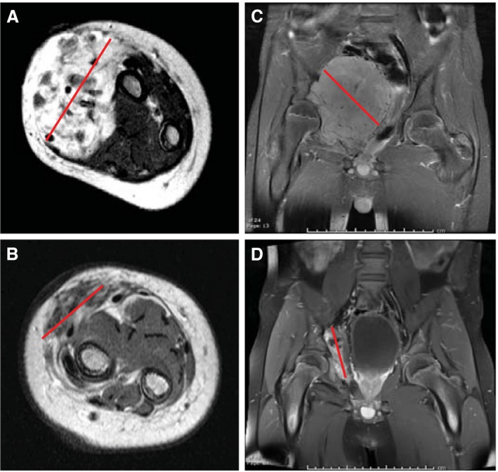
Figure 1.

(A) Baseline T1‐weighted magnetic resonance imaging (MRI) with gadolinium obtained prior to treatment with larotrectinib in case 2 with an ETS variant 6 (ETV6)–neurotrophic receptor tyrosine kinase 3 (NTRK3) fusion infantile fibrosarcoma arising in the forearm. (B) Preoperative T1‐weighted MRI with gadolinium obtained after 6 cycles of larotrectinib in case 2. (C) Baseline T1‐weighted MRI with gadolinium obtained prior to treatment with larotrectinib in case 3 with a tropomyosin 3 (TPM3)–NTRK1 fusion spindle cell sarcoma arising in the pelvis. (D) Preoperative T1‐weighted MRI with gadolinium after 9 cycles of larotrectinib in case 3. The red line indicates the maximum dimension in each panel.
