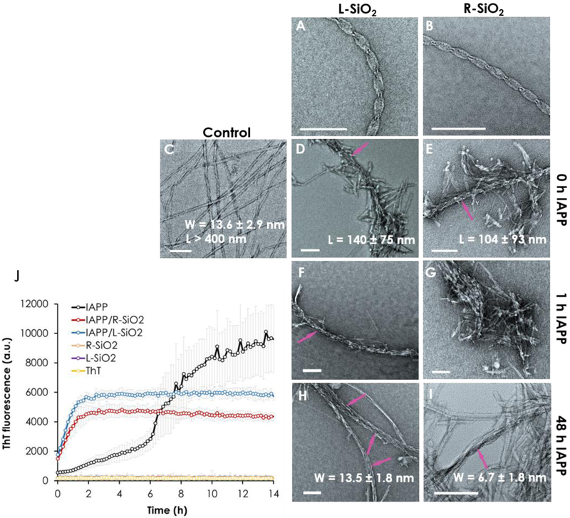
Figure 2.
Transmission electron microscopy images of (A, B) the L/R-SiO2 and (C) IAPP control. (D-G) Attractions between the peptide and the nanoribbons caused shortening and inhibition of the fibrils compared with the control. (H-I) The R-SiO2 was effective in remodeling the IAPP fibrils. All samples were incubated for 24 h. (J) ThT kinetic assay of IAPP fibrillization over 14 h show a shortened lag phase and inhibition of fibrillization in the presence of the L/R-SiO2. Arrows in panels D-F, H and I indicate discernible L/R-SiO2 in contact with IAPP protofibrils/fibrils. Scale bars: 100 nm. The experiments was carried out in triplicate and error bars show the standard deviations of the averaged data sets. IAPP concentration: 50 µM for the ThT assay and 20 µM for TEM, at room temperature and pH 7.

