Abstract
For a long time, it was believed that apoptosis and necrosis were the main pathways for cell death, but a growing body of research has shown that there are other pathways. Among these, necroptosis, a regulatory caspase-independent, programmed cell death pathway, is supposed to be of importance in the pathogenesis of many diseases. The mechanism of regulating, inducing and blocking necroptosis is a complex process that involves expression and regulation of a series of molecules including receptor interacting protein kinase 1 (RIPK1), RIPK3, and mixed lineage kinase like protein. By blocking or downregulating expression of key molecules in the necroptotic pathway, intestinal inflammation can be affected to some extent. In this paper, we introduce the concept of necroptosis, its main pathway, and its impact on the pathogenesis of inflammatory bowel disease (IBD) and other intestinal diseases, to explore new drug targets for intestinal diseases, including IBD.
Keywords: Inflammatory bowel disease, Necroptosis, Inflammation, Colorectal cancer, Intestinal infectious diseases, Drug therapy, Receptor interacting protein kinase 1 inhibitor
Core tip: This minireview is based on the currently available literature about necroptosis and aims to uncover the role of necroptosis in the pathogenesis of inflammatory bowel disease and other intestinal diseases, including colorectal cancer and intestinal infectious diseases. The main regulatory pathways for necroptosis are summarized. Drug therapy targeting of necroptosis is also described.
INTRODUCTION
The balance between cell death, proliferation and differentiation focuses on the maintenance of homeostasis and normal development. Cell death has long been thought to occur primarily in two ways: apoptosis and necrosis. The former refers to caspase-dependent programmed cell death. Due to the quick phagocytosis and degradation of apoptosomes by phagocytes[1], apoptosis is normally considered to be an immune silent cellular activity. The latter is mainly characterized by destruction of cell membrane integrity, release of a large number of intracellular components, and induction of inflammatory reactions in surrounding tissues[2]. However, more and more studies have proved that cell death is not only caused by apoptosis or necrosis. In 2005, Degterev et al[3] reported a new method of cell death: necroptosis. This new cell death pathway has similar morphological features to necrosis, but the mechanism of the regulation is caspase-independent programmed cell death. With the use of caspase inhibition, necroptosis can be triggered by the combination of death receptor and ligand.
TUMOR NECROSIS FACTOR-α PATHWAY AND OTHER PATHWAYS FOR NECROPTOSIS
Although studies have shown that there may be multiple pathways involved in necroptosis, the tumor necrosis factor (TNF)-α-mediating pathway is considered to be the most widely researched one. It can be activated by the combination of TNF-α and TNF receptor 1 (TNF-R1). Since the downstream of TNF-α and TNF-R1 pathways involves a variety of molecules that participate in several physiological processes including cell survival, apoptosis, necrosis, necroptosis, and inflammatory factor production, the combination can mediate different cell physiological processes through the formation of diverse compounds corresponding to specific physiological or pathological conditions.
Formation of Complex I
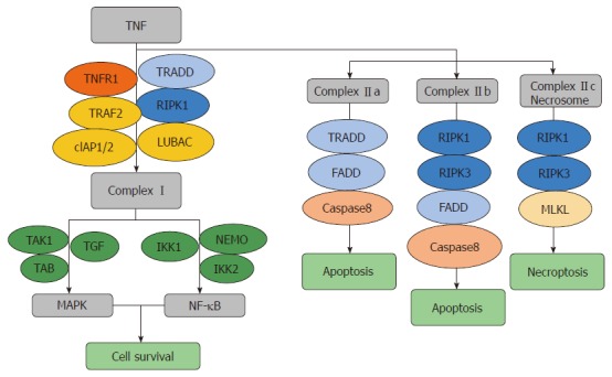
The combination of TNF-α and TNF-R1 can raise TNF-α receptor associated death domain protein (TRADD), receptor interacting protein kinase (RIPK)1, TNFR-associated factor 2, cellular inhibitors of apoptosis (cIAP1 or cIAP2) and linear ubiquitin chain assembly complex to form Complex I. Complex I can activate the mitogen-activated protein kinase (MAPK) pathway and nuclear factor (NF)-κB pathway by collecting TGF–TAK1–TAB complex and IKK1–IKK2–NEMO complex, then activating the proinflammatory pathway to avoid cell death[4].
Formation of Complex IIa
Complex IIa is composed of TRADD, Fas-associated death domain (FADD) and caspase-8, which forms when Complex I is unstable. Complex IIa mainly mediates cell apoptosis[4,5].
Formation of Complex IIb
In some particular cases, e.g., cIAP inhibitors exist or IAPs are knocked out[6], TAK1 inhibitors exist or TAK1 is knocked out[7], and NEMO is knocked out[8] the MAPK pathway and NF-κB pathway induced by Complex I cannot be activated. RIPK1, RIPK3, FADD and caspase-8 comprise Complex IIb. Since caspase-8 can break the interaction sites between RIPK1 and RIPK3, which leads to inactivation of the complex, Complex IIb typically activates the RIPK1-dependent apoptosis pathway rather than the necroptosis pathway mediated by the RIPK1–RIPK3 complex. Complex IIb may act through the formation of necrosomes when caspase-8 activity decreases or RIPK3 and mixed lineage kinase like protein (MLKL) levels are high enough. Intracellular FADD-like interleukin (IL)-1β converting enzyme (FLICE)-inhibitory protein (FLIPL) can form caspase-8-FLIPL heteromeric compounds, which have only caspase-8 catalytic activity. So, FLIPL participates in the regulation of both cell death pathways: apoptosis and necroptosis by controlling RIPK1 and RIPK3 level through proteolysis[4].
Formation of Complex IIc
When caspase-8 is inhibited or missing, Complex IIb will transfer to Complex IIc, forming necrosomes (the complex includes RIPK1, RIPK3 and MLKL). RIPK1 and RIPK3 interact and activate downstream MLKL to trigger the necroptosis pathways[4] (Figure 1).
Figure 1.

Tumor necrosis factor-α pathway for necroptosis. Combination of tumor necrosis factor (TNF)-α and TNF-R1 can raise TNF-α receptor associated death domain protein (TRADD), receptor interacting protein kinase 1 (RIPK1), TNFR-associated factor 2, cellular inhibitors of apoptosis (cIAP1 or cIAP2) and linear ubiquitin chain assembly complex to form Complex I, which can activate the Mitogen-activated protein kinase and NF-κB pathways by binding the TGF–TAK1–TAB and IKK1–IKK2–NEMO complexes, leading to cell survival. Complex IIa [consists of TRADD, Fas-associated death domain (FADD) and caspase-8] forms when Complex I is unstable and mediates apoptosis independent of RIPK1. Complex IIb (consists of RIPK1, RIPK3, FADD and caspase-8) forms when cIAP inhibitors are present or IAPs are knocked out, TAK1 inhibitors are present or TAK1 is knocked out, and NEMO is knocked out, and mediates apoptosis dependent on the binding of RIPK1 and RIPK3. Complex IIc (consists of RIPK1, RIPK3 and MLKL) forms when caspase-8 is inhibited or missing and leads to necroptosis. TRAF2: TNFR-associated factor 2; cIAP1 or cIAP2: Cellular inhibitors of apoptosis; LUBAC: Linear ubiquitin chain assembly complex; TRADD: TNF-α receptor associated death domain protein; MAPK: Mitogen-activated protein kinase; FADD: Fas-associated death domain.
Eventual executives for necroptosis
The eventual executives for necroptosis pathway are not yet clear. MLKL is one of the most widely researched molecules and a commonly used indicator assessing the effect of necroptosis in the pathogenesis of inflammatory bowel disease (IBD). MLKL is a fake kinase, which possesses a kinase domain structure but lacks kinase activity. RIPK3 can activate the corresponding threonine and serine site of MLKL and make it phosphorylate. The phosphorylation of MLKL changes its conformation to form oligomers. Oligomerized MLKL exposes the N-terminal domain structure, which promotes necrosome transfer from the cytoplasm to the cell or organelle membrane. This destroys membrane integrity and leads to cell death[9,10].
Other pathways for necroptosis
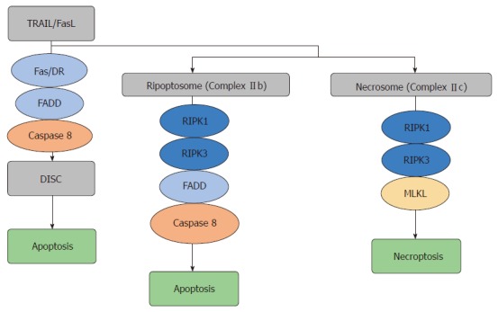
TNF ligand superfamily member 10 (TRAIL) and TNF ligand superfamily member 6 (FasL)-mediated pathways share many similarities with the TNF-α pathway, and activate caspase-8 and induce apoptosis. If caspase-8 is inhibited, the necroptosis pathway is activated. However, these two pathways also have their own characteristics. First, the TNF-α pathway forms a prosurvival signaling complex first and then a death-inducing complex. The TRAIL/FasL pathways form a death-inducing signaling complex, which consists of Fas/DR4,5 (also known as TRAILR1,2), FADD and caspase-8[11] and subsequently induce apoptosis. Second, RIPK1 can directly bind to TNF-R1 through its DD domain. The binding of FasL and TRAIL receptors to RIPK1 requires FADD as a bridge[12,13]. Therefore, FADD is necessary for TRAIL and FasL signaling pathways, but is not necessary for the TNF-α associated pathway[11]. The main function of FADD is to provide binding sites for caspase-8 and caspase-10 precursors and the activated combination subsequently mediates downstream caspase-3, caspase-6 and caspase-7, and finally induces apoptosis[14,15] (Figure 2).
Figure 2.

Tumor necrosis factor ligand superfamily member 10/tumor necrosis factor ligand superfamily member 6 pathways for necroptosis. Tumor necrosis factor (TNF) ligand superfamily member 10 and TNF ligand superfamily member 6 pathways induce apoptosis independent of receptor interacting protein kinase 1 (RIPK1) with the formation of death-inducing signaling complex [consists of Fas/DR, Fas-associated death domain (FADD) and caspase-8]. Ripoptosome (consists of RIPK1, RIPK3, FADD and caspase-8) forms in particular situations such as presence of cellular inhibitors of apoptosis (IAP) antagonists or IAP knock out and leads to apoptosis. Necrosomes (consists of RIPK1, RIPK3 and mixed lineage kinase like protein) form when caspase-8 is inhibited or blocked, and induces necroptosis. DISC: Death-inducing signaling complex; MLKL: Mixed lineage kinase like protein; TRAIL: TNF ligand superfamily member 10; FasL: TNF ligand superfamily member 6; FADD: Fas-associated death domain.
NECROPTOSIS AND INFLAMMATION
The relationship between necroptosis and inflammation remains unclear. Some scholars believe that cells release damage-associated molecular patterns (DAMPs) through pyrolysis in the process of necroptosis. DAMPs can activate the immune system by itself or combining with pathogen-associated molecular patterns. It can promote macrophages and dendritic cells and other sentinel cells activate and increase the level of abundant cytokines including interleukin (IL)-1 superfamily (IL-1α, IL-1β, IL-18, IL-33, IL-36α, IL-36β, and IL-36γ), which mediates the pathogenesis of various acute and chronic inflammatory diseases[4,16,17]. Duprez et al[18] have shown that necroptosis mediated by RIPK1–RIPK3 can increase the mortality from systemic inflammatory response syndrome induced by TNF. With RIPK3 gene knockout or preventive use of RIPK1 inhibitors, necrostain-1 can effectively alleviate the levels of DAMPs in the circulation. In the model of myocardial ischemia/reperfusion, phosphorylation of RIPK1 and RIPK3 is always accompanied by infiltration of a large number of neutrophils. Necrostain-1 dramatically reduces inflammation. It presents a lower level of TNF-α and oxidative stress, reduction of adverse myocardial remodeling, and improvement of cardiac function[19]. In addition, in the liver injury model induced by ethanol, the loss of RIPK3 can avoid the pathogenesis of liver cirrhosis, the increase in inflammatory factors, and therefore prevent liver tissue injury[20]. The dual deletion of RIPK1 and FADD induces necroptosis, which is dependent on RIPK3 in colon intestinal epithelial cells, decreases the number of Paneth cells, and causes localized inflammatory damage[21].
However, there are also researchers who consider that some pathogens may terminate the signaling cascade of proinflammatory factors through necroptosis to limit the cytokine storm. Necroptosis can inhibit excessive release of inflammatory factors induced by TNF/Toll-like receptor (TLR) in a specific environment and therefore plays an anti-inflammatory role[16]. The TNF-α pathway, for example, participates in inflammation, apoptosis and necroptosis, but has different outcomes under disparate physiological conditions. Therefore, the result of transformation from an inflammatory response dependent on TNF to necroptosis is not just cell death (although DAMPs released during the process can also promote inflammation), but the cascade induced by TNF itself, a powerful inflammatory factor. The end of the cascade avoids stimulation of cell synthesis and the release of more cytokines and chemokines, which limit the inflammatory response[22]. According to the study of Alvarez-Diaz et al [23], mice with FADD/MLKL or caspase-8/MLKL double knockout develop severe systemic autoimmune diseases and die within a short period of time due to the dramatic increase in a variety of cytokines and chemokines. In addition, Newton et al[24] have shown that RIPK3 deficiency does not improve sepsis induced by lipopolysaccharide (LPS), colitis induced by dextran sulfate sodium (DSS), pancreatitis induced by frosin, and brain injury induced by hypoxia. The absence of MLKL cannot protect against kidney injury caused by ischemia–reperfusion[24]. Bozec et al[25] have shown that expression of RIPK3 decreases in colorectal cancer (CRC) patients with IBD, which further demonstrate the anti-inflammatory effect of necroptosis.
NECROPTOSIS AND IBD
IBD is a kind of chronic, nonspecific intestinal inflammatory disease whose pathogenesis is unclear. The main pathological types of IBD are ulcerative colitis (UC) and Crohn’s disease (CD). Despite the lack of epidemiological data for IBD in developing countries, the prevalence of IBD is on the rise globally[26]. Although the cause of IBD remains unknown, environmental, infectious, immune and genetic factors are involved in the pathogenesis of excessive apoptosis of intestinal epithelial cells, damage to the intestinal mucosal barrier, and higher permeability of intestinal epithelial cells, which are considered to contribute to the development of IBD[27]. Necroptosis is a new mode of cell death. The process of necroptosis is regulated as well as apoptosis. So, some scholars think that necroptosis plays an important role in the pathogenesis of IBD.
In situ end labeling technology cannot distinguish between apoptosis and necroptosis, and early research about the effect of apoptosis in the pathogenesis of IBD cannot prove whether necroptosis plays the same role[4]. Recent studies based on animal models and patients with IBD have suggested that necroptosis plays a role in the development of intestinal inflammation.
Welz et al[28] found in mice that intestinal epithelial cells with FADD gene knock out presented with necrosis dependent on RIPK3, reduced Paneth cells, enteritis and severe colitis. The absence of RIPK3 or the use of CYLD inhibitors can inhibit this spontaneous pathological process. Mice with intestinal epithelial cell caspase-8 gene knock out also had inflammatory lesions in the terminal ileum (increased Paneth cell death and reduced goblet cells), and high susceptibility to colitis. These findings were related to increased levels of RIPK3[29]. RIPK3 knock down can reduce intestinal inflammation in caspase-8 knockout mice to a certain extent[30]. All these studies based on mouse models have shown that necroptosis of intestinal epithelial cells induces intestinal inflammation.
Günther et al[29] have confirmed that in adult CD patients, the level of RIPK3 expressed by Paneth cells increases along with necroptosis in the terminal ileum. By comparison between adult penetration type CD patients, adult narrow type CD patients and healthy controls, serum level of TNF-α and mucosal tissue level of RIPK3 are obviously increased in both types of CD[10]. Pierdomenico et al[31] investigated biopsy samples from 33 children with CD and 30 with UC and found increased expression of RIPK3 and MLKL in inflammatory tissue and dramatically decreased expression of caspase-8. There was no significant difference in expression of all three proteins in non-inflammatory tissues, which confirm that necroptosis can magnify the inflammatory response and induce pathological conditions in the intestine. Negroni et al[32] have confirmed that necroptosis induced by RIPK3 can activate MLKL and increase expression of IL-8, IL-1β, IL-33 and high-mobility group protein 1. Necroptosis then induces the translocation of NF-κB p65 and the assemblage of inflammasome NLRP3. At the same time, the integrity of the intestinal epithelial barrier induced by various intercellular connective proteins, including Cadherin E and occludin can also be influenced, which mediates the occurrence of intestinal inflammation[32].
NECROPTOSIS AND CRC
Incoercible proliferation of intestinal epithelial cells and decreased cell death are the main pathological processes of CRC. Therefore, necroptosis plays an important role in the occurrence and development of CRC. Expression levels of RIPK1 and RIPK3 were lower in mucosal tissue of CRC patients compared to normal mucosal tissues, which indicates that necroptosis plays a role in the induction of cell death and inhibition of CRC development. Bozec et al[25] have shown that RIPK3 has a suppressive effect on CRC, while tumor cells without RIPK3 are often more invasive. RIPK3 knockout mice are more susceptible to CRC related enteritis. The lack of RIPK3 gene leads to excessive activation of the NF-κB, signal transducer and activator of transcription (STAT) 3, AKT and Wnt-β–catenin pathways, which results in abnormal proliferation of intestinal epithelial cells and the occurrence of tumor.
The induction of TNF-α-related necroptosis by RIPK1 and NF-κB can overcome the apoptosis tolerance in CRC and significantly suppress tumor growth[33]. In vivo experiments have confirmed that combination of a new type of pan-caspase inhibitor IDN-7314 and 5-fluorouracil (5-FU) is beneficial in CRC patients with 5-FU resistance, through increased cell necroptosis[33]. Induction of necroptosis also increases intestinal epithelial cell death in CRC patients with apoptosis tolerance induced by absence of caspase-8[34]. Sun et al[35] have shown that 3-bromine pyruvate (hexokinase inhibitor) causes organelle swelling, damaged cell membrane integrity, exudation of cell contents and increased necroptosis in SW480 and HT29 colon cancer cells. Necrostin-1 significantly recovers cell survival. A recent study also showed that resibufogenin suppresses growth and metastasis of CRC through RIPK3-mediated necroptosis. This kind of effect can be reduced by RIPK3 deficiency rather than the use of z-VAD, a pan-caspase general inhibitor, which confirms the role of necroptosis in the treatment of CRC[36].
NECROPTOSIS AND INTESTINAL INFECTIOUS DISEASES
The balance between death and survival of intestinal epithelial cells is essential to maintain integrity of the intestinal barrier and intestinal homeostasis. Excessive death of intestinal epithelial cells damages the function of the intestinal barrier, leading to weakened resistance to pathogenic bacteria. Hefele et al[37] have shown that caspase-8 knockout mice present with more severe mucosal injury and intestinal epithelial cell death after infection with Salmonella typhimurium compared to wild-type mice. Knock out of RIPK3 or MLKL can significantly relieve the mucosal injury and intestinal epithelial cell death. Qi et al[38] have also shown that infection with S. typhimurium without sopB induces phosphorylation of MLKL and therefore promotes necroptosis of goblet cells. This induces more severe intestinal damage, including increased levels of cytokines and chemokines and decreased mucus and mucin-2. However, this kind of physiological or pathological process can be effectively alleviated in MLKL knockout mice. TLRs expressed on the surface of intestinal epithelial cells can promote the identification of microorganisms. Stimulation of TLRs leads to intestinal epithelial cell loss, which mediates epithelial antibacterial and antiviral activity in the host and helps to remove exogenous pathogens.
Caspase-8 knockout mice show more severe injury of intestinal epithelial cells with the activation of the necroptosis pathway induced by RIPK3 rather than the extrinsic apoptotic pathway after stimulation of TLRs. TLR-3 mediate necroptosis through TRIF and TLR-4 mediates necroptosis through TNF-α. RIPK inhibitor can improve intestinal injury induced by LPS and maintain the function of the intestinal barrier. These results provide new ideas for the treatment of intestinal inflammation and infectious diseases[39] (Table 1).
Table 1.
Effects of necroptosis in intestinal diseases
| Disease | Expression of necroptosis moleculars | Pathological processes may involve after the activation of Necroptosis |
| IBD | RIPK3 upregulated MLKL upregulated, caspase-8 downregulated | (1) Impair intestinal epithelial barrier and membrane permeability |
| (2) Commensal bacteria invade the mucosa through TLR signaling | ||
| (3) Reduce number of Paneth cell | ||
| (4) Increase expression of cytokines (e.g., IL-8, IL-1β, IL-33, HMGB1) | ||
| (5) Translocation of NF-κB p65 and assembly of inflammasomes NALRP3 | ||
| CRC | RIPK3 downregulated, | (1) Suppress excessive activation of NF-κB, STAT3, AKT and Wnt-β–catenin pathways |
| RIPK1 downregulated | (2) Overcome the apoptosis tolerance in CRC and suppress tumor growth and metastasis | |
| Intestinal infectious diseases | RIPK3 and MLKL upregulated when caspase-8 is deficient | (1) Impair the integrity of intestinal epithelial barrier |
| (2) Increase levels of cytokines and chemokines, and decrease levels of mucus and mucin-2 | ||
| (3) TLR-3 mediate necroptosis through TRIF and TLR-4 mediates necroptosis through TNF-α |
IBD: Inflammatory bowel disease; CRC: Colorectal cancer; RIPK: Receptor interacting protein kinase; IL: Interleukin; MLKL: Mixed lineage kinase like protein; TLR: Toll-like receptor; TNF: Tumor necrosis factor; STAT: Signal transducer and activator of transcription.
DRUG THREAPY TARGETING IN NECROPTOSIS
Since necroptosis may play a partial role in the pathogenesis of IBD, regulation of the key molecules during necroptosis may provide a new target for the treatment of IBD. The most frequently studied among these is necrostatin-1, an RIPK1-specific inhibitor whose protective effect in IBD has been widely reported[29,32,40]. Liu et al[40] have confirmed that when the structure of RIPK1–RIPK3 complex is destroyed[41,42] by necrostatin-1, expression of RIPK1 and RIPK3 is decreased and expression of caspase-8 is increased in a mouse model of intestinal inflammation induced by DSS. The use of necrostatin-1 thus restores weight loss, decreased colon length, damage to the intestinal mucosa, and excessive release of inflammatory factors like IL-6. As an effective necroptosis inhibitor, necrostatin-1 has the following characteristics: inhibition of necroptosis without affecting apoptosis and autophagy; no effect on the physiological function of normal cells[43].
Apart from necrostatin-1, autophagy protein ATG16L1 reported by Matsuzawa-Ishimoto et al[44] also alleviates necroptosis in an IBD model induced by virus infection, through TNF-α and RIPK1, and maintains integrity of the intestinal barrier. Dong et al[45] have confirmed that necrosulfonamide reverses the histological abnormality induced by TNF-α and Z-VAD-fmk and reduces the degree of necroptosis.
Other recent studies also reported drug therapy targeting necroptosis in other diseases. Nikseresht et al[46] revealed that ex-527, a selective inhibitor of Sirt1, reduced expression of critical regulators of necroptosis in a model of cerebral ischemia–reperfusion injury, which resulted in alleviation of infarction volume and increased survival. Rats with cognitive impairment induced by severe acute pancreatitis show increased levels of RIPK3 and RIPK1, and berberine can partly treat this cognitive impairment through attenuation of neuronal necroptosis[47]. Oliveira et al[48] have found that Oxa12, a new oxazolone, can reduce TNF-α-induced necroptosis in mouse L929 fibrosarcoma cells and zVAD-fmk-induced necroptosis in murine BV2 microglial cells. They have confirmed that Oxa12 disturbs the phosphorylation of MLKL and formation of necrosome complexes, which provides a new insight into the treatment of other inflammatory diseases. Another interesting study has shown that AdipoRon, an antidiabetic adiponectin receptor agonist, can lead to the death of MIAPaCa-2 cells (human pancreatic carcinoma cell line) through necroptosis via the activation of RIPK1 and extracellular signal-regulated kinase 1/2[49].
CONCLUSION
Although current research has confirmed that necroptosis is an important pathway of programmed cell death, many problems remain to be explored and resolved. How cells select survival, apoptosis, necrosis and necroptosis according to different physiological and pathological conditions is unclear. The interaction between various terminal states also needs to be further explored. In the necroptosis pathway, how RIPK3 induces necroptosis and inhibits apoptosis at the same time, the molecular mechanisms of MLKL, and the regulation of RIPK family activity and its substrates are still controversial. In the pathogenesis of IBD, the role of necroptosis in the process of initiation, amplification and extension of inflammation needs to be confirmed. The TNF-α pathway is the most widely studied necroptotic pathway, but which downstream effects are activated, necroptosis, apoptosis or inflammation, in the pathogenesis of IBD needs further investigation. TNF monoclonal antibody is currently the main drug for the treatment of IBD, although up to 50% of patients show no response, and an increased risk of infection[50]. This suggests that there is an urgent need to explore new treatments for IBD. We believe that with further investigation of the molecular mechanism of necroptosis, we will be more aware of cell death and develop new ideas for exploration of the pathogenesis of various human diseases.
Footnotes
Conflict-of-interest statement: The authors declare that there is no conflict of interest related to this study.
Manuscript source: Unsolicited manuscript
Peer-review started: August 19, 2018
First decision: October 5, 2018
Article in press: November 1, 2018
Specialty type: Medicine, research and experimental
Country of origin: China
Peer-review report classification
Grade A (Excellent): A
Grade B (Very good): B
Grade C (Good): C
Grade D (Fair): 0
Grade E (Poor): 0
P- Reviewer: Horje CSHT, Bosca L, Takagi T S- Editor: Dou Y L- Editor: Filipodia E- Editor: Song H
Contributor Information
Sha Li, Department of Gastroenterology, the First Affiliated Hospital, College of Medicine, Zhejiang University, Hangzhou 310003, Zhejiang Province, China.
Long-Gui Ning, Department of Gastroenterology, the First Affiliated Hospital, College of Medicine, Zhejiang University, Hangzhou 310003, Zhejiang Province, China.
Xin-He Lou, Department of Gastroenterology, the First Affiliated Hospital, College of Medicine, Zhejiang University, Hangzhou 310003, Zhejiang Province, China.
Guo-Qiang Xu, Department of Gastroenterology, the First Affiliated Hospital, College of Medicine, Zhejiang University, Hangzhou 310003, Zhejiang Province, China. 1193065@zju.edu.cn.
References
- 1.Ravichandran KS. Beginnings of a good apoptotic meal: the find-me and eat-me signaling pathways. Immunity. 2011;35:445–455. doi: 10.1016/j.immuni.2011.09.004. [DOI] [PMC free article] [PubMed] [Google Scholar]
- 2.Proskuryakov SY, Konoplyannikov AG, Gabai VL. Necrosis: a specific form of programmed cell death? Exp Cell Res. 2003;283:1–16. doi: 10.1016/S0014-4827(02)00027-7. [DOI] [PubMed] [Google Scholar]
- 3.Degterev A, Huang Z, Boyce M, Li Y, Jagtap P, Mizushima N, Cuny GD, Mitchison TJ, Moskowitz MA, Yuan J. Chemical inhibitor of nonapoptotic cell death with therapeutic potential for ischemic brain injury. Nat Chem Biol. 2005;1:112–119. doi: 10.1038/nchembio711. [DOI] [PubMed] [Google Scholar]
- 4.Pasparakis M, Vandenabeele P. Necroptosis and its role in inflammation. Nature. 2015;517:311–320. doi: 10.1038/nature14191. [DOI] [PubMed] [Google Scholar]
- 5.Micheau O, Tschopp J. Induction of TNF receptor I-mediated apoptosis via two sequential signaling complexes. Cell. 2003;114:181–190. doi: 10.1016/s0092-8674(03)00521-x. [DOI] [PubMed] [Google Scholar]
- 6.Wang L, Du F, Wang X. TNF-alpha induces two distinct caspase-8 activation pathways. Cell. 2008;133:693–703. doi: 10.1016/j.cell.2008.03.036. [DOI] [PubMed] [Google Scholar]
- 7.Dondelinger Y, Aguileta MA, Goossens V, Dubuisson C, Grootjans S, Dejardin E, Vandenabeele P, Bertrand MJ. RIPK3 contributes to TNFR1-mediated RIPK1 kinase-dependent apoptosis in conditions of cIAP1/2 depletion or TAK1 kinase inhibition. Cell Death Differ. 2013;20:1381–1392. doi: 10.1038/cdd.2013.94. [DOI] [PMC free article] [PubMed] [Google Scholar]
- 8.Legarda-Addison D, Hase H, O’Donnell MA, Ting AT. NEMO/IKKgamma regulates an early NF-kappaB-independent cell-death checkpoint during TNF signaling. Cell Death Differ. 2009;16:1279–1288. doi: 10.1038/cdd.2009.41. [DOI] [PMC free article] [PubMed] [Google Scholar]
- 9.Zhao ZJ, Chen M, Yu ZM. Research progress on the regulation mechanism of necroptosis. Jilin Yixue. 2015;36:2116–2117. [Google Scholar]
- 10.Xia BB. Hangzhou (China): Zhejiang University; 2016. The role of Necroptosis Signal Pathway in Intestinal Inflammation of Adults Crohn’s disease [D] [Google Scholar]
- 11.Jin Z, El-Deiry WS. Distinct signaling pathways in TRAIL- versus tumor necrosis factor-induced apoptosis. Mol Cell Biol. 2006;26:8136–8148. doi: 10.1128/MCB.00257-06. [DOI] [PMC free article] [PubMed] [Google Scholar]
- 12.Festjens N, Vanden Berghe T, Vandenabeele P. Necrosis, a well-orchestrated form of cell demise: signalling cascades, important mediators and concomitant immune response. Biochim Biophys Acta. 2006;1757:1371–1387. doi: 10.1016/j.bbabio.2006.06.014. [DOI] [PubMed] [Google Scholar]
- 13.Festjens N, Vanden Berghe T, Cornelis S, Vandenabeele P. RIP1, a kinase on the crossroads of a cell’s decision to live or die. Cell Death Differ. 2007;14:400–410. doi: 10.1038/sj.cdd.4402085. [DOI] [PubMed] [Google Scholar]
- 14.Lavrik IN. Regulation of death receptor-induced apoptosis induced via CD95/FAS and other death receptors. Mol Biol (Mosk) 2011;45:173–179. [PubMed] [Google Scholar]
- 15.Gonzalvez F, Ashkenazi A. New insights into apoptosis signaling by Apo2L/TRAIL. Oncogene. 2010;29:4752–4765. doi: 10.1038/onc.2010.221. [DOI] [PubMed] [Google Scholar]
- 16.Kearney CJ, Martin SJ. An Inflammatory Perspective on Necroptosis. Mol Cell. 2017;65:965–973. doi: 10.1016/j.molcel.2017.02.024. [DOI] [PubMed] [Google Scholar]
- 17.Martin SJ. Cell death and inflammation: the case for IL-1 family cytokines as the canonical DAMPs of the immune system. FEBS J. 2016;283:2599–2615. doi: 10.1111/febs.13775. [DOI] [PubMed] [Google Scholar]
- 18.Duprez L, Takahashi N, Van Hauwermeiren F, Vandendriessche B, Goossens V, Vanden Berghe T, Declercq W, Libert C, Cauwels A, Vandenabeele P. RIP kinase-dependent necrosis drives lethal systemic inflammatory response syndrome. Immunity. 2011;35:908–918. doi: 10.1016/j.immuni.2011.09.020. [DOI] [PubMed] [Google Scholar]
- 19.Oerlemans MI, Liu J, Arslan F, den Ouden K, van Middelaar BJ, Doevendans PA, Sluijter JP. Inhibition of RIP1-dependent necrosis prevents adverse cardiac remodeling after myocardial ischemia-reperfusion in vivo. Basic Res Cardiol. 2012;107:270. doi: 10.1007/s00395-012-0270-8. [DOI] [PubMed] [Google Scholar]
- 20.Roychowdhury S, McMullen MR, Pisano SG, Liu X, Nagy LE. Absence of receptor interacting protein kinase 3 prevents ethanol-induced liver injury. Hepatology. 2013;57:1773–1783. doi: 10.1002/hep.26200. [DOI] [PMC free article] [PubMed] [Google Scholar]
- 21.Dannappel M, Vlantis K, Kumari S, Polykratis A, Kim C, Wachsmuth L, Eftychi C, Lin J, Corona T, Hermance N, et al. RIPK1 maintains epithelial homeostasis by inhibiting apoptosis and necroptosis. Nature. 2014;513:90–94. doi: 10.1038/nature13608. [DOI] [PMC free article] [PubMed] [Google Scholar]
- 22.Kalliolias GD, Ivashkiv LB. TNF biology, pathogenic mechanisms and emerging therapeutic strategies. Nat Rev Rheumatol. 2016;12:49–62. doi: 10.1038/nrrheum.2015.169. [DOI] [PMC free article] [PubMed] [Google Scholar]
- 23.Alvarez-Diaz S, Dillon CP, Lalaoui N, Tanzer MC, Rodriguez DA, Lin A, Lebois M, Hakem R, Josefsson EC, O’Reilly LA, et al. The Pseudokinase MLKL and the Kinase RIPK3 Have Distinct Roles in Autoimmune Disease Caused by Loss of Death-Receptor-Induced Apoptosis. Immunity. 2016;45:513–526. doi: 10.1016/j.immuni.2016.07.016. [DOI] [PMC free article] [PubMed] [Google Scholar]
- 24.Newton K, Dugger DL, Maltzman A, Greve JM, Hedehus M, Martin-McNulty B, Carano RA, Cao TC, van Bruggen N, Bernstein L, et al. RIPK3 deficiency or catalytically inactive RIPK1 provides greater benefit than MLKL deficiency in mouse models of inflammation and tissue injury. Cell Death Differ. 2016;23:1565–1576. doi: 10.1038/cdd.2016.46. [DOI] [PMC free article] [PubMed] [Google Scholar]
- 25.Bozec D, Iuga AC, Roda G, Dahan S, Yeretssian G. Critical function of the necroptosis adaptor RIPK3 in protecting from intestinal tumorigenesis. Oncotarget. 2016;7:46384–46400. doi: 10.18632/oncotarget.10135. [DOI] [PMC free article] [PubMed] [Google Scholar]
- 26.Molodecky NA, Soon IS, Rabi DM, Ghali WA, Ferris M, Chernoff G, Benchimol EI, Panaccione R, Ghosh S, Barkema HW, et al. Increasing incidence and prevalence of the inflammatory bowel diseases with time, based on systematic review. Gastroenterology. 2012;142:46–54.e42; quiz e30. doi: 10.1053/j.gastro.2011.10.001. [DOI] [PubMed] [Google Scholar]
- 27.Buret AG, Bhargava A. Modulatory mechanisms of enterocyte apoptosis by viral, bacterial and parasitic pathogens. Crit Rev Microbiol. 2014;40:1–17. doi: 10.3109/1040841X.2012.746952. [DOI] [PubMed] [Google Scholar]
- 28.Welz PS, Wullaert A, Vlantis K, Kondylis V, Fernández-Majada V, Ermolaeva M, Kirsch P, Sterner-Kock A, van Loo G, Pasparakis M. FADD prevents RIP3-mediated epithelial cell necrosis and chronic intestinal inflammation. Nature. 2011;477:330–334. doi: 10.1038/nature10273. [DOI] [PubMed] [Google Scholar]
- 29.Günther C, Martini E, Wittkopf N, Amann K, Weigmann B, Neumann H, Waldner MJ, Hedrick SM, Tenzer S, Neurath MF, et al. Caspase-8 regulates TNF-α-induced epithelial necroptosis and terminal ileitis. Nature. 2011;477:335–339. doi: 10.1038/nature10400. [DOI] [PMC free article] [PubMed] [Google Scholar]
- 30.Weinlich R, Oberst A, Dillon CP, Janke LJ, Milasta S, Lukens JR, Rodriguez DA, Gurung P, Savage C, Kanneganti TD, et al. Protective roles for caspase-8 and cFLIP in adult homeostasis. Cell Rep. 2013;5:340–348. doi: 10.1016/j.celrep.2013.08.045. [DOI] [PMC free article] [PubMed] [Google Scholar]
- 31.Pierdomenico M, Negroni A, Stronati L, Vitali R, Prete E, Bertin J, Gough PJ, Aloi M, Cucchiara S. Necroptosis is active in children with inflammatory bowel disease and contributes to heighten intestinal inflammation. Am J Gastroenterol. 2014;109:279–287. doi: 10.1038/ajg.2013.403. [DOI] [PubMed] [Google Scholar]
- 32.Negroni A, Colantoni E, Pierdomenico M, Palone F, Costanzo M, Oliva S, Tiberti A, Cucchiara S, Stronati L. RIP3 AND pMLKL promote necroptosis-induced inflammation and alter membrane permeability in intestinal epithelial cells. Dig Liver Dis. 2017;49:1201–1210. doi: 10.1016/j.dld.2017.08.017. [DOI] [PubMed] [Google Scholar]
- 33.Oliver Metzig M, Fuchs D, Tagscherer KE, Gröne HJ, Schirmacher P, Roth W. Inhibition of caspases primes colon cancer cells for 5-fluorouracil-induced TNF-α-dependent necroptosis driven by RIP1 kinase and NF-κB. Oncogene. 2016;35:3399–3409. doi: 10.1038/onc.2015.398. [DOI] [PubMed] [Google Scholar]
- 34.He GW, Günther C, Thonn V, Yu YQ, Martini E, Buchen B, Neurath MF, Stürzl M, Becker C. Regression of apoptosis-resistant colorectal tumors by induction of necroptosis in mice. J Exp Med. 2017;214:1655–1662. doi: 10.1084/jem.20160442. [DOI] [PMC free article] [PubMed] [Google Scholar]
- 35.Sun YM. Bengbu (China): Bengbu Medical College; 2016. Mechanisms underlying 3-bromopyruvate induced cell death in colon cancer [D] [DOI] [PMC free article] [PubMed] [Google Scholar]
- 36.Han Q, Ma Y, Wang H, Dai Y, Chen C, Liu Y, Jing L, Sun X. Resibufogenin suppresses colorectal cancer growth and metastasis through RIP3-mediated necroptosis. J Transl Med. 2018;16:201. doi: 10.1186/s12967-018-1580-x. [DOI] [PMC free article] [PubMed] [Google Scholar]
- 37.Hefele M, Stolzer I, Ruder B, He GW, Mahapatro M, Wirtz S, Neurath MF, Günther C. Intestinal epithelial Caspase-8 signaling is essential to prevent necroptosis during Salmonella Typhimurium induced enteritis. Mucosal Immunol. 2018;11:1191–1202. doi: 10.1038/s41385-018-0011-x. [DOI] [PubMed] [Google Scholar]
- 38.Qi S. Changchun (China): Jilin University; 2017. The role of Salmonella sopB gene in bacteria colitis [D] [Google Scholar]
- 39.Günther C, Buchen B, He GW, Hornef M, Torow N, Neumann H, Wittkopf N, Martini E, Basic M, Bleich A, et al. Caspase-8 controls the gut response to microbial challenges by Tnf-α-dependent and independent pathways. Gut. 2015;64:601–610. doi: 10.1136/gutjnl-2014-307226. [DOI] [PMC free article] [PubMed] [Google Scholar]
- 40.Liu ZY, Wu B, Guo YS, Zhou YH, Fu ZG, Xu BQ, Li JH, Jing L, Jiang JL, Tang J, et al. Necrostatin-1 reduces intestinal inflammation and colitis-associated tumorigenesis in mice. Am J Cancer Res. 2015;5:3174–3185. [PMC free article] [PubMed] [Google Scholar]
- 41.Cho YS, Challa S, Moquin D, Genga R, Ray TD, Guildford M, Chan FK. Phosphorylation-driven assembly of the RIP1-RIP3 complex regulates programmed necrosis and virus-induced inflammation. Cell. 2009;137:1112–1123. doi: 10.1016/j.cell.2009.05.037. [DOI] [PMC free article] [PubMed] [Google Scholar]
- 42.Smith CC, Davidson SM, Lim SY, Simpkin JC, Hothersall JS, Yellon DM. Necrostatin: a potentially novel cardioprotective agent? Cardiovasc Drugs Ther. 2007;21:227–233. doi: 10.1007/s10557-007-6035-1. [DOI] [PubMed] [Google Scholar]
- 43.Chen BN, Du GH. Necroptosis: A new way for cell death. Shenglixue Jinzhan. 2010;41:95–102. [Google Scholar]
- 44.Matsuzawa-Ishimoto Y, Shono Y, Gomez LE, Hubbard-Lucey VM, Cammer M, Neil J, Dewan MZ, Lieberman SR, Lazrak A, Marinis JM, et al. Autophagy protein ATG16L1 prevents necroptosis in the intestinal epithelium. J Exp Med. 2017;214:3687–3705. doi: 10.1084/jem.20170558. [DOI] [PMC free article] [PubMed] [Google Scholar]
- 45.Dong W, Zhang M, Zhu Y, Chen Y, Zhao X, Li R, Zhang L, Ye Z, Liang X. Protective effect of NSA on intestinal epithelial cells in a necroptosis model. Oncotarget. 2017;8:86726–86735. doi: 10.18632/oncotarget.21418. [DOI] [PMC free article] [PubMed] [Google Scholar]
- 46.Nikseresht S, Khodagholi F, Ahmadiani A. Protective effects of ex-527 on cerebral ischemia-reperfusion injury through necroptosis signaling pathway attenuation. J Cell Physiol. 2018;234:1816–1826. doi: 10.1002/jcp.27055. [DOI] [PubMed] [Google Scholar]
- 47.Ou X, Hua Y, Liao X, Gong C, Kang Y. Cognitive impairments induced by severe acute pancreatitis are attenuated by berberine treatment in rats. Mol Med Rep. 2018;18:3437–3444. doi: 10.3892/mmr.2018.9313. [DOI] [PubMed] [Google Scholar]
- 48.Oliveira SR, Dionísio PA, Brito H, Franco L, Rodrigues CAB, Guedes RC, Afonso CAM, Amaral JD, Rodrigues CMP. Phenotypic screening identifies a new oxazolone inhibitor of necroptosis and neuroinflammation. Cell Death Discov. 2018;5:10. doi: 10.1038/s41420-018-0067-0. [DOI] [PMC free article] [PubMed] [Google Scholar]
- 49.Akimoto M, Maruyama R, Kawabata Y, Tajima Y, Takenaga K. Antidiabetic adiponectin receptor agonist AdipoRon suppresses tumour growth of pancreatic cancer by inducing RIPK1/ERK-dependent necroptosis. Cell Death Dis. 2018;9:804. doi: 10.1038/s41419-018-0851-z. [DOI] [PMC free article] [PubMed] [Google Scholar]
- 50.Chowers Y, Sturm A, Sans M, Papadakis K, Gazouli M, Harbord M, Jahnel J, Mantzaris GJ, Meier J, Mottet C, et al. Report of the ECCO workshop on anti-TNF therapy failures in inflammatory bowel diseases: biological roles and effects of TNF and TNF antagonists. J Crohns Colitis. 2010;4:367–376. doi: 10.1016/j.crohns.2010.05.011. [DOI] [PubMed] [Google Scholar]


