Abstract
Patient: Male, 54
Final Diagnosis: Cerebral infarction
Symptoms: Amnesia
Medication: —
Clinical Procedure: MRI (magnetic resonance imaging)
Specialty: Neurology
Objective:
Rare disease
Background:
The fornix is a white matter tract bundle that acts as the major output of the hippocampus and is an important component of the Papez circuit. We present an instructive imaging case of sudden onset of persistent amnesia due to selective ischemic damage of the anterior fornix.
Case Report:
A 54-year-old Japanese male came to our attention for a sudden onset of retrograde amnesia, associated with severe anterograde amnesia. The brain magnetic resonance imaging demonstrated a bright diffusion restriction, which was associated with swollen fornices bilaterally. His symptoms gradually improved, but episodic memory impairment still persisted after 1 month. The coronal T1-weighted MPRAGE (magnetization-prepared rapid acquisition with gradient echo) sequence clearly showed disruption of the left anterior fornix. Diffusion tensor tracking showed decrease in the density of entire fiber tracts on the Papez circuit as well as location of the left fornix.
Conclusions:
When dealing with sudden, persistent amnesia associated with small fornix infarction, it is prudent to consider the possibility of tract damage along with limbic system damage using MPRAGE sequence.
MeSH Keywords: Amnesia; Cerebral Infarction; Diffusion Tensor Imaging; Fornix, Brain; Limbic System; Stroke
Background
The fornix is a white matter tract bundle that acts as the major output of the hippocampus and constitutes a core element of the Papez circuit [1]. Involvement of the fornix is associated with clinical presentations such as transient/persistent amnesia and delirium. Although transient global amnesia is a common disease that presents as a sudden onset of amnesia over a period of several hours (average 4–6 hours, up to 12 hours) [2], there is a rare but remarkable entity of persistent amnesia due to selective ischemic damage of the anterior fornix [3]. The present case described the existence of broader functional damages in the limbic system, even with a small fornix infarction.
Case Report
A 54-year-old Japanese male presented to the outpatient clinic at our affiliated hospital for sudden onset of retrograde amnesia for events of the previous day, along with severe anterograde amnesia. Although he was driving home after his day at work, he could not recall any of the events that happened after stopping his car in the parking lot of the grocery store. He repeatedly asked his family the date and what they had done that day, despite just having heard the answer. He was able to recall other recent events.
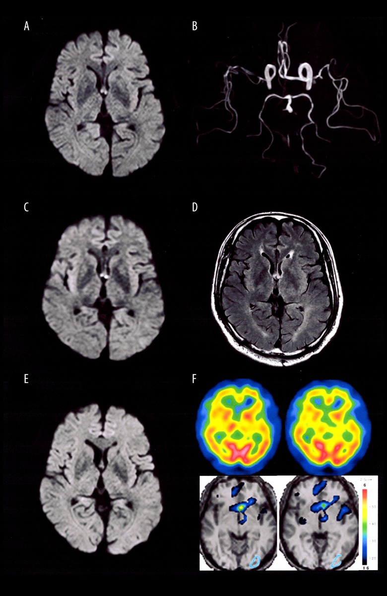
He had a previous history of diabetes mellitus type II and hypertension, which were controlled by oral antidiabetic and antihypertensive drugs. Physical examination was notable for mild hypertension (150/91 mmHg). Neurological examination was unremarkable for focal signs. His cognition was mildly impaired, with a Japanese version of Montreal Cognitive Assessment (MoCA-J) score of 20/30 (optimal cutoff of <26 was taken to indicate cognitive impairment) [4]. Laboratory tests were unremarkable except for HbA1c level of 7.0% (post meal 2-hour blood glucose level of 157 mg/dL). Initial head computed tomography (CT) showed no evidence of intracranial hemorrhage or mass effect. The magnetic resonance imaging (MRI) performed within 24 hours after the estimated onset time showed an isolated small faint focus of restricted diffusion involving the bilateral column of the anterior fornix (Figure 1A). Intracranial MR angiography revealed irregularity and stenosis of middle and anterior cerebral arteries indicating arteriosclerosis (Figure 1B). Transient global amnesia was clinically suspected, and 100 mg of aspirin was administered, based on the suspicion of an acute infarction.
Figure 1.
Brain imaging data on admission (upper row: A, B), 5 (middle row: C, D) and 14 days (bottom row: E, F) after the symptom onset. Diffusion-weighted (left column) and FLAIR (D) sequences, and MR angiography (B), and 99mTc-ECD brain SPECT perfusion images (upper panel) using the easy Z-score imaging system (eZIS) (lower panel) (F). MR – magnetic resonance; SPECT – single photon emission computed tomography; FLAIR – fluid-attenuated inversion recovery.
However, his symptoms persisted, and a repeat MRI was performed 5 days after symptom onset. The MRI demonstrated a brighter, persistent diffusion restriction (Figure 1C), which was associated with swollen fornices bilaterally as evidenced by fluid-attenuated inversion recovery (FLAIR) hyperintensities (Figure 1D). No remarkable findings suggestive of epileptiform activities were detected by electroencephalography.
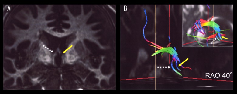
Follow-up MRI performed 14 days later, revealed the disappearance of the abnormal diffusion restriction (Figure 1E). A 99mTc-ECD brain single photon emission computed tomography (SPECT) demonstrated a relative decrease in regional cerebral blood flow around the left anterior fornix and thalamus, which could be corresponding to mild functional deficit of left Papez circuit (Figure 1F). His symptoms gradually improved but he still had mild episodic memory impairment after a month, with a MoCA-J score of 24/30. The coronal 3-dimentional T1-weighted magnetization-prepared rapid acquisition with gradient echo (MRAGE) sequence using a 3T MRI scanner clearly showed a disruption of the column of the left fornix (Figure 2A). Diffusion tensor tracking processed by a dTV. II SR software [5] showed decrease in the density of entire fiber tracts on the Papez circuit as well as location of the left anterior fornix (Figure 2B).
Figure 2.
Detailed MRI evaluation after a month. (A) Coronal T1-weighted MPRAGE sequence showed disruption of the left anterior fornix. (B) Diffusion tensor imaging tractography revealed reduced fiber tracts of the left fornix predominant in the limbic system. Yellow and dotted white arrows indicate left and right anterior fornix, respectively. Inset: Control subject. RAO – right anterior oblique view; MRI – magnetic resonance imaging; MPRAGE – magnetization-prepared rapid acquisition with gradient echo.
Discussion
Although various pathologies might affect the fornix (e.g., trauma, iatrogenic injury after anterior communicating artery aneurysm surgery, brain tumors, microangiopathy, and Wernicke encephalopathy), isolated ischemic infarction of the anterior fornix without an involvement of the corpus callosum and anterior cingulate gyrus has rarely been described [2,3,6,7]. Vascular etiology involving perforating branches of subcallosal artery arising from the anterior cerebral artery or anterior communicating artery has been hypothesized [1], but in the current clinical setting, it is still difficult to visualize the culprit vessels using conventional MRI.
The symptoms attributable to isolated fornix stroke, also known as “amnestic syndrome of the subcallosal artery” [3,8] might be, at least in part, reversible [9]. Gradual clinical improvement might be explained either by an incomplete lesion of the fornices, with preservation of residual functions or the recruitment of alternative memory networks bypassing the fornix [3]. On the other hand, anterograde topographical amnesia and persistent mild episodic memory impairment might be due to a temporary functional disconnection between hippocampal structures and diencephalon [10]. Taken together, in depth MRI examination through the MPRAGE and tractography images provides interesting clinic-anatomical data about the fornix fibers and involvement of adjacent structures consisting of the limbic system to ascribe a sequela of isolated fornix infarction. Especially, MPRAGE is characterized by high-resolution 3-dimensional anatomical sequence and has widely been available to both clinical and experimental neuroimaging communities. The feasibility of visualizing disruption of Papez circuit assisted by these imaging modalities would help us make more accurate diagnosis of amnestic disorders associated with the limbic system.
Conclusions
When dealing with sudden, persistent amnesia associated with small fornix infarction, it is prudent to consider the possibility of tract damage along with the limbic system impairment using MPRAGE sequence and tractography visualization.
Acknowledgments
We are grateful to Dr. Fusako Ono (Furukawa Seiryo Hospital, Miyagi, Japan) for referring this case. We also thank Daisuke Ito (Division of Radiology, Tohoku University Hospital) for technical support. Patient gave written and verbal consent for submit to journal.
Footnotes
Department and Institution where work was done
Department of Geriatric Medicine and Neuroimaging, Tohoku University Hospital, Sendai, Miyagi, Japan
Conflicts of interest
None.
References:
- 1.Fujii T. Perforating branches of the anterior communicating artery: Anatomy and infarction. In: Takahashi S, editor. Neurovascular Imaging. London: Springer; 2011. pp. 189–96. [Google Scholar]
- 2.Gupta M, Kantor MA, Tung CE, et al. Transient global amnesia associated with a unilateral infarction of the fornix: Case report and review of the literature. Front Neurol. 2014;5:291. doi: 10.3389/fneur.2014.00291. [DOI] [PMC free article] [PubMed] [Google Scholar]
- 3.Salvalaggio A, Cagnin A, Nardetto L, et al. Acute amnestic syndrome in isolated bilateral fornix stroke. Eur J Neurol. 2018;25:787–89. doi: 10.1111/ene.13592. [DOI] [PubMed] [Google Scholar]
- 4.Pendlebury ST, Mariz J, Bull L, et al. MoCA, ACE-R, and MMSE versus the National Institute of Neurological Disorders and Stroke-Canadian Stroke Network vascular cognitive impairment harmonization standards neuro-psychological battery after TIA and stroke. Stroke. 2012;43:464–69. doi: 10.1161/STROKEAHA.111.633586. [DOI] [PMC free article] [PubMed] [Google Scholar]
- 5.Masutani Y, Aoki S, Abe O, et al. MR diffusion tensor imaging: Recent advance and new techniques for diffusion tensor visualization. Eur J Radiol. 2003;46:53–66. doi: 10.1016/s0720-048x(02)00328-5. [DOI] [PubMed] [Google Scholar]
- 6.Meila D, Saliou G, Krings T. Subcallosal artery stroke: Infarction of the fornix and the genu of the corpus callosum. The importance of the anterior communicating artery complex. Case series and review of the literature. Neuroradiology. 2015;57:41–47. doi: 10.1007/s00234-014-1438-8. [DOI] [PubMed] [Google Scholar]
- 7.Zhu QY, Zhu HC, Song CR. Acute amnesia associated with damaged fiber tracts following anterior fornix infarction. Neurology. 2018;90:706–7. doi: 10.1212/WNL.0000000000005306. [DOI] [PubMed] [Google Scholar]
- 8.Turine G, Gille M, Druart C, et al. Bilateral anterior fornix infarction: The “amnestic syndrome of the subcallosal artery”. Acta Neurol Belg. 2016;116:371–73. doi: 10.1007/s13760-015-0553-6. [DOI] [PubMed] [Google Scholar]
- 9.Ren C, Yuan J, Tong S, et al. Memory impairment due to a small acute infarction of the columns of the fornix. J Stroke Cerebrovasc Dis. 2018;27:e138–43. doi: 10.1016/j.jstrokecerebrovasdis.2018.02.039. [DOI] [PubMed] [Google Scholar]
- 10.Aguirre GK, D’Esposito M. Topographical disorientation: A synthesis and taxonomy. Brain. 1999;122(Pt 9):1613–28. doi: 10.1093/brain/122.9.1613. [DOI] [PubMed] [Google Scholar]


