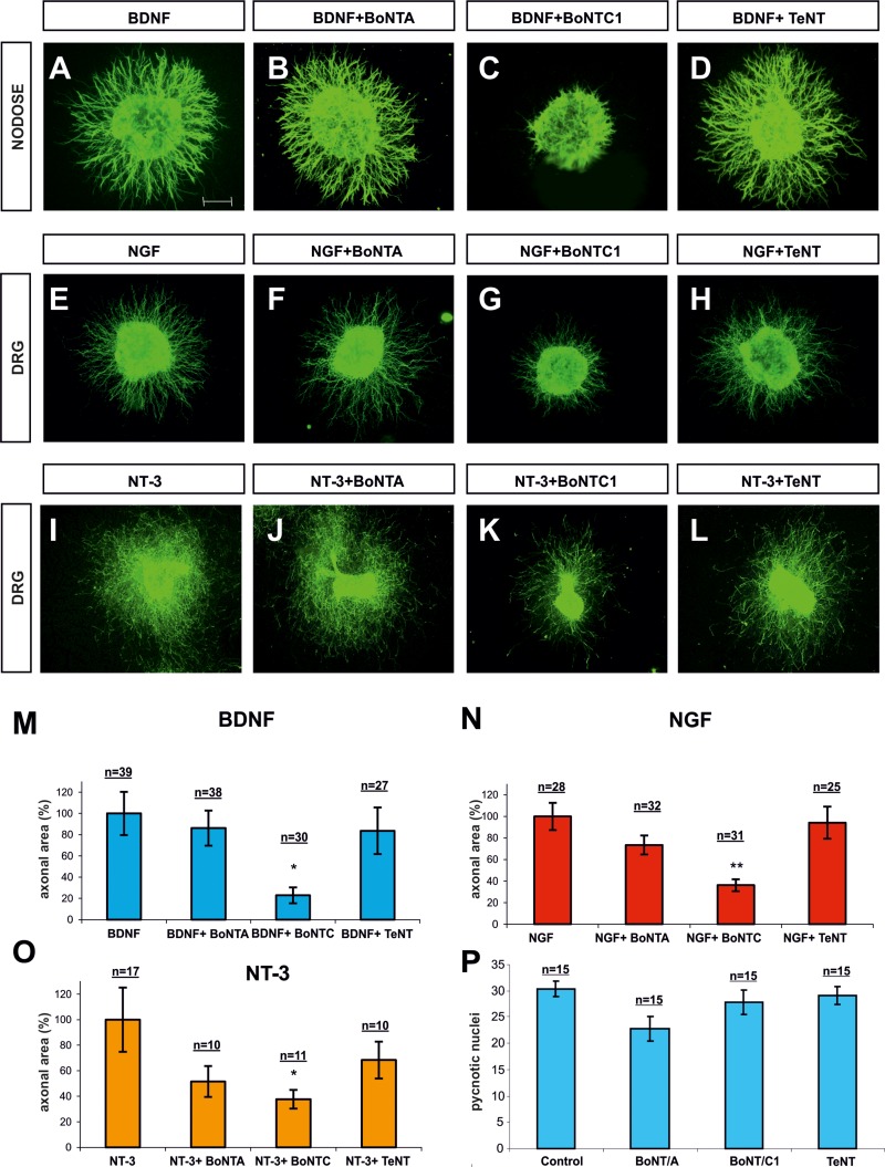
Figure 5. Confocal images of Nodose and DRG explants co-cultured in collagen gel.
Explants were immunolabeled for βIII-Tubulin and counterstained with fluorescent anti-mouse 488. Explants were co-cultured with BDNF (A–D), NGF (E–H) and NT3 (I–L). In each row, explants were treated with BoNT/A, BoNT/C1 and TeNT. Bar graph expressing the area of neurites in Nodose explants supplemented with BDNF (M) and in DRG explants supplemented with, NGF (N) and NT-3 (O), and treated with BoNT/A, BoNT/C1 and TeNT. Bar graph showing the number of cells with pycnotic nuclei, indicating that toxin treatment did not affect neuronal death (P). N indicates the number of explants obtained from different animals in different experiments. Nodose explants came from five experiments in which at least three embryos per condition were used. DRG explants were obtained from six experiments in which one embryo per condition was used. Significant differences are labeled by asterisks (*p ≤ 0.05; **p ≤ 0.01). Scale bar: A, 300 μm. Error bars indicate SEM.

