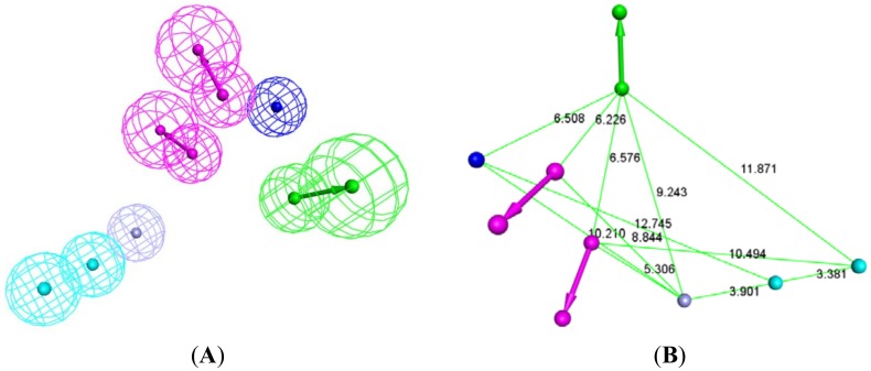
Figure 6.
Structure-based pharmacophore of the active site of Glo-1 enzyme. (A) 3D representation of this pharmacophore; HY: sky-blue; NI: blue; HBD: magenta; HBA: green; ZB: purple. (B) simplified structure-based pharmacophore with no tolerance spheres to facilitate visualization of inter feature distances; HY: sky-blue; NI: blue; HBD: purple; HBA: green; ZB: purple.

