Abstract
This is the case of a 66-year-old male with cervical myelopathy secondary to severe cervical stenosis manifesting as worsening dexterity and numbness in his right hand. The patient underwent C3–C6 laminoplasty with bilateral foraminotomies. During the procedure an incidental durotomy occurred which was patched intraoperatively with Duragen and Tisseel. At 1 month follow-up, the patient reported that he was doing well and skin sutures were removed. Two days later, the patient presented to the emergency department with postoperative wound dehiscence, cerebrospinal fluid (CSF) drainage, altered mental status and lethargy. At that time, a computed tomography (CT) scan confirmed a tension pneumocephalus which was treated with a cranial burr hole and revision durotomy repair. The patient improved and was discharged to a rehabilitation facility with intact motor and cognitive function. At the 1-year follow-up appointment, he continued to do well without sequelae.
Keywords: tension pneumocephalus, dural tear, durotomy
Introduction
Pneumocephalus is a condition in which gas is trapped in the cranial vault and is generally associated with trauma to the skull or face, neurosurgical procedures, and otolaryngology procedures. 1 2 Although uncommon, pneumocephalus after spinal surgery has been known to occur. 1 2 Many factors can contribute to pneumocephalus including the type of anesthesia used, positioning of the patient, and length of surgery. 1 3 When pneumocephalus is encountered, differentiating a tension pneumocephalus from an uncomplicated pneumocephalus is imperative to prevent patient morbidity and mortality. An uncomplicated pneumocephalus is not an uncommon finding on postoperative imaging after neurosurgical procedures and may not require treatment as the gas is slowly resorbed over time and rarely causes significant clinical morbidity. 4 Tension pneumocephalus occurs when gas enters through the dura and can't exit, creating compressive forces on the brain. 5 Tension pneumocephalus may require a burr hole or craniotomy to relieve the pressure on the brain and usually requires dural repair to prevent air from entering through the same route but may also resolve with conservative measures. 6 Patients who have sustained a tension pneumocephalus can present with headache, nausea, vomiting, photophobia, seizures, altered mental status, and decreased neurologic function. 1 2 7
There are two main theories on how tension pneumocephalus occurs after spinal surgery. The first involves the “inverted bottle theory,” where cerebrospinal fluid (CSF) leaks out creating negative pressure in the subarachnoid space allowing air to enter. 6 7 8 9 10 The second theory is described as a “ball valve mechanism,” where a dural tear resulting from a fracture allows air to enter but cannot exit, similar to a tension pneumothorax. 6 7 8 9 10
The incidence of pneumocephalus after spine surgery is unknown with few published case reports describing its occurrence. 1 However, the risk of dural tears, which is associated with pneumocephalus, was reported by Khan et al to occur in 1.8 to 17.4% of degenerative lumbar surgery cases. 11 A retrospective study by Guerin et al reports that the incidence of incidental durotomy during all spinal surgeries, including cervical, thoracic, and lumbar spine surgeries, was estimated to be 3.84% with most durotomies occurring in patients who had a posterior thoracolumbar surgery. 12 Furthermore, a retrospective study by OʼNeill et al reports incidental durotomies occurring in 1% of patients undergoing cervical spine surgery. 13 Most of the case reports of pneumocephalus after spine surgery have been associated with lumbar arthrodesis or similar lumbar surgeries causing a dural leak. 8 There was a case report by Goodwin et al where a tension pneumocephalus was discovered after an anterior cervical discectomy and fusion for traumatic C5/C6 subluxation with coincident esophageal perforation. 4 14 A case report by Sweni et al describes a case of tension pneumocephalus after cervical epidural injection. 15 However, delayed tension pneumocephalus following posterior cervical decompression complicated by intraoperative durotomy, wound dehiscence, and CSF leak has not been described to our knowledge. Therefore, we present the first case of delayed tension pneumocephalus following a posterior cervical spinal decompression surgery complicated by a durotomy.
Case Report
The patient is a 66-year-old male with cervical myelopathy secondary to severe cervical stenosis, most prominently at C3–C4 and C5–C6 with cord signal changes noted on preoperative magnetic resonance imaging (MRI; Fig. 1 ). He presented to clinic with complaints of dexterity issues, right-sided weakness, and numbness which he felt were progressively worsening. The patient was scheduled to undergo C3–C6 laminoplasty with bilateral C4/C5 and C5/C6 foraminotomies. Intra operatively, during the C3 laminoplasty, a right-sided durotomy occurred while creating the open gutter with the burr which was followed by brisk ventral epidural bleeding. The C3 laminoplasty was converted to a complete laminectomy for additional exposure to address the durotomy and ventral epidural bleeding. Brisk ventral epidural bleeding was controlled with surgifoam with thrombin. A small durotomy hole was noted on the lateral margin. The dural tear was repaired in a sutureless fashion with a collagen matrix and fibrin sealant. No CSF leak was noted at the end of the case and a lumbar drain was placed for 72 hours. Postoperatively, patients surgical site remained clean and dry, he denied postural headaches and was discharged.
Fig. 1.

( A–C ) Preoperative MRI. ( A ) Sagittal image demonstrating canal stenosis with cord signal changes. ( B and C ) T2 axial images demonstrating canal stenosis at C3–C4 and C5–C6 respectively. MRI, magnetic resonance imaging.
Two weeks postoperatively, patient presented to the emergency room with complaints of diarrhea. He was diagnosed with clostrium difficile colitis, placed on an appropriate antibiotic regimen which he responded well to and was subsequently discharged to a rehabilitation facility. One month postoperatively, the patient was seen in our clinic at which point he was ambulatory, denying postural headaches, and displaying normal motor strength and sensory function bilaterally. His posterior surgical site was clean, dry, and intact and skin sutures were removed without issue.
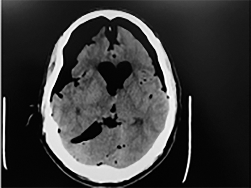
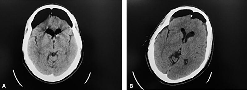
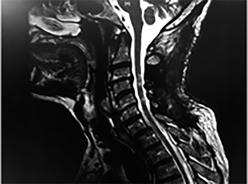
Two days following his suture removal and clinic appointment, the patient presented to the emergency room with the presentation of acute onset altered mental status, decreased mentation, lethargy, and a small 1 cm draining posterior cervical dehisced wound. CT scan obtained in the emergency department was notable for tension pneumocephalus with “Mount Fuji” sign ( Fig. 2 ). The patient underwent bedside burr hole placement and was then taken urgently to the operating room for wound exploration and revision durotomy repair. Intraoperatively, persistent CSF leak at the previous C3 durotomy site was noted. Dural repair was performed with 6–0 prolene simple interrupted sutures, myoplasty with local muscle flap, Duragen, and Tisseel. A lumbar drain was placed intraoperatively and the patient was monitored closely postoperatively with serial CT scans to monitor his pneumocephalus and a progressive decrease in the degree of pneumocephalus was noted ( Fig. 3 ). The patient was extubated and following commands on postoperative day 2 and progressed well postoperatively without sequela or deficits. The lumbar drain was removed on postoperative day 5. He was able to ambulate with physical therapy, demonstrated 5/5 strength in both his upper and lower extremities and was ultimately discharged to a rehabilitation facility. The patient was most recently seen in clinic for his 1-year follow-up and continues to do well. He has no neck pain, good neck range of motion, and normal neurological function without any new postoperative neurological deficits or residual neurological deficits after his tension penumocephalus. His most recent cervical MRI obtained shows no pseudomeningocele and good cord decompression with persistent pre-operative cord signal changes ( Fig. 4 ).l
Fig. 2.

Representative cut from CT scan obtained at presentation to emergency room with patient experiencing decreased mentation, altered mental status and lethargy. CT, computed tomography.
Fig. 3.

( A and B ) CT scans following burr hole placement and revision durotomy repair postoperative days 1 ( A ) and 6 ( B ), respectively. CT, computed tomography.
Fig. 4.

MRI C-spine 6-month postoperatively. MRI, magnetic resonance imaging.
Discussion
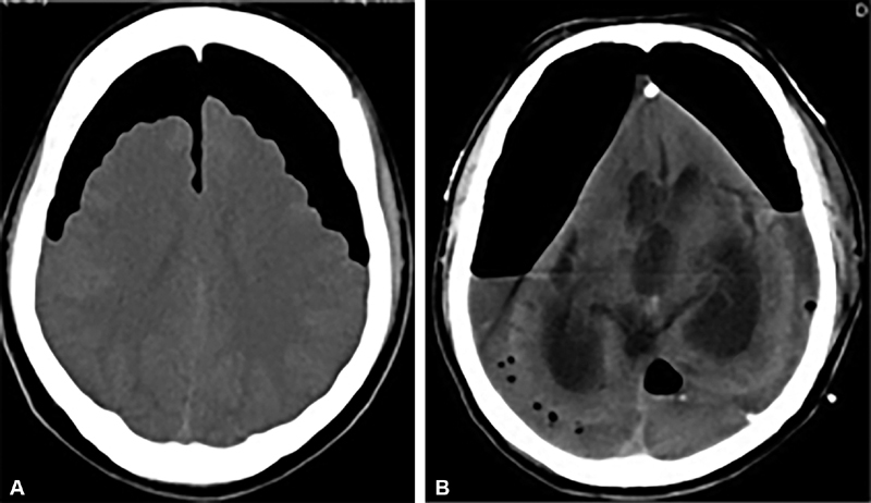
Pneumocephalus is defined as the presence of intracranial air and generally resolves spontaneously or with conservative treatment. 15 However, tension pneumocephalus can lead to clinical deterioration due to its effects within the cranial vault including the pressure exerted by the entrapped air causing a mass effect and elevating intracranial pressures. 15 This can lead to a multitude of neurological findings including cranial nerve palsies, hemiparesis, aphasia, and if not identified and treated early, brainstem herniation, coma, and death. Sweni et al elaborate on the Mount Fuji sign as an indicator of tension pneumocephalus and it should raise concern when noted on imaging ( Fig. 5A ). Named after the tallest mountain peak in Japan, Mount Fuji peaks at 3,776 m in elevation and rests as a dormant volcano on Honshu Island. The appearance of this famous landmark can be paralleled with CT findings in the case of tension pneumocephalus. Accumulation of trapped air in the subdural and interhemispheric spaces lead to both compression and separation of the frontal lobes, as was seen in the case of our patient. This is contrasted with the Peaking sign also described by Sweni et al ( Fig. 5B ) which lacks the interhemispheric space and is less commonly associated with a tension pneumocephalus.
Fig. 5.

( A ) Mount Fuji sign showing pneumocephalus with interhemispheric separation of the frontal lobes. ( B ) Peaking sign showing pneumocephalus without separation of the interhemispheric region (Source: reference 15 15 ).
The incidence of dural tears in cervical spine surgery is 1% as reported in two retrospective studies, consisting of a total of 5,842 patients, by O'Neill et al and Hannallah et al. 13 16 O'Neill et al. reports nine risk factors that were associated with an increased risk of dural tears during cervical spine surgery which include: older age, rheumatoid arthritis, ossification of the posterior longitudinal ligament, cervical deformity, longer operative time, greater number of surgical levels, worse neurological status, performance of a corpectomy, and revision laminectomy. 13 A study by Kalevski et al, found the overall incidence of lumbar dural tears to be 12.66%. They also specified the type of surgeries that were associated with an increased risk of dural tears which included: lumbar reoperations, surgeries for traumatic lumbar spine injuries, patients with degenerative spinal stenosis, lumbar spinal tumors, and surgeries for lumbar disc herniations. 17 Finally, a study by Deyo et al reports that the risk of dural tears was lowest for young patients and patients undergoing microdiscectomy procedures, while the highest risk of dural tears was in elderly patients and patients having reoperative procedures. 18
Although most dural tears are detected and immediately repaired intraoperatively, not all dural tears are identified during surgery. Postoperatively, symptoms include a postural headache, nausea, vomiting, dizziness, and can be difficult to distinguish from common side effects of anesthesia. 7 17 A less immediate but serious sequelae of a dural tear is pneumocephalus, as in our case, and other serious complications include fistula formation, pseudomeningocele, meningitis, and epidural abscess. 17 The presenting symptoms of a pneumocephalus are typically nonspecific, such as lethargy, headache, nausea, vomiting, and confusion. However, more concerning and severe symptoms of pneumocephalus include hemiparesis, seizures, and cranial nerve palsies. 1 2 7
Little is known about the prognosis of patients who have sustained pneumocephalus or tension pneumocephalus because, to our knowledge, there have been no large published studies regarding this rare complication. However, there are individual case reports discussing the treatment of such patients. As discussed in a case report by Simmons and Luks, the prognosis of patients with pneumocephalus is likely related to the time between the onset of pneumocephalus and treatment leading to the resolution of symptoms. 14 However, the outcomes of patients with dural tears causing a CSF leak was studied by Hannallah et al. Their study included 20 patients with CSF leak after cervical spine surgery and concluded that 100% of patients had no signs or symptoms at 4 months. They also report that 60% of patients had resolution of symptoms after just 3 days. Furthermore, they report no long-term sequelae due to the CSF leak at an average of 5.4 years of follow-up. 16 They did report three complications, including a pseudomeningocele requiring drainage, a draining wound, and hand weakness that resolved without treatment. A study by Kalevski et al included 66 patients with incidental dural tears in the lumbar spine and found patients reported lower level of function compared with patients without dural tears, based on Oswestry Disability Index at 2 years follow-up. 17
Dural tears can be managed by a variety of surgical techniques and products. A Study by Dafford and Anderson compared the use of 6-0 polypropylene monofilament suture and 5-0 coated braided nylon suture with random assignment of interrupted or continuous locked suture technique. 19 They concluded that 6-0 polypropylene monofilament had significantly less leakage flow rate than 5-0 coated braided nylon suture. However, they reported that the leakage they did have was from the needle holes rather than the original dural tear. Furthermore, they did not find a significant difference between the suture techniques used to repair the dura. Dafford and Anderson also compared various sealants used to prevent CSF leakage. They concluded that there was not a significant difference between hydrogel sealant, cyanocrylic sealant, and fibrin glue. However, they did note an 80% reduction in leak area with the hydrogel and cyanoacrylic sealants compared with a 38% reduction with fibrin glue. 19 Therefore, they concluded that leakage was significantly reduced with the use of a sealant after suture repair. Another study by Miscusi et al concluded that a bovine serum albumin glutaraldehyde surgical adhesive was also an effective addition to dural repair with sutures with results similar to that of fibrin glue. 20 A study by Narotam et al concluded that sutureless repair of dural tears using a collagen matrix was effective for cerebrospinal fluid containment in 95% of cases. 21 However, not all dural repairs in this study were due to incidental durotomies. Lastly, subarachnoid lumbar drains have been used as a means to aid in closure of complex and or tenuous dural tears. A study by Shapiro and Scully followed a cohort of patients that underwent subarachnoid lumbar drain placement for prevention or treatment of CSF fistulas and found that 38 of the patients in their study underwent drain placement for augmentation of a tenous durotomy closure and all 38 went on to successful closure. 22
Conclusion
In conclusion, we believe that despite tension pneumocephalus being a rare complication, it is one that we must all be aware of in the postoperative period, particularly in cases in which an incidental durotomy occurs. Even though sutureless repair of dural tears have a high success rate, failures may have devastating complications. Primary repair at the time of the durotomy, in this case, could have potentially prevented pneumocephalus. The majority of pneumocephalus cases appear to be spontaneous in origin but recognizing a tension pneumocephalus is important in both management and prognosis. Radiographic signs, such as the Mount Fuji sign can help to lead one to further suspect this complication. We present this case of tension pneumocephalus following cervical spine surgery to further add to the sparse body of literature on this topic. This case also elucidates how dramatic and devastating the symptoms of this condition can be. However, if tension pneumocephalus is identified and managed rapidly, a favorable prognosis can be obtained, as was seen in the case of our patient.
Conflict of Interest None.
Disclosures
Each author certifies that he or she, or a member of his or her immediate family, has no commercial associations (e.g., consultancies, stock ownership, equity interest, patent/licensing arrangements, etc.) that might pose a conflict of interest in connection with this submitted article.
This case report has been created in accordance with the ethical principles of research. Patient consent has been obtained for publication of this case and use of imaging.
References
- 1.Andarcia-Bañuelos C, Cortés-García P, Herrera-Pérez M U, Deniz-Rodríguez B. Pneumocephalus: an unusual complication of lumbar arthrodesis. A clinical case and literature review. Rev Esp Cir Ortop Traumatol. 2015;59(04):222–226. doi: 10.1016/j.recot.2014.04.007. [DOI] [PubMed] [Google Scholar]
- 2.Yun J H, Kim Y J, Yoo D S, Ko J H. Diffuse pneumocephalus : a rare complication of spinal surgery. J Korean Neurosurg Soc. 2010;48(03):288–290. doi: 10.3340/jkns.2010.48.3.288. [DOI] [PMC free article] [PubMed] [Google Scholar]
- 3.Badiger S V, Desai S N, Dasar S. Pneumocephalus following spinal anaesthesia for spine surgery. Indian J Anaesth. 2016;60(05):361–362. doi: 10.4103/0019-5049.181612. [DOI] [PMC free article] [PubMed] [Google Scholar]
- 4.Goodwin C R, Boone C E, Pendleton J et al. Pneumocephalus leading to the diagnosis of cerebrospinal fluid leak and esophageal perforation after cervical spine surgery. J Clin Neurosci. 2016;26:141–142. doi: 10.1016/j.jocn.2015.10.016. [DOI] [PubMed] [Google Scholar]
- 5.Pulickal G G, Sitoh Y Y, Ng W H. Tension pneumocephalus. Singapore Med J. 2014;55(03):e46–e48. doi: 10.11622/smedj.2014041. [DOI] [PMC free article] [PubMed] [Google Scholar]
- 6.Çelikoğlu E, Hazneci J, Ramazanoğlu A F. Tension pneumocephalus causing brain herniation after endoscopic sinus surgery. Asian J Neurosurg. 2016;11(03):309–310. doi: 10.4103/1793-5482.179646. [DOI] [PMC free article] [PubMed] [Google Scholar]
- 7.Gauthé R, Latrobe C, Damade C, Foulongne E, Roussignol X, Ould-Slimane M. Symptomatic compressive pneumocephalus following lumbar decompression surgery. Orthop Traumatol Surg Res. 2016;102(02):251–253. doi: 10.1016/j.otsr.2015.12.006. [DOI] [PubMed] [Google Scholar]
- 8.Karavelioglu E, Eser O, Haktanir A. Pneumocephalus and pneumorrhachis after spinal surgery: case report and review of the literature. Neurol Med Chir (Tokyo) 2014;54(05):405–407. doi: 10.2176/nmc.cr2013-0118. [DOI] [PMC free article] [PubMed] [Google Scholar]
- 9.Kizilay Z, Yilmaz A, Ismailoglu O. Symptomatic pneumocephalus after lumbar disc surgery: a case report. Open Access Maced J Med Sci. 2015;3(01):143–145. doi: 10.3889/oamjms.2015.028. [DOI] [PMC free article] [PubMed] [Google Scholar]
- 10.Pirris S M, Nottmeier E W. Symptomatic pneumocephalus associated with lumbar dural tear and reverse trendelenburg positioning: a case report and review of the literature. Case Rep Neurol Med. 2013;2013:792168. doi: 10.1155/2013/792168. [DOI] [PMC free article] [PubMed] [Google Scholar]
- 11.Khan M H, Rihn J, Steele G et al. Postoperative management protocol for incidental dural tears during degenerative lumbar spine surgery: a review of 3,183 consecutive degenerative lumbar cases. Spine. 2006;31(22):2609–2613. doi: 10.1097/01.brs.0000241066.55849.41. [DOI] [PubMed] [Google Scholar]
- 12.Guerin P, El Fegoun A B, Obeid I et al. Incidental durotomy during spine surgery: incidence, management and complications. A retrospective review. Injury. 2012;43(04):397–401. doi: 10.1016/j.injury.2010.12.014. [DOI] [PubMed] [Google Scholar]
- 13.OʼNeill K R, Neuman B J, Peters C, Riew K D. Risk factors for dural tears in the cervical spine. Spine. 2014;39(17):E1015–E1020. doi: 10.1097/BRS.0000000000000416. [DOI] [PubMed] [Google Scholar]
- 14.Simmons J, Luks A M. Tension pneumocephalus: an uncommon cause of altered mental status. J Emerg Med. 2013;44(02):340–343. doi: 10.1016/j.jemermed.2012.01.055. [DOI] [PubMed] [Google Scholar]
- 15.Sweni S, Senthilkumaran S, Balamurugan N, Thirumalaikolundusubramanian P. Tension pneumocephalus: a case report with review of literature. Emerg Radiol. 2013;20(06):573–578. doi: 10.1007/s10140-013-1135-7. [DOI] [PubMed] [Google Scholar]
- 16.Hannallah D, Lee J, Khan M, Donaldson W F, Kang J D. Cerebrospinal fluid leaks following cervical spine surgery. J Bone Joint Surg Am. 2008;90(05):1101–1105. doi: 10.2106/JBJS.F.01114. [DOI] [PubMed] [Google Scholar]
- 17.Kalevski S K, Peev N A, Haritonov D G. Incidental dural tears in lumbar decompressive surgery: incidence, causes, treatment, results. Asian J Neurosurg. 2010;5(01):54–59. [PMC free article] [PubMed] [Google Scholar]
- 18.Deyo R A, Cherkin D C, Loeser J D, Bigos S J, Ciol M A. Morbidity and mortality in association with operations on the lumbar spine. The influence of age, diagnosis, and procedure. J Bone Joint Surg Am. 1992;74(04):536–543. [PubMed] [Google Scholar]
- 19.Dafford E E, Anderson P A. Comparison of dural repair techniques. Spine J. 2015;15(05):1099–1105. doi: 10.1016/j.spinee.2013.06.044. [DOI] [PubMed] [Google Scholar]
- 20.Miscusi M, Polli F M, Forcato S et al. The use of surgical sealants in the repair of dural tears during non-instrumented spinal surgery. Eur Spine J. 2014;23(08):1761–1766. doi: 10.1007/s00586-013-3138-1. [DOI] [PubMed] [Google Scholar]
- 21.Narotam P K, José S, Nathoo N, Taylon C, Vora Y.Collagen matrix (DuraGen) in dural repair: analysis of a new modified technique Spine 200429242861–2867., discussion 2868–2869 [DOI] [PubMed] [Google Scholar]
- 22.Shapiro S A, Scully T. Closed continuous drainage of cerebrospinal fluid via a lumbar subarachnoid catheter for treatment or prevention of cranial/spinal cerebrospinal fluid fistula. Neurosurgery. 1992;30(02):241–245. doi: 10.1227/00006123-199202000-00015. [DOI] [PubMed] [Google Scholar]


