



Abstract
Background
Prinzmetal's angina is characterized by a transient reduction in luminal diameter of a coronary artery resulting in spontaneous and often recurrent episodes of angina with electrocardiography (ECG) findings consistent with ST elevation.
Case report
A 67-year-old male presented with complaint of intermittent chest discomfort during the previous 2–3 weeks. ECG obtained at presentation was consistent with ST elevation in inferior leads. The patient underwent urgent cardiac catheterization with uncomplicated balloon angioplasty to proximal and distal right coronary artery (RCA). At two-week follow up, the patient continued to complain of recurrent chest discomfort with repeat ECG showing ST elevation in inferior leads. He was given nitroglycerin with symptomatic improvement and gradual resolution of ST elevation. Given concern for reocclusion, the patient again underwent urgent cardiac catheterization showing subtotal occlusion of the proximal RCA. During the procedure, intracoronary nitroglycerin was administered with visible resolution of the occlusion via angiography consistent with coronary vasospasm. The patient was therefore initiated on therapy with amlodipine and isosorbide mononitrate and remained symptom free at subsequent follow-up visits.
Conclusion
Prinzmetal's angina is a well-documented but under-recognized etiology of recurrent ST elevation myocardial infarction which should be considered in those with both typical and atypical cardiovascular risk profiles.
Keywords: Variant angina, Prinzmetal angina, Coronary vasospasm, ST elevation myocardial infarction
Introduction
Prinzmetal's angina or variant angina (VA) is characterized by a transient reduction in the luminal diameter of a coronary artery resulting in spontaneous and often recurrent episodes of angina with transient ST elevation on electrocardiogram (ECG). The attack of VA occurs at rest or during ordinary activity and is not precipitated by strenuous exercise [1]. ECG findings with ST elevation often result in urgent cardiac catheterization especially for those with notable cardiovascular risk factors in which VA may not be initially suspected. Here, we present a case of VA resulting in recurrent cardiac catheterization to evaluate chest pain with ST elevation.
Case report
A 67-year-old African American male with past medical history significant for hypertension, hyperlipidemia, chronic obstructive pulmonary disease, and tobacco use presented to his primary care physician with complaint of intermittent chest discomfort during the previous 2–3 weeks. The pain was described as a substernal pressure which was non-radiating and occurred daily lasting for 5–15 min per episode. The pain occurred spontaneously and was not associated with exertion. Home medications included hydrochlorothiazide and lisinopril. He did not have a significant family history for premature coronary artery disease. ECG at presentation was consistent with ST elevation in the inferior leads. The patient was immediately transferred to the emergency department with subsequent urgent cardiac catheterization notable for 90% proximal and 90% distal right coronary artery (RCA) lesions and underwent uncomplicated balloon angioplasty without stenting. Of note, the patient had a peak troponin of 0.02 μg/L and ejection fraction (EF) of 65% with mild inferior wall hypokinesis by left ventriculography. He was discharged on aspirin, clopidogrel, metoprolol, simvastatin, lisinopril, and nitroglycerin as needed for chest pain.
At two-week follow up with cardiology, the patient complained of having recurrent chest pain mostly in the morning since the time of discharge. He also developed similar chest discomfort in the morning on the day of his appointment. It was 7/10 in intensity and reduced to 5/10 in clinic. His blood pressure was 143/85 mmHg and ECG showed 3 mm ST elevation in leads II, III, and aVF and reciprocal ST depression in leads I and aVL. The patient was given nitroglycerin after which his blood pressure reduced to 123/78 mmHg and repeat ECG showed gradual decrease in ST elevations with symptomatic improvement in the chest discomfort (Fig. 1, Fig. 2). The patient was taken back for cardiac catheterization due to concern for reocclusion of RCA.
Fig. 1.

Electrocardiogram 1: on initial presentation showing ST elevation in leads II, III, and aVF.
Fig. 2.

Electrocardiogram 2: 30 min after sublingual nitroglycerin showing gradual resolution of ST segments in inferior leads.
Repeat cardiac catheterization showed subtotal occlusion of proximal RCA. During the procedure, he was given intracoronary nitroglycerin with visible resolution of the occlusion consistent with coronary vasospasm (Fig. 3, Fig. 4). EF was 65% with no wall motion changes on cardiac catheterization. As the repeat catheterization had occlusion at the same location, it was perceived that vasospasm must have been the cause during the first event. No intracoronary nitroglycerin was used during the first catheterization. Following this, he was diagnosed with VA and was started on amlodipine and isosorbide mononitrate as vasodilator therapy. The patient remained symptom free on subsequent follow-up visits.
Fig. 3.
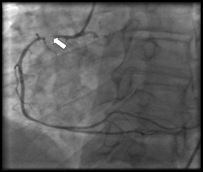
Coronary angiogram with evidence of luminal narrowing of the right coronary artery.
Fig. 4.
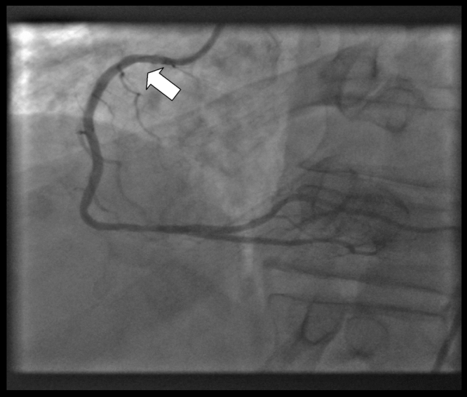
Coronary angiogram showing normalization of the right coronary artery following intracoronary nitroglycerin suggesting coronary vasospasm.
Discussion
The key finding for the diagnosis of coronary vasospasm includes simultaneous resolution of ST segment changes and anginal symptoms spontaneously or with nitroglycerin, or reversibility of coronary occlusions upon intraluminal injections of nitroglycerin during cardiac catheterization. Yasue et al. [2] suggested the diagnostic criteria for VA without performing coronary angiography if anginal attacks disappear quickly upon administration of nitroglycerin and if any one of following criteria were met: (1) attacks appear at rest, particularly between night and early morning; (2) reduction in exercise capacity in the early morning; (3) attacks accompanied by ST segment elevation on ECG; (4) attacks are induced by hyperventilation; (5) attacks are suppressed by calcium channel blockers but not by beta-blockers. This has been included in diagnostic criteria for VA by the Japanese Society of Circulation 2008 [3].
Female gender, relatively younger age, smoking, magnesium deficiency, lipid abnormalities, stress, and genetic factors are important risk factors [4]. Genetic factors play an important role as seen by three times higher prevalence of VA in Asian countries like Japan compared to Western countries. The exact pathogenesis is unclear but various studies have shown increased vagal activity and hyperactivity to adrenergic system before the attacks [5]. Also endothelial dysfunction and decrease in smooth muscle relaxation may result in vasospasm.
Patients with VA show ST segment elevation and reciprocal depression on ECG during attacks of chest pain. Ambulatory ECG monitoring in patients with VA showed circadian variation with most of the ischemic episodes occurring between midnight to early morning hours with peak frequency around 05.00 h [6]. Another important feature of VA is the frequency of asymptomatic ischemic episodes being much higher than symptomatic episodes. One study revealed that 82% (872 out of 1062) of episodes were asymptomatic [6]. Severe arrhythmias, including ventricular tachycardia, high-degree atrioventricular block, and bradyarrythmias resulting in syncopal episodes may be occasionally seen during an attack of VA. Patients with recurrent chest pain with normal coronaries on angiography should be considered for Holter or ambulatory ECG monitoring to detect asymptomatic ischemic changes from VA [4].
Medical treatment for VA includes cardiovascular risk factor reduction and vasodilators including calcium-channel blockers and nitrates. In resistant cases, addition of magnesium supplements, statins, antioxidants, and fasudil, a Rho-kinase inhibitor, prevent coronary vasospasm [7].
Conclusion
Printzmetal's angina/VA is a well-documented etiology of recurrent ST elevation myocardial infarction which should be considered in patients with and without typical cardiovascular risk profiles to avoid repeat invasive studies.
Conflict of interest
All authors declare that they have no conflicts of interest in this case report.
Footnotes
DOI of commentary article: http://dx.doi.org/10.1016/j.jccase.2012.08.006.
References
- 1.Prinzmetal M., Kennamer R., Merliss R., Wada T., Bor N. Angina pectoris. I. A variant form of angina pectoris; preliminary report. Am J Med. 1959;27:375–388. doi: 10.1016/0002-9343(59)90003-8. [DOI] [PubMed] [Google Scholar]
- 2.Yasue H., Nakagawa H., Itoh T., Harada E., Mizuno Y. Coronary artery spasm—clinical features, diagnosis, pathogenesis, and treatment. J Cardiol. 2008;51:2–17. doi: 10.1016/j.jjcc.2008.01.001. [DOI] [PubMed] [Google Scholar]
- 3.Guidelines for diagnosis and treatment of patients with vasospastic angina (coronary spastic angina) (JCS 2008): digest version. Circ J. 2010;74:1745–1762. doi: 10.1253/circj.cj-10-74-0802. [DOI] [PubMed] [Google Scholar]
- 4.Kusama Y., Kodani E., Nakagomi A., Otsuka T., Atarashi H., Kishida H., Mizuno K. Variant angina and coronary artery spasm: the clinical spectrum, pathophysiology, and management. J Nihon Med Sch. 2011;78:4–12. doi: 10.1272/jnms.78.4. [DOI] [PubMed] [Google Scholar]
- 5.Saitoh T., Kishida H., Hanashi A., Tsukada Y., Fukuma Y., Sano J., Fukuma N., Kusama Y., Hayakawa H. Coronary hyperreactivity to adrenergic stimulation and increased nocturnal vagal tone trigger coronary vasospasm. Jpn Circ J. 1998;62:721–726. doi: 10.1253/jcj.62.721. [DOI] [PubMed] [Google Scholar]
- 6.Kishida H., Tada Y., Fukuma N., Saitoh T., Kusama Y., Sano J. Significant characteristics of variant angina patients with associated syncope. Jpn Heart J. 1996;37:317–326. doi: 10.1536/ihj.37.317. [DOI] [PubMed] [Google Scholar]
- 7.Masumoto A., Mohri M., Shimokawa H., Urakami L., Usui M., Takeshita A. Suppression of coronary artery spasm by the Rho-kinase inhibitor fasudil in patients with vasospastic angina. Circulation. 2002;105:1545–1547. doi: 10.1161/hc1002.105938. [DOI] [PubMed] [Google Scholar]