Abstract
Two-dimensional (2D) magnetism has been long sought-after and only very recently realized in atomic crystals of magnetic van der Waals materials. So far, a comprehensive understanding of the magnetic excitations in such 2D magnets remains missing. Here we report polarized micro-Raman spectroscopy studies on a 2D honeycomb ferromagnet CrI3. We show the definitive evidence of two sets of zero-momentum spin waves at frequencies of 2.28 terahertz (THz) and 3.75 THz, respectively, that are three orders of magnitude higher than those of conventional ferromagnets. By tracking the thickness dependence of both spin waves, we reveal that both are surface spin waves with lifetimes an order of magnitude longer than their temporal periods. Our results of two branches of high-frequency, long-lived surface spin waves in 2D CrI3 demonstrate intriguing spin dynamics and intricate interplay with fluctuations in the 2D limit, thus opening up opportunities for ultrafast spintronics incorporating 2D magnets.
Characteristics of spin waves in recently discovered two-dimensional (2D) Ising ferromagnets are still lacking. Here, Jin and Kim et al. report Raman resonance evidence of two sets of surface spin waves in the 2D honeycomb ferromagnet CrI3.
Introduction
The recent discovery of two-dimensional (2D) ferromagnetism1,2 proves that magnetic anisotropy can overcome thermal fluctuations and stabilize long-range magnetic orders in the 2D limit at finite temperatures. This has immediately triggered tremendous interest3–12 in potential 2D magnet-based applications, ranging from ultrathin magnetic sensors to high-efficiency spin filter devices. Naturally, a complete description of the 2D magnetic phase is now needed, which requires not only the identification of the ordered ground state, but equally important, the understanding of the excitations, i.e., spin waves, or equivalently, magnons13,14. To date, there have been no direct comprehensive experimental studies on the full characteristics of spin waves in these 2D Ising ferromagnets, aside from the quasiparticle excitation spectra from the inelastic tunneling measurements in ref. 11.
A spin wave describes the spin dynamics of magnetic ordering when excited, and its frequency determines the ultimate switching speed of state-of-the-art ultrafast spintronic devices15–18. Generally speaking, the well-established spintronic devices based on Heisenberg ferromagnets have speeds in the gigahertz (GHz) regime due to the weak magnetic anisotropy19,20, while the speeds of the recently proposed antiferromagnet-based spintronic devices fall into the terahertz (THz) range owing to the exchange interaction between the two sublattices of the antiferromagnets21–23. Remarkably, the newly discovered 2D Ising honeycomb ferromagnet CrI3 possesses both merits for realizing high-frequency spin waves: the strong magnetic anisotropy from the Ising-type spin interactions24 and the large exchange coupling between the two Cr3+ sublattices within the honeycomb framework25.
In this work, we study the spin wave excitations in the 2D Ising honeycomb ferromagnet CrI3 using polarized micro-Raman spectroscopy. Based on Raman symmetry analysis, we uniquely distinguish two spin wave modes at 2.28 and 3.75 THz from the rest of the optical phonon modes. The doubling of the spin wave mode number is a direct consequence of the underlying non-Bravais honeycomb lattice, while the exceptionally high THz frequency for a ferromagnet reflects the strong magnetic anisotropy and exchange interactions in good agreement with theoretical expectations. From the temperature and thickness dependence of the spin wave characteristics, we show that short-range magnetic correlations are set in prior to the formation of a long-range static magnetic order for every thickness, and that stronger fluctuation effects appear in thinner flakes. Remarkably, we found that the integrated intensities (I. I.) of the two spin wave modes exhibit nearly no thickness dependence, in stark contrast to the fact that the I. I. for all optical phonons scale linearly with their sample thickness, as expected for all bulk modes in quasi-2D-layered materials. This observation on the two spin waves, however, shows striking analogy to the surface modes whose I. I. is independent of the thickness. Moreover, we show that, from more than ten-layer to monolayer CrI3, the spin wave frequencies (2.28 and 3.75 THz) and the onset temperatures (45 K) remain nearly constant, while their lifetimes decrease significantly from 50 and 100 ps to 15 ps, but remain an order of magnitude longer than their corresponding spin wave temporal periods.
Results
Spin wave dispersion calculations in 2D CrI3
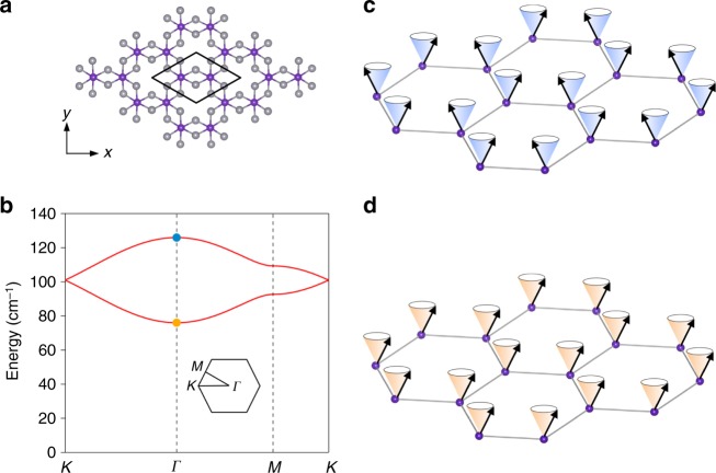
We first performed the spin wave dispersion relation calculations for the monolayer ferromagnet CrI3, in which the Cr3+ cations form a honeycomb structure with the edge-sharing octahedral coordination formed by six I− anions and the magnetic moment of the two Cr3+ cations (S = 3/2) per unit cell aligns in the same out-of-plane direction (Fig. 1a). The minimum model to describe this ferromagnetism in the monolayer CrI3 is the Ising spin Hamiltonian, , where is the spin operator along the X (Y, Z) direction at the Cr3+ site i (j); JZ and JXY are the exchange coupling constants for the out-of-plane and the in-plane spin components, respectively, and satisfy JZ>JXY> 0 for Ising ferromagnetism; and 〈i,j〉 denotes the approximation of the nearest-neighbor exchange coupling. Figure 1b shows the calculated spin wave dispersion relation along the high-symmetry directions (Κ–Γ–Μ–Κ) of the first Brillouin zone (Fig. 1b, inset). Because there are two magnetic Cr3+ cations per primitive cell, there are two spin wave branches whose eigenstates contain in-phase (lower branch) and out-of-phase (upper branch) spin precessions between the two sublattices26. Of particular interest to Raman scattering, as well as the tunneling geometry of the magnetic filter devices, are the spin wave states at the Brillouin zone center (Γ point, zero-momentum) describing the uniform precession of the spins about the easy axis. The energy barrier from the ground state to the lower branch is proportional to the magnetic anisotropy, , which is the energy cost for the spins to uniformly tilt off the easy axis in this excited state (Fig. 1d). The energy barrier for the upper branch results from a combined effect of both the magnetic anisotropy and the in-plane exchange coupling, , corresponding to the energy needed for tilting the spins at the two Cr3+ sublattices in the opposite directions upon this excitation (Fig. 1c).
Fig. 1.
Magnetic excitations in the 2D Ising ferromagnet CrI3. a Top view of the atomic structure of monolayer CrI3, with Cr3+ in purple and I– in gray. Cr3+ ions form a honeycomb lattice with two Cr3+ ions per unit cell (black enclosure). b Calculated magnon dispersion relation in monolayer CrI3. The inset shows the Brillouin zone. Magnon modes at the zone center (Γ point, zero-momentum) are highlighted by yellow and blue solid circles. c, d Schematic of the zone center magnon modes. The cones represent the precession trajectories of the spins (black arrows). The precession of spins on two Cr3+ sublattices are out-of-phase (c) and in-phase (d), which corresponds to the high- and low-energy modes, respectively, in b
Raman signature of two sets of zero-momentum spin waves
To study both zero-momentum spin waves in 2D CrI3 crystals, we fabricated CrI3 thin flakes fully encapsulated by hexagonal boron nitride (hBN) (see sample fabrication details in Methods and thickness characterization in Supplementary Note 1) and performed micro-Raman spectroscopy measurements on them as a function of layer number and temperature, as frequency-resolved magnetic Raman scattering cross section is directly proportional to the time-domain magnetic correlation function27. In contrast to phonon Raman scattering which preserves time-reversal symmetry (TRS) and thus has symmetric Raman tensors28, the leading-order one-magnon Raman scattering involves spin flipping (ΔS = ±1) that breaks TRS, and consequently corresponds to antisymmetric Raman tensors29,30. Based on the difference in the Raman tensors, we can therefore readily distinguish magnon Raman modes from phonon modes via polarization selection rules. In our Raman measurements with the backscattering geometry, the polarizations of the incident and the scattered light were kept to be either parallel or perpendicular to each other, and could be rotated together with respect to the in-plane crystal axis by any arbitrary angle ϕ. The incident photon energy of 1.96 eV was chosen to be on resonance with the charge transfer and the Cr3+ 4A2 to 4T1 transitions of CrI3 in order to increase the Raman sensitivity of magnon scattering4 (see Supplementary Note 2 for a comparison with the nonresonant Raman spectra).
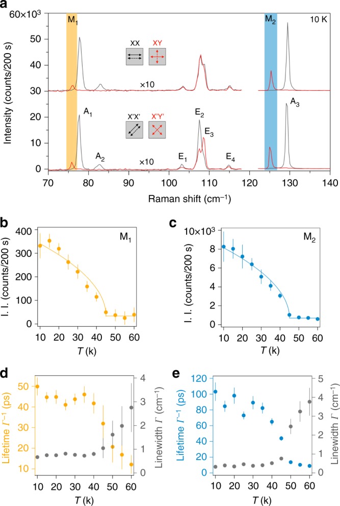
Figure 2a shows low-temperature (10 K) Raman spectra taken on a 13-layer (13 L) CrI3 flake in both parallel and cross-polarization selection channels at ϕ = 0o and ϕ = 45o, denoted as XX, XY, X’X’, and X’Y’, respectively. In total, there are nine Raman-active modes observed, which can be categorized into three groups based on their selection rules. First and most remarkably, the M1 and M2 modes are only present in the cross-channels (XY and X’Y’) and are absent in the parallel channels (XX and X’X’) within our detection resolution. This leads to the unique identification of purely antisymmetric Raman tensors for these two modes, evidence of them arising from the two zero-momentum magnons depicted in Fig. 1c and 1d. Of equal interest are their high frequencies at 76 cm–1 (2.28 THz, or 9.4 meV) and 125 cm–1 (3.75 THz, or 15.5 meV), respectively, which are three orders of magnitude higher than those of the conventional ferromagnets used in most spintronic devices today (in the GHz range)19,20. Even though our measured magnon frequencies are in a similar energy scale as those reported in Ref. 11 (3 and 7 meV, and possibly 17 meV), their quantitative difference is significant and invites further investigations on the spin dynamics in 2D CrI3. Furthermore, by substituting ΔL and ΔU with the two magnon frequencies above, the exchange coupling constants for the in-plane (JXY) and the out-of-plane (JZ) spins are determined to be 11 cm–1 and 44 cm–1, respectively, in good agreement with that obtained from generalized calculations of magnetic coupling constants for bulk CrI331. Second, the A1, A2, and A3 modes show up only in the parallel channels (XX and X’X’) without any ϕ dependence, and therefore these are the Ag phonon modes under the rhombohedral crystal point group C3i (space group )32. Third, the E1, E2, E3, and E4 modes are the Eg phonon modes of C3i, because of their appearance in both parallel and cross-channels, as well as the rotational anisotropy of their intensities (see detailed analysis for all Raman modes in Supplementary Note 3).
Fig. 2.

Detection of the zero-momentum magnons in thick CrI3. a Raman spectra of a thick CrI3 flake (13 layers) at low temperature (10 K) in the parallel and cross-channels at ϕ = 0o (XX and XY) and at ϕ = 45o (X’X’ and X’Y’). Magnon modes, M1 and M2, appearing only in the cross-channels (XY and X’Y’), are highlighted in yellow and blue. Phonon modes are labeled as A1, A2, E1–E4, and A3. The spectral intensities in the 70–120 cm–1 range are multiplied by a factor of 10. The spectra in the XX and XY channels are vertically offset for clarity. The spectra are acquired using a 633-nm excitation laser. b, c Temperature dependence of I. I. of the M1 and M2 magnon modes, respectively. Solid curves are fits to . d, e Temperature dependence of the lifetime (Γ−1, left axis) and the linewidth (Γ, right axis) of the M1 and M2 magnon modes, respectively. Error bars in b–e represent the two standard errors of fitting parameters in the Lorentzian fits to individual temperature-dependent Raman spectra
Temperature dependence of spin waves
Having identified the two single-magnon modes in the Raman spectra at low temperature, we proceed to evaluate their temperature dependence. To see the results quantitatively, we fit the magnon modes with a Lorentzian function of the form , where ωo, Γ, and A are the central frequency, linewidth, and peak intensity of the magnon mode, respectively. Figure 2b, c show the temperature dependence of the I. I., , of the two magnon modes. Clearly, both traces exhibit a clear upturn below a critical temperature TC = 45 K (the same value for bulk CrI3), that is, however, lower than the bulk Curie temperature of 60 K determined by the magnetic susceptibility measurements under a magnetic field of 0.1 T (see the magnetic susceptibility data and Raman data on bulk CrI3 in Supplementary Note 4). The temperature dependence of the magnon lifetimes (inverse of the linewidths, Γ−1, Fig. 2d, e), on the other hand, shows that a short lifetime of < 10 ps sets in at 60 K and saturates at about 50 ps (100 ps) around 45 K for the M1 (M2) magnon. This, together with the divergent behavior of the linewidth temperature dependence (see Fig. 2d, e), indicates that strong magnetic fluctuations are present before the static magnetic order is established at 45 K. It is therefore likely that 60 K marks the onset of the field-stabilized magnetic correlations, while 45 K denotes the intrinsic transition to the spontaneous ferromagnetism, reconciling the difference between the critical temperatures measured by the two different experimental techniques. This is also consistent with the temperature-dependent magneto-optical Kerr effect2 and tunneling7,12 measurements of thin samples at zero fields, and explains the difference between the tunneling resistance with11 and without7,12 a magnetic field appearing above TC.
Thickness dependence of spin waves
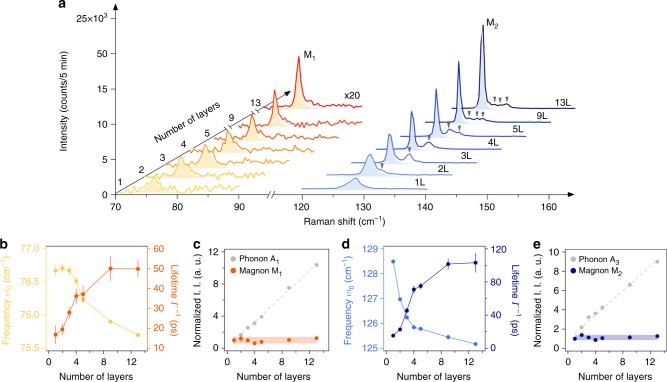
To investigate how thermal fluctuations impact the intrinsic 2D ferromagnetism and its excitations, we performed a systematic Raman study of the M1 and M2 magnon modes measured on atomically thin CrI3 crystals ranging from 13 L to monolayer (1 L). It is clear from the data taken at 10 K in Fig. 3a that both main magnon modes (M1 and M2) and their satellite modes (highlighted with gray triangles in Fig. 3a) have a notable layer number dependence. First, the satellite magnon modes arise from the finite thickness effect in thin layers, in which broken translational symmetry perpendicular to the basal plane makes single-magnon modes with finite out-of-plane momenta accessible in Raman scattering33,34 (see the same effect for phonons in Supplementary Note 5). The small energy separation between the main mode and the nearest satellite, on the order of 3 cm–1, indicates the weak interlayer magnetic coupling strength26, consistent with the small training magnetic field of less than 1 T reported in literature2,8,11. Moreover, as the layer number decreases, there are fewer but stronger observable satellite magnon modes in the Raman spectra. Second, the two main magnon modes, M1 and M2, persist down to the monolayer with symmetric lineshapes, while their peak intensities drop and their linewidths broaden with decreasing layer numbers.
Fig. 3.
Layer number dependence of the zero-momentum magnon characteristics. a Raman spectra of the M1 (intensity multiplied by 20) and M2 magnons for 1–5 L, 9 L, and 13 L samples. Light yellow and blue shaded areas are the Lorentzian fits for the M1 and M2 magnon modes, respectively. The gray triangles highlight the satellite magnon peaks. b Frequency (ω0, left axis) and lifetime (Γ–1, right axis) of the M1 magnon as a function of layer number. c I. I. of the M1 magnon as a function of layer number with I. I. of the A1 phonon shown in gray for comparison. d, e Plots for M2 that are similar to b, c, with I. I. of the A3 phonon plotted in gray in e for comparison. Error bars in b–e represent the two SEs of fitting parameters in the Lorentzian fits to individual Raman spectra at different thicknesses
To understand the layer number dependence of the M1 and M2 magnons in greater detail, we extracted the magnon mode frequencies (ωo), lifetimes (Γ−1), and I. I. (), from fitting their Raman spectra with the Lorentz function (), and the results are summarized in Fig. 3b–e. The frequencies of the M1 and M2 magnons increase slightly, by about 0.8 cm–1 (1.1%) and 3 cm–1 (2.4%), respectively, as the layer number decreases from 13 L to 1 L, possibly because the reduced electronic screening in thinner samples enhances the exchange coupling. In sharp contrast, their lifetimes drop significantly, from about 50 ps (100 ps) in 13 L to 15 ps in 1 L for M1 (M2), which is consistent with the increased thermal fluctuations in 2D. Despite this decrease, even in the monolayer, the lifetimes are still more than one order of magnitude larger than the corresponding magnon temporal periods, about 30 times for M1 and 50 times for M2. This ratio of magnon lifetime to the temporal period in 2D CrI3 is significantly higher than that of the Heisenberg ferromagnets19,20 and at least comparable to, if not greater than, that of the antiferromagnets20–22, making coherent control of both THz spin waves in the time domain feasible down to the monolayer limit of CrI3. Remarkably, I. I. of both magnons, which is known to be proportional to the magnon density, remain nearly constant and independent of the layer number, while that of the phonons scale linearly with the thickness (Fig. 3c, e). This observation on the M1 and M2 magnons is consistent with surface magnons whose density is thickness-independent. Considering that surface magnons have been theoretically predicted in 2D honeycomb ferromagnets26, although mainly at different wave vectors K points, it might not be unreasonable to speculate the surface origin of the two THz magnon modes that we have detected here.
Phase diagram of 2D CrI3
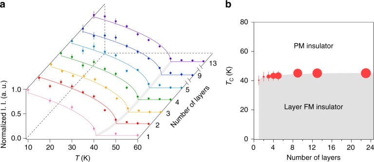
By carrying out temperature-dependent Raman measurements and analysis similar to Fig. 2b, c for different thickness samples, we tracked the layer dependence of the 2D ferromagnetism onset temperature. Figure 4a displays the temperature-dependent traces of the normalized I. I. of the M2 magnon plotted as a function of layer number, with the onset temperature (TC) for each trace determined by fitting with an order-parameter-like function for a ferromagnet (see the similar plot of M1 in Supplementary Note 6). As the layer number decreases from 13 L to 1 L, the extracted TC has an observable decline from 45 K to 40 K. This approximately 12% suppression in TC is in sharp contrast to the slight enhancement of the magnon frequencies, i.e., 1.1% increase of ΔL for M1 and 2.4% of ΔU for M2, which then suggests that the drop in TC is due to stronger thermal fluctuations in thinner samples35. Nevertheless, the finite TC for all samples with various thicknesses establishes a phase boundary for the intrinsic transition to the intralayer ferromagnetism in 2D CrI3 (see Fig. 4b).
Fig. 4.
Temperature versus layer number phase diagram of the 2D layer ferromagnetic CrI3. a Temperature dependence of I. I. of the M2 magnon normalized to the value at 10 K as a function of layer number. The colored solid curves are fits to . The gray curve is the guide to the eye of the evolution of TC. Error bars correspond to the two SEs of normalized I. I. in the Lorentzian fits at individual temperatures for different thicknesses of 2D CrI3. b Temperature versus layer number phase diagram (PM for paramagnetism and FM for ferromagnetism). The size of the data points represents the M2 magnon lifetime. Error bars correspond to the two SEs of TC in the fits in a
Discussion
We have identified two branches of THz spin waves with their lifetime on the order of 10–100 ps in a 2D Ising ferromagnet, CrI3, whose magnetic onset temperature TC remains close to that of their bulk crystal. The robust THz magnons in 2D CrI3 are in stark contrast to spin waves in conventional metallic ferromagnetic thin films that occur at relatively low, GHz frequencies16 and also show significant substrate-dependence36–38. Similar to many antiferromagnets, 2D CrI3 is a semiconductor that possesses high-frequency, long-lived spin waves and is free of stray magnetic fields within 2D domains39,40. Different from bulk antiferromagnets, 2D CrI3 couples efficiently with external magnetic fields2–4,8,11,12 and can be tailored in various device geometries with definitive thicknesses6,10. We envision that these unique characteristics of spin waves in 2D CrI3 will provide unprecedented opportunities for applications in ultrafast and ultracompact spintronic devices.
Methods
Magnon dispersion calculations
The Ising Hamiltonian with anisotropic exchange coupling is transformed by applying the Holstein–Primakoff transformation. The single-site spin operators, and , are related to the momentum space magnon creation and annihilation operators, and αk, as , , and where ϕ = a or b, corresponding to the two Cr3+ sublattices (n = 2 and S = 3/2). The bosonic Hamiltonian is then diagonalized using wavefunction to extract the magnon dispersion relations and the eigenvectors.
Growth of CrI3 single crystals
The single crystals of CrI3 were grown by the chemical vapor transport method. Chromium powder (99.99% purity) and iodine flakes (99.999%) in a 1:3 molar ratio were put into a silicon tube with a length of 200 mm and an inner diameter of 14 mm. The tube was pumped down to 0.01 Pa and sealed under vacuum, and then placed in a two-zone horizontal tube furnace. The two growth zones were raised up slowly to 903 K and 823 K for 2 days, and were then held there for another 7 days. Shiny, black, plate-like crystals with lateral dimensions of up to several millimeters can be obtained from the growth. In order to avoid degradation, the CrI3 crystals were stored in a glovebox filled with nitrogen.
Fabrication of few-layer samples
CrI3 samples were exfoliated in a nitrogen-filled glovebox and the thickness of the flakes was first estimated by the optical contrast. Using a polymer-stamping technique inside the glove box, CrI3 flakes were sandwiched between two few-layer hBN flakes to avoid surface reaction with oxygen and moisture in the ambient environment. The encapsulated CrI3 samples were then moved out of the glove box for Raman spectroscopy measurements. After Raman spectroscopy measurements, the thicknesses of the encapsulated CrI3 flakes were determined by the atomic force microscopy (AFM) measurements.
Raman spectroscopy
Raman spectroscopy measurements were carried out using both a 633-nm and a 532-nm excitation laser with a beam spot size of ~3 μm. The laser power was kept at 80 μW, corresponding to a similar fluence used in literature (10 μW over an ~1-μm-diameter area), to minimize the local heating effect. Backscattering geometry was used. The scattered light was dispersed by a Horiba Labram HR Raman spectrometer and detected by a thermoelectric cooled CCD camera. Selection rule channels XX and XY denote the parallel and cross-polarizations of incident and scattered light at ϕ = 0o; X’X’ and X’Y’ represent the parallel and cross-channels at ϕ = 45o. A closed-cycle helium cryostat was interfaced with the micro-Raman system for the temperature-dependent measurements. All thermal cycles were performed at a base pressure lower than 7 × 10⁻⁷ Torr.
Electronic supplementary material
Source Data Supplementary Figure 8 (1L data)
Source Data Supplementary Figure 8 (2L data)
Source Data Supplementary Figure 8 (3L data)
Source Data Supplementary Figure 8 (4L data)
Source Data Supplementary Figure 8 (5L data)
Source Data Supplementary Figure 8 (9L data)
Source Data Supplementary Figure 8 (13L data)
Acknowledgements
We acknowledge useful discussions with Roberto Merlin, Lu Li, Andrew Millis, and Allan McDonald. We thank Gaihua Ye for his technical assistance. R.H. acknowledges support by NSF CAREER Grant No. DMR-1760668 (Z.Y., F.D., S.S., E.W., and R.H.), and NSF MRI Grant No. DMR-1337207 (low-temperature equipment). L.Z. acknowledges support by NSF CAREER Grant No. DMR-1749774 (S.L. and L.Z.). A.W.T. acknowledges support from NSERC Discovery grant RGPIN-2017-03815 and the Korea–Canada Cooperation Program through the National Research Foundation of Korea (NRF) funded by the Ministry of Science, ICT and Future Planning (NRF-2017K1A3A1A12073407). This research was undertaken, thanks in part to funding from the Canada First Research Excellence Fund. H.L. acknowledges support by the National Key R&D Program of China (Grant No. 2016YFA0300504), the National Natural Science Foundation of China (Grant No. 11574394, No. 11774423), and the Fundamental Research Funds for the Central Universities, and the Research Funds of Renmin University of China (15XNLF06, 15XNLQ07, and 18XNLG14). K.S. acknowledges support via NSF Grant No. NSF-EFMA-1741618 and the Alfred P. Sloan Foundation.
Author contributions
W.J. and L.Z. conceived and initiated this project; C.L., S.T., and H.L. synthesized and characterized the bulk CrI3 single crystals; H.H.K., B.Y., and A.W.T. fabricated and characterized the few-layer samples; Z.Y., P.R., F.D., S.S., E.W., and R.H. performed the Raman measurements on CrI3 thin layers, and W.J. and S.L. took the Raman spectra on bulk CrI3; S.L. carried out the magnon dispersion calculations under the guidance of K.S. and L.Z.; W.J., R.H., and L.Z. analyzed the data; W.J., R.H., and L.Z. wrote the paper and all authors participated in the discussions of the results.
Data availability
The data that support the plots of this paper are available from the corresponding authors upon reasonable request. And the source data underlying Figs. 2b–e, 3b–e, and 4a, b and Supplementary Figs 5b–g, 6b–g, and 8 are provided as a Source Data file.
Competing interests
The authors declare no competing interests.
Footnotes
Publisher’s note: Springer Nature remains neutral with regard to jurisdictional claims in published maps and institutional affiliations.
These authors contributed equally: Wencan Jin, Hyun Ho Kim.
Contributor Information
Liuyan Zhao, Email: lyzhao@umich.edu.
Rui He, Email: rui.he@ttu.edu.
Electronic supplementary material
Supplementary Information accompanies this paper at 10.1038/s41467-018-07547-6.
References
- 1.Gong C, et al. Discovery of intrinsic ferromagnetism in two-dimensional van der Waals crystals. Nature. 2017;546:265–269. doi: 10.1038/nature22060. [DOI] [PubMed] [Google Scholar]
- 2.Huang B, et al. Layer-dependent ferromagnetism in a van der Waals crystal down to the monolayer limit. Nature. 2017;546:270–273. doi: 10.1038/nature22391. [DOI] [PubMed] [Google Scholar]
- 3.Zhong D, et al. Van der Waals engineering of ferromagnetic semiconductor heterostructures for spin and valleytronics. Sci. Adv. 2017;3:e1603113. doi: 10.1126/sciadv.1603113. [DOI] [PMC free article] [PubMed] [Google Scholar]
- 4.Seyler KL, et al. Ligand-field helical luminescence in a 2D ferromagnetic insulator. Nat. Phys. 2018;14:277–281. doi: 10.1038/s41567-017-0006-7. [DOI] [Google Scholar]
- 5.Huang B, et al. Electrical control of 2D magnetism in bilayer CrI3. Nat. Nanotechnol. 2018;13:544–548. doi: 10.1038/s41565-018-0121-3. [DOI] [PubMed] [Google Scholar]
- 6.Jiang S, Shan J, Mak KF. Electric-field switching of two-dimensional van der Waals magnets. Nat. Mater. 2018;17:406–410. doi: 10.1038/s41563-018-0040-6. [DOI] [PubMed] [Google Scholar]
- 7.Wang Z, et al. Very large tunneling magnetoresistance in layered magnetic semiconductor CrI3. Nat. Commun. 2018;9:2516. doi: 10.1038/s41467-018-04953-8. [DOI] [PMC free article] [PubMed] [Google Scholar]
- 8.Song Tiancheng, Cai Xinghan, Tu Matisse Wei-Yuan, Zhang Xiaoou, Huang Bevin, Wilson Nathan P., Seyler Kyle L., Zhu Lin, Taniguchi Takashi, Watanabe Kenji, McGuire Michael A., Cobden David H., Xiao Di, Yao Wang, Xu Xiaodong. Giant tunneling magnetoresistance in spin-filter van der Waals heterostructures. Science. 2018;360(6394):1214–1218. doi: 10.1126/science.aar4851. [DOI] [PubMed] [Google Scholar]
- 9.Deng Y, et al. Gate-tunable room-temperature ferromagnetism in two-dimensional Fe3GeTe2. Nature. 2018;563:94–99. doi: 10.1038/s41586-018-0626-9. [DOI] [PubMed] [Google Scholar]
- 10.Jiang S, Li L, Wang Z, Mak KF, Shan J. Controlling magnetism in 2D CrI3 by electrostatic doping. Nat. Nanotechnol. 2018;13:549–553. doi: 10.1038/s41565-018-0135-x. [DOI] [PubMed] [Google Scholar]
- 11.Klein D. R., MacNeill D., Lado J. L., Soriano D., Navarro-Moratalla E., Watanabe K., Taniguchi T., Manni S., Canfield P., Fernández-Rossier J., Jarillo-Herrero P. Probing magnetism in 2D van der Waals crystalline insulators via electron tunneling. Science. 2018;360(6394):1218–1222. doi: 10.1126/science.aar3617. [DOI] [PubMed] [Google Scholar]
- 12.Kim HH, et al. One million percent tunnel magnetoresistance in a magnetic van der Waals heterostructure. Nano. Lett. 2018;18:4885–4890. doi: 10.1021/acs.nanolett.8b01552. [DOI] [PubMed] [Google Scholar]
- 13.Herring C, Kittel C. On the theory of spin waves in ferromagnetic media. Phys. Rev. 1951;81:869–880. doi: 10.1103/PhysRev.81.869. [DOI] [Google Scholar]
- 14.Patton CE. Magnetic excitations in solids. Phys. Rep. 1984;103:251–315. doi: 10.1016/0370-1573(84)90023-1. [DOI] [Google Scholar]
- 15.Chumak A, Vasyuchka V, Serga A, Hillebrands B. Magnon spintronics. Nat. Phys. 2015;11:453–461. doi: 10.1038/nphys3347. [DOI] [Google Scholar]
- 16.Walowski J, Münzenberg M. Perspective: ultrafast magnetism and THz spintronics. J. Appl. Phys. 2016;120:140901. doi: 10.1063/1.4958846. [DOI] [Google Scholar]
- 17.Bossini D, Rasing T. Femtosecond optomagnetism in dielectric antiferromagnets. Phys. Scr. 2017;92:024002. doi: 10.1088/1402-4896/aa54d4. [DOI] [Google Scholar]
- 18.Stupakiewicz A, Szerenos K, Afanasiev D, Kirilyuk A, Kimel A. Ultrafast nonthermal photo-magnetic recording in a transparent medium. Nature. 2017;542:71–74. doi: 10.1038/nature20807. [DOI] [PMC free article] [PubMed] [Google Scholar]
- 19.Koopmans B, Ruigrok J, Dalla Longa F, De Jonge W. Unifying ultrafast magnetization dynamics. Phys. Rev. Lett. 2005;95:267207. doi: 10.1103/PhysRevLett.95.267207. [DOI] [PubMed] [Google Scholar]
- 20.Kirilyuk A, Kimel AV, Rasing T. Ultrafast optical manipulation of magnetic order. Rev. Mod. Phys. 2010;82:2731–2784. doi: 10.1103/RevModPhys.82.2731. [DOI] [Google Scholar]
- 21.Jungwirth T, Marti X, Wadley P, Wunderlich J. Antiferromagnetic spintronics. Nat. Nanotechnol. 2016;11:231–241. doi: 10.1038/nnano.2016.18. [DOI] [PubMed] [Google Scholar]
- 22.Baltz V, Manchon A, Tsoi M, Moriyama T, Ono T, Tserkovnyak Y. Antiferromagnetic spintronics. Rev. Mod. Phys. 2018;90:015005. doi: 10.1103/RevModPhys.90.015005. [DOI] [Google Scholar]
- 23.Bossini D, et al. Macrospin dynamics in antiferromagnets triggered by sub-20 femtosecond injection of nanomagnons. Nat. Commun. 2016;7:10645. doi: 10.1038/ncomms10645. [DOI] [PMC free article] [PubMed] [Google Scholar]
- 24.McGuire MA, Dixit H, Cooper VR, Sales BC. Coupling of crystal structure and magnetism in the layered, ferromagnetic insulator CrI3. Chem. Mater. 2015;27:612–620. doi: 10.1021/cm504242t. [DOI] [Google Scholar]
- 25.Lado JL, Fernandez-Rossier J. On the origin of magnetic anisotropy in two dimensional CrI3. 2D Mater. 2017;4:035002. doi: 10.1088/2053-1583/aa75ed. [DOI] [Google Scholar]
- 26.Pershoguba SS, et al. Dirac magnons in honeycomb ferromagnets. Phys. Rev. X. 2018;8:011010. doi: 10.1103/PhysRevX.8.011010. [DOI] [Google Scholar]
- 27.Cooper SL, et al. Optical Techniques for Solid-state Materials Characterization. Boca Raton, Florida: CRC Press; 2011. Raman scattering as a tool for studying complex materials. [Google Scholar]
- 28.Loudon R. The Raman effect in crystals. Adv. Phys. 1964;13:423–482. doi: 10.1080/00018736400101051. [DOI] [Google Scholar]
- 29.Fleury P, Loudon R. Scattering of light by one-and two-magnon excitations. Phys. Rev. 1968;166:514–530. doi: 10.1103/PhysRev.166.514. [DOI] [Google Scholar]
- 30.Wettling W, Cottam M, Sandercock J. The relation between one-magnon light scattering and the complex magneto-optic effects in YIG. J. Phys. C: Solid State Phys. 1975;8:211–228. doi: 10.1088/0022-3719/8/2/014. [DOI] [Google Scholar]
- 31.Feldkemper S, Weber W. Generalized calculation of magnetic coupling constants for Mott-Hubbard insulators: application to ferromagnetic Cr compounds. Phys. Rev. B. 1998;57:7755–7766. doi: 10.1103/PhysRevB.57.7755. [DOI] [Google Scholar]
- 32.Larson DT, Kaxiras E. Raman spectrum of CrI3: An ab initio study. Phys. Rev. B. 2018;98:085406. doi: 10.1103/PhysRevB.98.085406. [DOI] [Google Scholar]
- 33.Nemanich RJ, Solin SA. First-and second-order Raman scattering from finite-size crystals of graphite. Phys. Rev. B. 1979;20:392–401. doi: 10.1103/PhysRevB.20.392. [DOI] [Google Scholar]
- 34.Saito R, Takeya T, Kimura T, Dresselhaus G, Dresselhaus M. Finite-size effect on the Raman spectra of carbon nanotubes. Phys. Rev. B. 1999;59:2388–2392. doi: 10.1103/PhysRevB.59.2388. [DOI] [Google Scholar]
- 35.Mermin N, Wagner H. Absence of ferromagnetism or antiferromagnetism in one- or two-dimensional isotropic Heisenberg models. Phys. Rev. Lett. 1966;17:1133–1136. doi: 10.1103/PhysRevLett.17.1133. [DOI] [Google Scholar]
- 36.Huang F, Kief M, Mankey G, Willis R. Magnetism in the few-monolayers limit: a surface magneto-optic Kerr-effect study of the magnetic behavior of ultrathin films of Co, Ni, and Co-Ni alloys on Cu (100) and Cu (111) Phys. Rev. B. 1994;49:3962–3971. doi: 10.1103/PhysRevB.49.3962. [DOI] [PubMed] [Google Scholar]
- 37.Back C, Würsch C, Vaterlaus A, Ramsperger U, Maier U, Pescia D. Experimental confirmation of universality for a phase transition in two dimensions. Nature. 1995;378:597–600. doi: 10.1038/378597a0. [DOI] [Google Scholar]
- 38.Elmers HJ, Hauschild J, Gradmann U. Critical behavior of the uniaxial ferromagnetic monolayer Fe (110) on W (110) Phys. Rev. B. 1996;54:15224–15233. doi: 10.1103/PhysRevB.54.15224. [DOI] [PubMed] [Google Scholar]
- 39.Van Schendel P, Hug H, Stiefel B, Martin S, Güntherodt HJ. A method for the calibration of magnetic force microscopy tips. J. Appl. Phys. 2000;88:435–445. doi: 10.1063/1.373678. [DOI] [Google Scholar]
- 40.Park S, Horibe Y, Choi Y, Zhang C, Cheong SW, Wu W. Pancakelike Ising domains and charge-ordered superlattice domains in LuFe2O4. Phys. Rev. B. 2009;79:180401. doi: 10.1103/PhysRevB.79.180401. [DOI] [Google Scholar]
Associated Data
This section collects any data citations, data availability statements, or supplementary materials included in this article.
Supplementary Materials
Source Data Supplementary Figure 8 (1L data)
Source Data Supplementary Figure 8 (2L data)
Source Data Supplementary Figure 8 (3L data)
Source Data Supplementary Figure 8 (4L data)
Source Data Supplementary Figure 8 (5L data)
Source Data Supplementary Figure 8 (9L data)
Source Data Supplementary Figure 8 (13L data)
Data Availability Statement
The data that support the plots of this paper are available from the corresponding authors upon reasonable request. And the source data underlying Figs. 2b–e, 3b–e, and 4a, b and Supplementary Figs 5b–g, 6b–g, and 8 are provided as a Source Data file.


