Abstract
The aim of our study was to test the feasibility of a new developed RFA probe made especially for EUS use and also capable of injecting iron oxide nanoparticles within the targeted liver area. The procedures were performed on domestic pigs, divided in groups: A.liver RFA was performed; B –IONs were injected in the liver followed by EUS-RFA in the same area; C.local EUS-guided liver IONs injection were performed. After EUS measurements for the ablation areas, group A had a mean of 4.9 cm, while group B had a mean of 5.2 cm (Fig.3, 4). IONs exposure was on a median area of 3.1 cm. EUS imaging pointed out a regular oval shape in group A, and a slightly irregular outline on group B, with more echo bubbles around. MRI sections revealed different patterns for each group separately. In group A and B, RFA lesions were easily identified with specific liver parenchyma changes. Group B revealed few deposits of nanoparticles further away from the targeted point. The last group pointed out a large amount of IONs within the injection region and a larger amount of dispersed IONs within the liver than group B.
Keywords: endoscopy, ablation probe, experimental, in vivo
Introduction
Radiofrequency ablation (RFA) represents an evolutionary step for the therapeutic management of liver tumors [1, 2, 3, 4], both primary and secondary malignancies. The need of innovative minimal invasive approaches has led to the appearance of new techniques that may supersede open surgery, especially hepatic resection. RFA is thus considered for the therapy of primary hepatic tumors such as small diameter hepatocellular carcinoma (HCC), with superior cost-effectiveness results than resection. However, in larger tumors, RFA has its boundaries, mainly because of the difficulty in achieving safety margins, and sometimes to include the entire lesion [5]. So far, depending on tumor size, studies position surgery above RFA techniques with a slightly lower 3 year survival rate of 74.8% for RFA compared 79.8% for surgery [6].
Several RFA designs are currently used for liver tumors, starting from percutaneous to surgical and even an endoscopic ultrasound (EUS) approach [7]. Different electrodes were developed from a simple needle to an umbrella shaped probe in order to cover a larger area or to enhance the ablative interventions. Well controlled ablation margins may provide more precise and better results in treating liver tumors.
Iron oxide nanoparticles (IONs) have been widely studied in terms of diagnosis and therapeutic management of liver disease. With personalised features, IONs have attracted extensive attention and have been incresingly used as potential new options in imaging related techniques such as magnetic resonance imaging (MRI), as well as targeted therapy or magnetic hyperthermia [8]. Several MRI contrast agents containing magnetic nanoparticles have been approved for commercial and clinical use: ferumoxide (Endorem, Guerbet) and ferucarbotran (Resovist, Bayer Healthcare Pharmaceuticals) [9]. IONs properties provide a proper environment to accumulate within the mononuclear phagocyte system and due to their nanometric size are absorbed by Kupffer cells, process which does not take place in HCC cases where there is an alteration within the normal hepatic tissue, therefore highlighting the tumor.
The aim of our study was to test the feasibility of a new developed RFA probe made especially for EUS use and also capable of injecting iron oxide nanoparticles within the targeted liver area.
Material and methods
The procedures were performed on 6 domestic pigs with an average weight of 31.7 kg and a height of 50-70 cm. Animals were divided in two groups: A.liver RFA was performed; B –IONs were injected in the liver followed by EUS-RFA in the same area; C.local EUS-guided liver IONs injection were performed. Animals from all three groups were maintained in special conditions, with controlled temperature and in separate cages. Needle insertion was performed under EUS imaging and the tip length was the same for every group. All procedures required general anesthesia, which was maintained with propofol (Fresenius Kabi, Brasov Romania) 0.5 mg/kg continously, Fentanyl (Gedeon Richter, România, SA Târgu Mureș) 3 μg/kgc, and pavulone (pancuronium bromide, N.V. Organon, Oss, Netherland) 0,1 mg/kgc.
Figure 1.
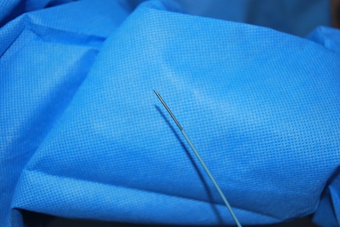
EUS-RFA needle design which allowed IONs injection
Figure 2.
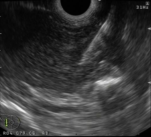
EUS-RFA needle inserted in the pig’s liver
Electrocardiographic and SpO2 levels were being monitored.
IONs synthesis was described in a previous study [10], as they were developed through the co-precipitation method and covered with citric acid. The resulted ferrofluid had a hydrodynamic diameter between 20.150 nm.
The RFA interventions were performed with dedicated animal medical equipment. For all groups, a linear array EUS scope (GFUCT140-AL5, Olympus, America), with a large interventional channel, coupled with a corresponding Evis Exera System (Olympus, America) and an AlokaPro-Sound 5500 Ultrasound System (Hitachi-Aloka, Tokio, Japan) were used. For group B, the first step was to locally inject IONs in a selected area (1 ml each) followed by 2 minutes of 20 W RFA exposure. Group A consisted of RFA exposure with the same power within the liver, as for group C, only local IONs injection were performed.
Biological samples such as aspartate transaminase (AST), alanine transaminase (ALT), gama-glutamyl transpeptidase (GGT), alkaline phosphatase (AP) were also performed 3 day after interventions. Second-look EUS was performed to examine the ablation area as well as any sign of potential complications.
After closely evaluating during one week any change in their behaviour, , the experimental models were sacrificed with pentobarbital and their liver was harvested. Macroscopic examination was performed and organs were sent for imaging evaluation with a 3T MRI (Philips Ingenia 3T, Netherlands).
The next step consisted of pathological analysis, with the tissues fixed in 10% phosphate-buffered formalin and paraffin embedding. Hematoxylin-eosin and Prusian Blue staining were used.
Results
All generated lesions did not induce any complications during the procedure or at any moment in time. With a defined time for each intervention, no differences were noted between the mean electric RFA impedances. Necropsy did not show any collections, adhesions, or any vascular lesions. Harvested biological samples showed elevated AST, ALT levels in all groups, with no significant changes after IONs exposure.
After EUS measurements for the ablation areas, group A had a mean of 4.9 cm, while group B had a mean of 5.2 cm (Fig.3, 4). IONs exposure was on a median area of 3.1 cm (Fig.5). EUS imaging pointed out a regular oval shape in group A, and a slightly irregular outline on group B, with more echo bubbles around. MRI sections revealed different patterns for each group separately. In group A and B, RFA lesions were easily identified with specific liver parenchyma changes. Group B revealed few deposits of nanoparticles further away from the targeted point. In contrast, the last group pointed out a large amount of IONs within the injection region and a larger amount of dispersed IONs within the liver than group B.
Figure 3.
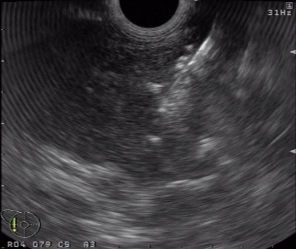
EUS-RFA procedure describing a hyperchoic mass which extends around the needle
Figure 4.
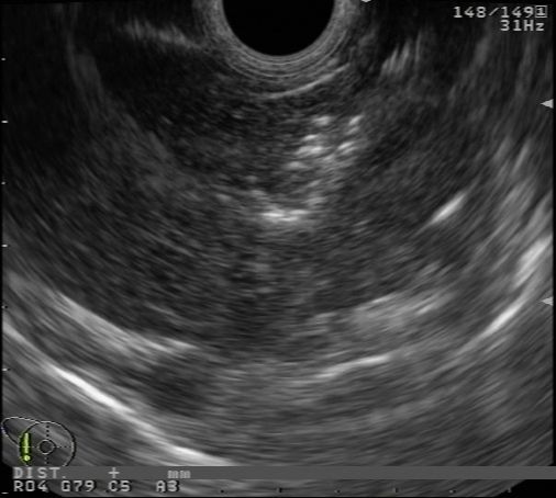
EUS-RFA area obtained after IONs local injection in the pigs liver
Figure 5.
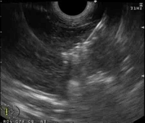
EUS IONs injection in the liver lobe forming a hyperechoic mass at needle’s tip
Table 1.
EUS imaging of the exposed area
| Group | A (cm) | B (cm) | C (cm) |
| 5.1 | 5.6 | 2.9 | |
| 4.7 | 4.8 | 3.3 | |
| Median | 4.9 | 5.2 | 3.1 |
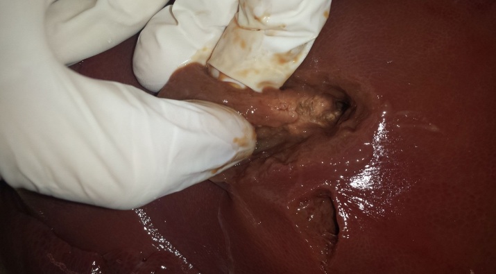
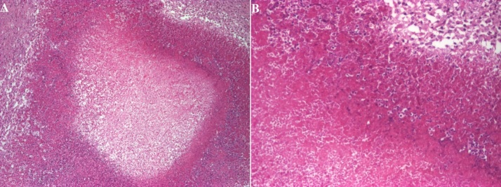
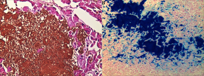
Macroscopically, RFA lesions were easily identified with the entry point being exposed. A whitish oval shape area was seen in all ablated zones, while group C did not show significant findings (Fig.6). Pathological assessment did not show any relevant difference between group A and B. The RFA lesions in HE showed signs of coagulation necrosis, followed by a peripheral area with neutrophils, macrophages and granulated tissue (Fig.7), whereas the group with IONs injection revealed on Prusian Blue staining intravascular dissemination as well as magnetic nanoparticles infiltration among the hepatocytes within the hepatic lobules. The group C did not show any signs of necrosis but degenerative-like lesions post injection (Fig.8).
Figure 6.
Macroscopic view of the ablation area
Figure 7.
A. Ablation area image of the hepatic tissue, HE staining x40; B. Detailed image of the ablation area with coagulation necrosis (1), with neutrophils deposits (2), and granulation tissue at the peripheral area (3), HE staining x100
Figure 8.
A. ION deposits within the hepatic lobule, Col HE x200; B. ION deposits within the hepatic lobule, Prusian Blue staining, x100
When comparing the solitary RFA lesions with the ones in group B there was a slight difference in the ablation area size in favor of IONs addition.
Discussion
Liver RFA is among the most popular ablation therapies for solid hepatic tumors with established guidelines for HCC and liver metastases [11]. However, controlling the exact area seems to be rather difficult as tumors size and shape may differ and therefore may become more challengeable in assuring that the right amount of tissue is destroyed. Over the years, new potential therapies of enhancing the ablation area have been assessed with most of them being used in an experimental environment. Even more, the need of acquiring an ablation area while using a less invasive method than open or laparoscopic surgery seems the focus point for patient’s wellbeing. This has led to the development of new therapeutic EUS-RFA needles which have the potential of extending in hard-to-reach liver regions [12].
Several techniques have been studied for controlling or enhancing the ablation area having RFA as a fundamental method. Fong et al [13] has associated the use of hepatic arterial embolization in patients with colorectal liver metastases with minimal morbidity and noting a 5 year survival rate in 10% of the patients. Also, saline infusion for RFA has been proven to enlarge the ablation lesion [14]. An experimental study on VX2 liver tumor rabbits which compared several fluids in association with RFA to maximize hepatic lesions necrosis showed that preoperative use of hypertonic saline, lidocaine and anhydrous ethanol may result in a larger and wider necrosis coagulation area. Even more, combining them was more effective than their single use [15].
IONs are currently used in different regims according to their intrinsec properties. Their use as a possible enhancing option of RFA represents an interesting objective in research. A recent study proved that metal nanoparticle infiltration resulted in significantly larger ablation areas when exposing to an RFA probe. The experiments were performed on ex vivo myocardial tissue and on an in vivo porcine thigh revealing that magnetic-guided localization of IONs during RFA led to wider ablative areas than RFA alone [16].
Our research study suggested that previous injection of ferrofluid solution in a certain area followed by RFA may lead to a wider ablation than RFA alone. We injected 1 ml of ION solution in a precise spot, while controlling the depth of the needle. The main visual aspect which confirmed the area differences was that after IONs injection and RFA, the ablation zone had a more irrgular form than RFA alone. An important advantage was the needle design which has the capability of both injecting and ablating without damaging to much of the tissue or changing its position. This may come in hand in case of a hepatic tumor as it may avoid tumor dissemination. Also, all ablation procedures were performed aproximately one minute after injection in order to prevent IONs loss, as a large amount may spread further away our target. The injected solution intervenes in cell destruction by direct injection in the precise location, and it amplifies as RFA is performed, due to its conductivity.
The RFA range was larger in IONs injection group, appearance which was confirmed on second-look EUS procedures and further on pathological examination. Coagulation necrosis was observed on a more wider area in group B with granulation tissue at the edge and also with several intact IONs deposit on Prusian Blue staining at distance from the injection point. This showed the ferrofluid needs to be controlled when injected and therefore is limited in action to the desirable target. Since IONs have the capability of ablating a region themselves when heated, exposing them to a magnetic field while also using RFA may become more useful in covering an even wider area. During the experiment we did not measure the temperature in group A or B, so we could not establish if the IONs had a precise influence on the RFA exposure.
In spite of all that, a larger number of procedures on experimental animals should be performed. Also, the IONs conductivity should be broader studied, because their properties are relevant of RFA enhancing power. The EUS needle design represents a new step of reaching difficult positioned lesions and it also may be more helpful for the patient condition as it is less invasive.
Conclusions
Controlling and enhacing RFA area represents a next step in the evolution therapy of hepatic tumors. Nonetheless, the need of developing new medical devices, capable of providing better control of tumor destruction are more than necessary as they may be embedded as new options for cancer patients.
Acknowledgments
All authors had equal contribution.
This article was financed by the Partnership program in priority areas.PN II, implemented with support from National Authority of Scientific Research (ANCS), CNDI.UEFISCDI, project nr. 2011-3.1-0252 (NANO-ABLATION).
Conflict of interests
The authors declare that they have no conflict of interests.
References
- 1.European Association for the Study of the Liver European Organisation for Research and Treatment of Cancer. EASL-EORTC clinical practice guidelines: management of hepatocellular carcinoma. J Hepatol. 2012;56:908–943. doi: 10.1016/j.jhep.2011.12.001. [DOI] [PubMed] [Google Scholar]
- 2.Solbiati L, Ahmed M, Cova L, Ierace T, Brioschi M, Goldberg SN. Small liver colorectal metastases treated with percutaneous radiofrequency ablation: local response rate and long-term survival with up to 10-year follow-up. Radiology. 2012;265:958–968. doi: 10.1148/radiol.12111851. [DOI] [PubMed] [Google Scholar]
- 3.Gervais DA, Arellano RS, Mueller PR. Percutaneous radiofrequency ablation of ovarian cancer metastasis to the liver: indications, outcomes, and role in patient management. AJR Am J Roentgenol. 2006;187:746–750. doi: 10.2214/AJR.05.1106. [DOI] [PubMed] [Google Scholar]
- 4.Kim HR, Cheon SH, Lee KH, Ahn JR, Jeung HC, Lee SS, et al. Efficacy and feasibility of radiofrequency ablation for liver metastases from gastric adenocarcinoma. Int J Hyperthermia. 2010;26:305–315. doi: 10.3109/02656730903555696. [DOI] [PubMed] [Google Scholar]
- 5.Ungureanu BS, Sandulescu L, Surlin V, Sp�rchez Z, Saftoiu A. Surgical hepatic resection vs. ultrasonographic guided radiofrequency ablation in colorectal liver metastases: what should we choose? Med Ultrason. 2014;16(2):145–151. doi: 10.11152/mu.201.3.2066.162.bsu1. [DOI] [PubMed] [Google Scholar]
- 6.Cucchetti A, Piscaglia F, Cescon M, Colecchia A, Ercolani G, Bolondi L, et al. Cost-effectiveness of hepatic resection versus percutaneous radiofrequency ablation for early hepatocellular carcinoma. J Hepatol. 2013;59:300–307. doi: 10.1016/j.jhep.2013.04.009. [DOI] [PubMed] [Google Scholar]
- 7.Goldberg SN, Mallery S, Gazelle GS, et al. EUS-guided radiofrequency ablation in the pancreas: Results in a porcine model. Gastrointest Endosc. 1999;50:392–401. doi: 10.1053/ge.1999.v50.98847. [DOI] [PubMed] [Google Scholar]
- 8.Ungureanu BS, Teodorescu CM, Saftoiu A. Magnetic Nanoparticles for Hepatocellular Carcinoma Diagnosis and Therapy. J Gastrointestin Liver Dis. 2016;25(3):375–383. doi: 10.15403/jgld.2014.1121.253.nan. [DOI] [PubMed] [Google Scholar]
- 9.Vogl TJ, Hammerstingl R, Schwarz W, et al. Magnetic resonance imaging of focal liver lesions. Comparison of the superparamagnetic iron oxide resovist versus gadolinium-DTPA in the same patient. Invest Radiol. 1996;31:696–708. doi: 10.1097/00004424-199611000-00004. [DOI] [PubMed] [Google Scholar]
- 10.Ungureanu BS, Pirici D, Margaritescu C, Gheonea IA, Trincu FN, Fifere A, Saftoiu A. Endoscopic ultrasound guided injection of iron oxide magnetic nanoparticles for liver and pancreas: a feasibility study in pigs. Med Ultrason. 2016;18(2):157–162. doi: 10.11152/mu.2013.2066.182.eus. [DOI] [PubMed] [Google Scholar]
- 11.Feng K, Ma KS. Value of radiofrequency ablation in the treatment of hepatocellular carcinoma. World J Gastroenterol. 2014;20(20):5987–5998. doi: 10.3748/wjg.v20.i20.5987. [DOI] [PMC free article] [PubMed] [Google Scholar]
- 12.Ungureanu BS, Pirici D, Margaritescu C, Sandulescu L, Fronie S, Patrascu S, Surlin V, Saftoiu A. Endoscopic ultrasound-guided radiofrequency ablation of the pancreas: An experimental study with pathological correlation. Endosc Ultrasound. 2015;4(4):330–335. doi: 10.4103/2303-9027.170426. [DOI] [PMC free article] [PubMed] [Google Scholar]
- 13.Fong ZV, Palazzo F, Needleman L, Brown DB, Eschelman DJ, Chojnacki KA, Yeo CJ, Rosato EL. Combined hepatic arterial embolization and hepatic ablation for unresectable colorectal metastases to the liver. Am Surg. 2012;78(11):1243–1248. [PubMed] [Google Scholar]
- 14.Yull Park J, Young Park C, Min Lee J. Estimation of saline-mixed tissue conductivity and ablation lesion size. Comput Biol Med. 2013;43(5):504–512. doi: 10.1016/j.compbiomed.2013.02.012. [DOI] [PubMed] [Google Scholar]
- 15.Sun YX, Cheng W, Han X, Liu Z, Wang QC, Shao H. In vivo experimental study on the effects of fluid in increasing the efficiency of radiofrequency ablation. Asian Pac J Cancer Prev. 2014;15(14):5799–5804. doi: 10.7314/apjcp.2014.15.14.5799. [DOI] [PubMed] [Google Scholar]
- 16.Nguyen DT, Tzou WS, Zheng L, Barham W, Schuller JL, Shillinglaw B, Quaife RA, Sauer WH. Enhanced Radiofrequency Ablation With Magnetically Directed Metallic Nanoparticles. Circ Arrhythm Electrophysiol. 2016;9(5):e003820–e003820. doi: 10.1161/CIRCEP.115.003820. [DOI] [PubMed] [Google Scholar]


