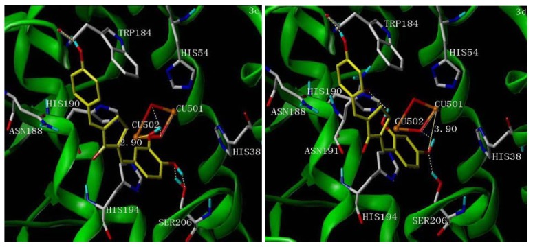
Figure 3.
The proposed binding modes of 3c (left picture) and 3d (right picture) in the active site of tyrosinase (PDB access code 2ZWE). The inhibitor molecules are colored in yellow for carbon atoms. The dashed lines show hydrogen-bonding, the real lines show the distance of metal-coordination interactions. The docking models are generated using Surflex-Dock.

