Abstract
Chemical investigation of an acetonitrile fraction from the endophytic fungus Phomopsis sp. led to the isolation of the new natural product 2-hydroxy-alternariol (7) together with the known compounds cytochalasins J (1) and H (2), 5'-epialtenuene (3) and the mycotoxins alternariol monomethyl ether (AME, 4), alternariol (AOH, 5) and cytosporone C (6). The structure of the new compound was elucidated by using 1-D and 2-D NMR (nuclear magnetic resonance) and high resolution mass spectrometry. The cytochalasins J (1) and H (2) and AOH (5) exhibited potent inhibition of the total ROS (reactive oxygen species) produced by stimulated human neutrophils and acted as potent potential anti-inflammatory agents. Moreover, cytochalasin H (2) demonstrated antifungal and acetylcholinesterase enzyme (AChE) inhibition in vitro.
Keywords: secondary metabolites, bioactivities, endophytic fungi, Phomopsis sp., Senna spectabilis
1. Introduction
Endophytes are microorganisms that inhabit plant interiors, especially the leaves, stems and roots, with no apparent harm to their host [1,2]. These microorganisms have received considerable attention over the last 20 years since their ability to protect a host against insects and pathogens was noted [3]. This protection is associated with biologically active compounds isolated from the endophytes. Special attention has been given to the presence of mycotoxins in grains that carry endophytes primarily because some of these secondary metabolites, such as alternariol (AOH) and alternariol monomethyl ether (AME), are toxic to both humans and animals [4] and are responsible for spoilage in grains, fruits and vegetables [5]. The presence of mycotoxins in the natural environment and in foodstuffs has been reported as an agricultural problem for several decades [4].
Brazilian Cerrado trees are well-known sources of bioactive secondary metabolites [6,7,8,9], of which Senna spectabilis (Fabaceae) was chosen for a detailed microbiological investigation. This plant presented several endophytes, including Phomopsis sp., which were chemically and biologically investigated.
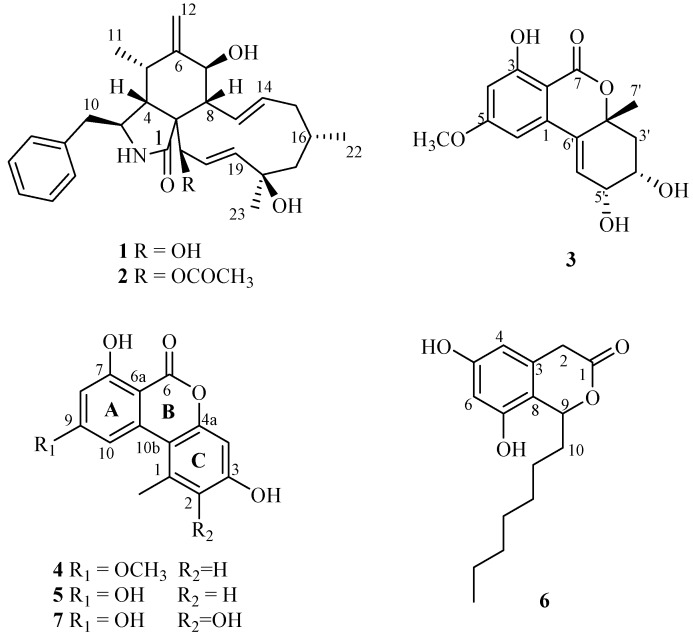
We report the isolation of cytochalasin J (1) and H (2), 5'-epialtenuene (3), alternariol monomethyl ether (4), alternariol (5), cytosporone C (6) and the new natural product 2-hydroxyalternariol (7) (Figure 1). Furthermore, the antioxidant, anti-inflammatory, antifungal and cytotoxic activities of these compounds were evaluated.
Figure 1.
Chemical structures of 1–7 produced by Phomopsis sp.
2. Results and Discussion
Column chromatography and preparative HPLC were used to isolate the six known compounds: cytochalasin J (1), cytochalasin H (2) [10,11,12], 5'-epialtenuene (3) [13,14], alternariol monomethyl ether (AME) (4) alternariol (AOH) (5) [15,16,17], cytosporone C (6) [18,19], and the new natural product 2-hydroxyalternariol (7).
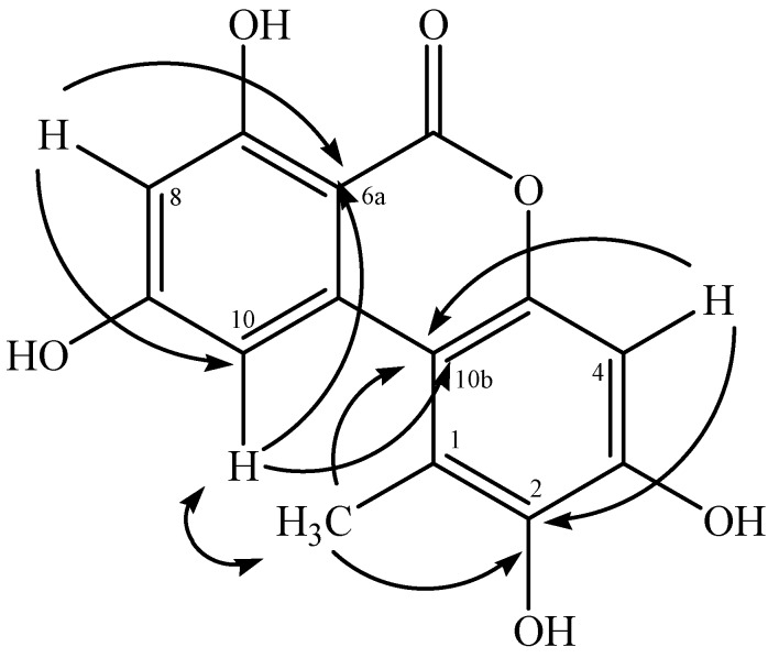
Compound 7 was obtained as a white amorphous powder, and its HRMS-ESI data indicated the molecular formula C14H10O6 ([M−H]−) from m/z 273.0363 (calcd for C14H9O6 273.0420). The 1H-NMR (Table 1) spectrum indicated the presence of two aromatic signals at δH 6.34 (d, 1H, J = 2.5 Hz, H-8) and at δH 7.26 (d, 1H, J = 2.5 Hz, H-10). The 1H-1H COSY spectrum showed correlation between both signals, suggesting a tetrasubstituted aromatic ring system. Additionally, it was observed the proton signal at δH 6.68 (1H, s, H-4), suggesting the second aromatic ring system was pentasubstituted. HMBC (Figure 2) spectrum provided the connection of H-8 to C-6a/C-10, suggesting the ring A. The correlation of H-4 and 1-CH3 to C-10b/C-2, suggesting the ring C. HMBC correlation of H-10 with C-10b indicated the connection for both aromatic rings. The signals of aromatic ring A obtained in 7 was consistent with NMR data reported to AOH [15,16], suggesting its derivative. Thus, compound 7 was identified as 2-hydroxy-alternariol, a new natural product.
Table 1.
.1H (500 MHz) and 13C-NMR (125 MHz) spectral data for 7 in DMSO-d6.
| 7 | ||
|---|---|---|
| Position | δH,mult. (J in Hz) | δC |
| 1 | - | 122.0 |
| 2 | - | 141.7 |
| 3 | - | 146.5 |
| 4 | 6.68 ( s) | 100.7 |
| 5 | - | * |
| 6 | - | * |
| 6a | - | 97.4 |
| 7 | - | 164.4 |
| 8 | 6.34 ( d, 2.5 Hz) | 100.5 |
| 9 | - | 165.0 |
| 10 | 7.26 ( d, 2.5 Hz) | 104.1 |
| 10a | - | * |
| 10b | - | 109.0 |
| 1-CH3 | 2.58 ( s) | 18.8 |
* not detected.
Figure 2.
Key HMBC (→) and NOESY (↔) correlations for 2-hydroxy-alternariol (7).
Compound 7 was previously reported by Pfeiffer et al. [20] as a biotransformation compound from AOH originating in the microsomes of rat, human and porcine liver. The mass spectrum of compound 7 was compared with the crude extract obtained from Phomopsis sp. by HPLC-MS, which confirmed that 7 is a new natural product and not an artifact. This report is the first to present NMR data for this compound.
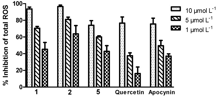
Compounds 1, 2 and 5 exhibited a potent inhibitory effect on ROS produced by stimulated neutrophils (Figure 3). Compound 2 exhibited the most significant inhibitory effect (IC50 = 0.91 ± 0.26 µmol L−1) with an inhibitory concentration exceeding those of apocynin (IC50 = 3.90 ± 0.30 µmol L−1), which is an efficient inhibitor of the NADPH oxidase complex [21], and quercetin (IC50 = 4.86 ± 0.36 µmol L−1), a flavonoid with potent antioxidant activity [22]. Compounds 1, 2 and 5 were able to modulate the neutrophilic ROS generation triggered by the zymosan opsonized stimuli in a concentration-dependent manner.
Figure 3.
The ability of the tested compounds and standards to inhibit the total ROS produced by stimulated neutrophils.
Of the ROS generated by the neutrophils, HOCl is the most potent oxidant. The product of the myeloperoxidase (MPO)-catalyzed oxidation of chloride in neutrophils, HOCl, plays an important role in the immune system by killing invading bacteria, and its excessive production has been linked to the progression of a variety of diseases including atherosclerosis, rheumatoid arthritis, certain inflammatory cancers, kidney diseases [23,24,25].
Compounds 1, 2 and 5 were HOCl scavengers, with IC50 values of 17.05 ± 0.67, 16.25 ± 0.67 and 16.48 ± 0.79 µmol L−1, respectively. Quercetin, an antioxidant used as a reference, exhibited high scavenging capacity (IC50 = 3.631 ± 0.29 µmol L−1), which can be attributed to the presence of the 3'- and 4'-hydroxyl groups (catechol groups) [22]. Compound 4 showed an IC50 exceeding 50 µmol L−1; compounds 3, 6 and 7 were not tested because an insufficient quantity was isolated.
Compounds 1, 2, 4 and 5 were tested for their MPO enzymatic activity and as scavengers of the superoxide anion (the first ROS produced through NADPH oxidase activation in neutrophils) but were inactive, showing IC50 values exceeding 50 µmol L−1.
Compounds 1, 2 and 5 did not act by inhibiting the MPO chlorinating activity or by scavenging the superoxide anion, but efficiently inhibited the ROS produced by neutrophils, which indicates that these compounds may act as NADPH oxidase complex inhibitors, thus, reducing the ability of neutrophils to produce HOCl, which could prove beneficial for the prevention of oxidative damage [23].
To investigate whether a possible toxic effect of 1, 2, 4 and 5 caused the decrease in neutrophil function, the cytotoxicity of these compounds was evaluated using a trypan blue exclusion assay (Table 2). Compounds 1, 2, 4 and 5 were nontoxic to human neutrophils at concentrations below 100 µmol L−1 at 30 min and after 60 min. At a concentration of 10 µmol L−1 (at which a high inhibition of oxidative neutrophil metabolism has been verified), the viability of the cells exceeded 98%. These results indicate that the inhibitory effects were not mediated through cell death.
Table 2.
Evaluation of the cytotoxicity of compounds 1, 2, 4 and 5 at different times and concentrations.
| Compounds | Concentration (µmol L−1) | Viable cells (%) * | |
|---|---|---|---|
| 30 min | 60 min | ||
| Control | 99.0 ± 0.60 | 99.0 ± 1.41 | |
| 1 | 100 | 80.0 ± 2.80 | 50.0 ± 2.80 |
| 1 | 10 | 97.0 ± 0.70 | 97.0 ± 0.70 |
| 2 | 100 | 85.0 ± 1.40 | 60.0 ± 2.80 |
| 2 | 10 | 96.0 ± 0.70 | 96.0 ± 1.41 |
| 4 | 100 | 85.0 ± 3.50 | 50.0 ± 2.80 |
| 4 | 10 | 95.0 ± 1.41 | 95.0 ± 0.70 |
| 5 | 100 | 70.0 ± 2.80 | 45.0 ± 2.80 |
| 5 | 10 | 96.0 ± 0.70 | 96.0 ± 0.70 |
* The data are expressed as the means ± the standard deviation; n = 3. Each compound was assayed in triplicate.
A classic assay for DPPH● radical scavenger capacity was performed, but all compounds tested were inactive, with IC50 values exceeding 50 µmol L−1.
Cytochalasin H (2) exhibited activity against C. Cladosporioides and C. sphaerosphermum (the minimum quantities required to inhibit fungal growth were 10.0 and 25.0 µg, respectively) with nystatin used as the reference (the minimum quantity required to inhibit fungal growth was 1.0 µg). Cytochalasin H (2) exhibited acetylcholinesterase activity (the minimum quantity required to inhibit acetylcholinesterase was 25.0 µg), and the physostigmine was used as reference (the minimum quantity required to inhibit fungal growth was 1.0 µg). The other compounds were inactive in these assays.
The cytochalasins are a group of secondary fungal metabolites structurally complex. They have been found in several fungal genus, such as Ascochyta sp., Aspergillus sp., Phomopsis sp., Turbercularia sp., Xylaria sp., among others. These substances have a wide range of biological activities including inhibition of HIV-1 protease 2, as well as antibiotic and cytotoxic activities [26,27].
The chemical and biological study of the CH3CN fraction from Phomopsis sp. led to the isolation of mycotoxins with potent bioactivity. AOH (5) and AME (4) are mycotoxins commonly found in foods contaminated with Alternaria alternate [4]. A previous study demonstrated that consuming food contaminated with these toxins can increase the incidence of esophageal cancer, and there have been several reports concerning the mutagenicity and genotoxicity of AOH and AME [5,20,28].
3. Experimental Section
3.1. General Experimental Procedures
The 1H-NMR (500 MHz), 13C-NMR (125 MHz), gHMBC, gHMQC and gCOSY experiments were recorded on a Varian DRX-500 spectrometer using the non-deuterated residual signal as the reference. The mass spectra were measured on a LC–MS Q-TOF Micromass spectrometer in the ESI mode with MeOH as the eluent (cone voltage = 25 V). The TLC was performed using Merck silica gel 60 (230 mesh). The spots on the TLC plates were observed under UV light by spraying with anisaldehyde-H2SO4 followed by heating to 100 °C. Preparative HPLC was performed on a Shimadzu system coupled with a UV SPD detector using a Phenyl preparative column (250 mm × 2.0 mm). Analytical HPLC was performed on a Shimadzu system coupled with a UV SPD detector system using a phenyl column (25.0 cm × 3.0 cm). Column chromatography (CC) was performed using silica gel (0.060–0.200 mm; Acros Organics, Fair Lawn, NJ, USA). Optical rotations were measured on a PerkinElmer polarimeter with a sodium lamp operating at 589 nm at 25 °C. IR spectra were obtained with a PerkinElmer FTIR-1600 spectrophotometer using KBr pellets.
Taurine, calcium chloride, magnesium chloride, glucose, catalase (EC 1.11.1.6), dimethyl sulfoxide (DMSO), luminol (5-amino-2,3-dihydro-1,4-phthalazinedione), 2,2-diphenyl-1-picrylhydrazyl (DPPH●), 5,5'-tetramethylbenzidine (TMB), zymosan, Histopaque®-1077 and Histopaque®-1119 were purchased from Sigma–Aldrich Chemical Co. (St. Louis, MO, USA). 2-(4-Iodophenyl)-3-(4-nitrophenyl)-5-(2,4-disulfophenyl)-2H-tetrazolium monosodium salt (WST-1) was purchased from Santa Cruz (Santa Cruz, CA, USA). MPO (EC 1.11.1.7) was purchased from Planta Natural Products (Vienna, Austria), and its concentration was determined from its absorption at 430 nm (ɛ = 89,000 mol−1 cm−1 per heme) [29]. Hydrogen peroxide was prepared by diluting a 30% stock solution purchased from Peroxidos do Brazil (Sao Paulo, SP, Brazil), and its concentration was calculated using its absorption at 240 nm (ɛ = 43.6 mol−1 cm−1) [30]. Hypochlorous acid was prepared by diluting a concentrated commercial bleach solution, and its concentration was calculated from its absorption at 292 nm (ɛ = 350 mol−1 cm−1) [29]. All solutions were prepared with water purified in a Milli-Q system (Millipore, Bedford, MA, USA). All reagents used to prepare the buffers were of analytical grade.
3.2. Plant Material
Authenticated Senna spectabilis (Fabaceae) was collected next to the Chemistry Institute—UNESP, Araraquara, São Paulo, Brazil, in February, 2007. A voucher specimen was deposited at the Herbarium of the Institute of Botany of São Paulo, Brazil (voucher No. SP 384109).
3.3. Isolation and Identification of the Endophytes
The endophytic fungus Cs-c2 was isolated from healthy adult leaves of Senna spectabilis according to reported methods [31]. The fungus was identified by morphology analysis by the André Tosello Foundation, Campinas—SP and deposited in the NuBBE collection under the number Cs-c2 for Phomopsis sp.
3.4. Fermentation, Extraction and Isolation
The endophytic fungus strain Phomopsis sp. was cultivated in eight Erlenmeyer flasks (500 mL), each containing 90 g of corn meal and 75 mL of H2O. The medium was autoclaved three times (on three consecutive days) at 121 °C for 20 min. After sterilization, the medium was inoculated with the endophyte and incubated while stationary at 26 °C for 21 days. At the end of the incubation period, the cultures were combined, ground and extracted with CH3OH (7 × 200 mL). The solvent was evaporated to yield a crude CH3OH extract (21.0 g). The CH3OH extract was dissolved in CH3CN (500 mL) and defatted with hexane via liquid partitioning. The CH3CN fraction was then evaporated to yield 2.90 g.
The CH3CN fraction was fractionated via column chromatography on a silica gel column (2.5 cm × 27.0 cm) and eluted with a CHCl3/CH3OH gradient (1%–100% MeOH) to yield 31 sub-fractions (Psp01-31), which were retained based on their similarity with the TLC profiles. The sub-fraction Psp18-20 (360.5 mg) was submitted to preparative HPLC separation using H2O/CH3OH (60:40 v/v until 0:100% over 40 min, 10 mL min−1, λmax = 235 nm) as the eluent to yield compounds 1 (15.1 mg, RT = 24.8 min), 2 (103.5 mg, RT = 28.1 min), 3 (1.2 mg, RT = 21.5 min) and 4 (40.0 mg, RT = 31.5 min). CHCl3 (15 mL) was added to the sub-fractions Psp25 and Psp30, the substances 5 + 6 (9.2 mg) and 7 (4.2 mg), respectively, were obtained after stirring and filtration of the mixture. Compounds 5 + 6 were purified via preparative HPLC using H2O/CH3OH (45:55 until 20:80 over 20 min at a rate of 10 mL min−1, λmax = 235 nm) as the eluent to yield 5 (6.0 mg) and 6 (1.2 mg).
3.5. Biological Activity
3.5.1. The Reactive Oxygen Species (ROS) Inhibitory Activity was Measured Using a Cellular Assay
The total ROS produced by the stimulated neutrophils was measured using luminol-enhanced chemiluminescence (LumCL) assays. The human neutrophils were isolated using blood samples obtained from healthy volunteers. The experiments were performed in accordance with the regulations of the Research Ethics Committee (29/2011, Faculty of Pharmaceutical Sciences, UNESP, São Paulo, Brazil). The neutrophils were isolated and the LumCL assay was performed according to previously reported methods [32,33].
3.5.2. Myeloperoxidase (MPO) Inhibitory Activity
The chlorination activity of the MPO was based on the reaction of HOCl with taurine to produce taurine chloramine, which oxidizes TMB [34]. The resulting oxidation product was detected spectrophotometrically at 655 nm using a microplate reader (Synergy 2 Multi-Mode, BioTek, Winooski, VT, USA), according to the procedure described by Zeraik et al. [35].
3.5.3. Antioxidant Capacity
The scavenging capacity of compounds 1, 2, 4 and 5 was evaluated using the potent oxidant HOCl (a compound generated by MPO), O2●− (produced during a respiratory burst by NADPH oxidase) and DPPH● (the classic method for measuring antioxidant capacity). The abilities of 1, 2, 4 and 5 to scavenge HOCl were determined by measuring the oxidation of TMB by taurine chloramine at 655 nm using a microplate reader (Synergy 2 Multi-Mode, BioTek) according to the procedure described by Ximenes et al. [36]. The superoxide anion radical generated from the xanthine/xanthine oxidase system (X/XO) was studied by reducing the tetrazolium salt, WST-1, to produce a soluble formazan superoxide according to the modified method proposed by Tan and Berridge [37]. The DPPH● method was also performed to verify the antioxidant capacity of 1, 2, 4 and 5. The method developed by Brand-Williams et al. [38] was followed with certain modifications [39]. Quercetin was used as a positive control under different concentrations in DMSO.
3.5.4. Cytotoxic Activity
The cytotoxic effect of compounds 1, 2, 4 and 5 on human neutrophils was studied using the trypan blue exclusion assay according to the method by Kitawaga et al. [33].
3.5.5. Antifungal Activity
Cladosporium cladosporioides (Fresen) de Vries CCIBt 140 and C. sphaerospermum (Penzig) CCIBt 491 were used in the antifungal assay. Compounds 1, 2, 4, 5 and 7 were applied to pre-coated Si-gel TLC plates using a solution (10 mL) containing 100.0, 50.0, 25.0, 10.0, 5.00 and 1.00 µg samples of the pure compounds. After eluting with CHCl3/CH3OH (9:1), the plates were sprayed with the fungal suspension [40]. Nystatin was used as the positive control at 1.0 µg.
3.5.6. Acetylcholinesterase Inhibitory Activity
Compounds 1, 2, 4, 5 and 7 were investigated (elution of the TLC with the CHCl3/CH3OH (9:1)) according to reported methods [41]. Galantamine was employed as the positive control at 1.0 µg.
4. Conclusions
The production of cytochalasins with known potent biological activity [10,26], such as cytochalasin H (2), with demonstrated activity against pathogenic fungi and the production of mycotoxins reinforce the hypothesis that symbiosis between the endophyte and the host plant can produce substances that show antifungal activity against possible phytopathogenic fungi or are harmful to predators of the plant. Additionally, cytochalasins J (1) and H (2) and alternariol were found to have potent inhibitory effects on human neutrophils by acting as potential inhibitors of NADPH oxidase and may be promising targets for the development of anti-inflammatory agents. This study of the fungus Phomopsis sp. revealed that the endophyte produces bioactive metabolites, thus justifying further chemical study of this class of microorganisms.
Acknowledgments
We gratefully acknowledge financial support from FAPESP (The State of São Paulo Research Foundation, grants # 2010/52327-5 and # 2013/07600-3. LMZ and VMC acknowledge the Aperfeiçoamento de Pessoal de Nível Superior (Capes), and MLZ acknowledges the FAPESP for scholarships.
Supplementary Materials
Supplementary materials can be accessed at: http://www.mdpi.com/1420-3049/19/5/6597/s1.
Author Contributions
ARA, VSB, DHSS, AJC, MNL designed research; VMC, MLZ, VFX, MCMY, LMZ and LMF performed research and analyzed the data; VMC, MLZ, MCMY and ARA wrote the paper. All authors read and approved the final manuscript.
Conflicts of Interest
The authors declare no conflict of interest.
Footnotes
Sample Availability: Samples are not available.
References
- 1.Aly H.A., Debbab A., Proksch P. Fungal endophytes: Unique plant inhabitants with great promises. Appl. Microbiol. Biotechnol. 2011;90:1829–1845. doi: 10.1007/s00253-011-3270-y. [DOI] [PubMed] [Google Scholar]
- 2.Gunatilaka A.A.L. Natural Products from plant-associated microorganisms: Distribution, structutal diversity, bioactivity, and implications of their occurrence. J. Nat. Prod. 2006;69:509–526. doi: 10.1021/np058128n. [DOI] [PMC free article] [PubMed] [Google Scholar]
- 3.Jalgaonwala R.E., Mohite B.V., Mahajan R.T. A review: Natural products from plant associated endophytic fungi. J. Microbiol. Biotech. Res. 2011;1:21–32. [Google Scholar]
- 4.Magnani R.F., de Souza G.D., Rodrigues-Filho E. Analysis of alternariol and alternariol monomethyl ether on flavedo and albedo tissues of tangerines (Citrus reticulata) with symptoms of Alternaria brown spot. J. Agric. Food Chem. 2007;55:4980–4986. doi: 10.1021/jf0704256. [DOI] [PubMed] [Google Scholar]
- 5.Watanabe I., Kakishima M., Adachi Y., Nakajima H. Potencial mycotoxin productivity of Alternaria alternata isolated from garden trees. Mycotoxins. 2007;57:3–9. [Google Scholar]
- 6.Bolzani V.S., Trevisan L.M.V., Young M.C.M. Caffeic acid esters and triterpenes of Alibertia macrophyla. Phytochemistry. 1991;30:2089–2091. [Google Scholar]
- 7.Young M.C.M., Braga M.R., Dietrich S.M.C., Gottlieb H.E., Trevisan L.M.V., Bolzani V.S. Fungitoxic non-glycosidic iridoids from Alibertia macrophylla. Phytochemistry. 1992;31:3433–3435. doi: 10.1016/0031-9422(92)83701-Y. [DOI] [Google Scholar]
- 8.Silva V.C., Faria A.O., Bolzani V.S., Lopes M.N. A new ent-kaurane diterpene from stems of Alibertia macrophylla K. Schum. (Rubiaceae) Helv. Chim. Acta. 2007;90:1781–1785. doi: 10.1002/hlca.200790187. [DOI] [Google Scholar]
- 9.Junior C.V., Pivatto M., Rezende A., Hamerski L., Silva D.H.S., Bolzani V.S. (–)-7-Hydroxycassine: A new 2,6-dialkylpiperidin-3-ol alkaloid and other constituents isolated from flowers and fruits of Senna spectabilis (Fabaceae) J. Braz. Chem. Soc. 2013;24:230–235. [Google Scholar]
- 10.Izawa Y., Hirose T., Shimizu T., Koyama K., Natori S. Six new 10-phenyl-[11]cytochalasans, cytochalasins N-S from Phompsis sp. Tetrahedron. 1989;45:2323–2335. doi: 10.1016/S0040-4020(01)83434-7. [DOI] [Google Scholar]
- 11.Tao Y., Zeng X., Mou C., Li J., Cai X., She Z., Zhou S., Lin Y. 1H and 13C NMR assignments of three nitrogen containing compounds from the mangrove endophytic fungus (ZZF08) Magn. Reson. Chem. 2008;46:501–505. doi: 10.1002/mrc.2194. [DOI] [PubMed] [Google Scholar]
- 12.Ondeyka J., Hensens O.D., Zink D., Ball R., Lingham R.B., Bills G., Dombrowski A., Goetz M. L-696,474, a novel cytochalasin as an inhibitor of HIV-1-protease. II. Isolation and structure. J. Antibiot. 1992;45:679–685. doi: 10.7164/antibiotics.45.679. [DOI] [PubMed] [Google Scholar]
- 13.Bradburn N., Coker R.D., Blunden G., Turner C.H., Crabb T.A. 5'-Epiltenuene and neoaltenuene, dibenzo-a-pyrones from Alternaria alternata cultured on rice. Phytochemistry. 1994;35:665–669. doi: 10.1016/S0031-9422(00)90583-1. [DOI] [Google Scholar]
- 14.Jiao P., Gloer J.B., Campbell J., Shearer C.A. Altenuene derivatives from an unidentified freshwater fungus in the family Tubeufiaceae. J. Nat. Prod. 2006;69:612–615. doi: 10.1021/np0504661. [DOI] [PMC free article] [PubMed] [Google Scholar]
- 15.Gu W. Bioactive metabolites from Alternaria brassicicola ML-P08, an endophytic fungus residing in Malus halliana. World J. Microbiol. Biotechnol. 2009;25:1677–1683. doi: 10.1007/s11274-009-0062-y. [DOI] [Google Scholar]
- 16.Aly A.H., Edrada-ebel R., Indriani I.D., Wray V., Muller W.E.G., Totzke F., Zirrgiebel U., Schachtele C., Kubbutat M.H.G., Lin W.H., et al. Cytotoxic metabolites from the fungal endophyte Alternaria sp. and their subsequent detection on its host plant Polygonum senegalense. J. Nat. Prod. 2008;71:972–980. doi: 10.1021/np070447m. [DOI] [PubMed] [Google Scholar]
- 17.Tan N., Tao Y., Pan J., Wang S., Xu F., She Z., Lin Y., Jones E.B.G. Isolation, structure elucidation, and mutagenicity of four alternariol derivatives produced by the mangrove endophytic fungus N. 2240. Chem. Nat. Comp. 2008;43:296–300. [Google Scholar]
- 18.Xu J., Kjer J., Sendker J., Wray V., Guan H., Edrada R., Muller V.E.G., Bayer M., Lin W., Wu J., et al. Cytosporones, coumarins, and an alkaloid from the endophytic fungus Pestalotiopsis sp. isolated from the Chinese mangrove plant Rhizophoramucronata. Bioorg. Med. Chem. 2009;17:7362–7367. doi: 10.1016/j.bmc.2009.08.031. [DOI] [PubMed] [Google Scholar]
- 19.Brady S.F., Wagenaar M.M., Singh M.P., Janso F.E., Cardy J. The cytosporones, new octaketide antibiotics isolated from an endophytic fungus. Organic Letters. 2000;25:4043–4046. doi: 10.1021/ol006680s. [DOI] [PubMed] [Google Scholar]
- 20.Pfeiffer E., Schebb N.H., Podlech J., Metzler M. Novel oxidative in vitro metabolites of the mycotoxins alternariol and alternariol methyl ether. Mol. Nutr. Food Res. 2007;51:307–316. doi: 10.1002/mnfr.200600237. [DOI] [PubMed] [Google Scholar]
- 21.Almeida A.C., Marques O.C., Arslanian C., Condino-Neto A., Ximenes V.F. 4-Fluoro-2-methoxyphenol, na apocynin analog with enhaced inhibitory effect on leukocyte oxidant production and phagocytosis. Eur. J. Pharmacol. 2011;660:445–453. doi: 10.1016/j.ejphar.2011.03.043. [DOI] [PubMed] [Google Scholar]
- 22.Jan A.T., Kamli M.R., Murtaza I., Singh J.B., Ali A., Haq Q.M.R. Dietary flavonoid quercetin and associated health benefits — an overview. Food Rev. Int. 2010;26:302–317. doi: 10.1080/87559129.2010.484285. [DOI] [Google Scholar]
- 23.Ford D.A. Lipid oxidation by hypochlorous acid: Chlorinated lipids in atherosclerosis and myocardial ischemia. Clin. Lipidol. 2010;5:835–852. doi: 10.2217/clp.10.68. [DOI] [PMC free article] [PubMed] [Google Scholar]
- 24.Summers F.A., Morgan P.E., Davies M.J., Hawkins C.L. Identification of plasma proteins that are susceptible to thiol oxidation by hypochlorous acid and N-chloramines. Chem. Res. Toxicol. 2008;21:1832–1840. doi: 10.1021/tx8001719. [DOI] [PubMed] [Google Scholar]
- 25.Klebanoff S.J. Myeloperoxiase: Friend and foe. J. Leukocyte Biol. 2005;77:598–625. doi: 10.1189/jlb.1204697. [DOI] [PubMed] [Google Scholar]
- 26.Xu S., Ge H.M., Song Y.C., Shen Y., Ding H., Tan R.X. Cytotoxic cytochalasin metabolites of endophytic Endothia gyrosa. Chem. Biodivers. 2009;6:739–745. doi: 10.1002/cbdv.200800034. [DOI] [PubMed] [Google Scholar]
- 27.Lin Z., Zhang G., Zhu T., Liu R., Wei H., Gu Q. Bioactive cytochalasins from Aspergillus flavipes, an endophytic fungus associated with the mangrove plant Acanthus ilicifolius. Helv. Chim. Acta. 2009;92:1538–1544. doi: 10.1002/hlca.200800455. [DOI] [Google Scholar]
- 28.Ostry V. Alternaria mycotoxins: An overview of chemical characterization, producers, toxicity, analysis and occurrence in foodstuffs. World Mycot. J. 2008;1:175–188. doi: 10.3920/WMJ2008.x013. [DOI] [Google Scholar]
- 29.Kettle A.J. Detection of 3-chlorotyrosine in proteins exposed to neutrophil oxidants. Meth. Enzymol. 1999;300:111–120. doi: 10.1016/S0076-6879(99)00119-6. [DOI] [PubMed] [Google Scholar]
- 30.Beers R.J., Sizer I.W. A spectrophotometric method for measuring the breakdown of hydrogen peroxide by catalase. J. Biol. Chem. 1952;195:133–140. [PubMed] [Google Scholar]
- 31.Silva G.H., Teles H.L., Zanardi L.M., Young M.C.M., Eberlin M.N., Haddad R., Pfenning L.H., Costa-Neto C., Castro-Gamboa I., Bolzani V.S., et al. Cadinane sesquiterpenoids of Phomopsis cassiae, an endophytic fungus associated with Cassia spectabilis (Leguminosae) Phytochemistry. 2006;67:1964–1969. doi: 10.1016/j.phytochem.2006.06.004. [DOI] [PubMed] [Google Scholar]
- 32.English D., Andersen B.R. Single-step separation of red blood cells, granulocytes and mononuclear leukocytes on discontinuous density gradients of Ficoll-Hypaque. J. Immunol. Methods. 1974;5:249–252. doi: 10.1016/0022-1759(74)90109-4. [DOI] [PubMed] [Google Scholar]
- 33.Kitagawa R.R., Raddi M.S.G., Khalil N.M., Vilegas W., Fonseca L.M. Effect of the isocoumarin paepalantine on the luminol and lucigenin amplified chemiluminescence of rat neutrophils. Biol. Pharm. Bull. 2003;26:905–908. doi: 10.1248/bpb.26.905. [DOI] [PubMed] [Google Scholar]
- 34.Malle E., Furtmuller P.G., Sattler W., Obinger C. Myeloperoxidase: A target for new drug development? Br. J. Pharmacol. 2007;152:838–854. doi: 10.1038/sj.bjp.0707358. [DOI] [PMC free article] [PubMed] [Google Scholar]
- 35.Zeraik M.L, Ximenes V.F., Regasini L.O., Dutra L.A., Silva D.H., Fonseca L.M., Coelho D., Machado S.A., Bolzani V.S. 4'-Aminochalcones as novel inhibitors of the chlorinating activity of myeloperoxidase. Curr. Med. Chem. 2012;19:5405–5413. doi: 10.2174/092986712803833344. [DOI] [PubMed] [Google Scholar]
- 36.Ximenes V.F., Kanegae M.P., Rissato S.R., Galhiane M.S. The oxidation of apocynin catalyzed by myeloperoxidase: Proposal for NADPH oxidase inhibition. Arch. Biochem. Biophys. 2007;457:134–141. doi: 10.1016/j.abb.2006.11.010. [DOI] [PubMed] [Google Scholar]
- 37.Tan A.S., Berridge M.V. Superoxide produced by active neutrophils efficiently reduces the tetrazolium salt, WST-1 to produce a soluble formazan: A simple colorimetric assay for measuring respiratory burst activation and for screening anti-inflammatory agents. J. Immunol. Methods. 2000;238:59–68. doi: 10.1016/s0022-1759(00)00156-3. [DOI] [PubMed] [Google Scholar]
- 38.Brand-Williams W., Cuvelier M.E., Berset C. Use of a free radical method to evaluate antioxidant activity. Lebensm.-Wiss. Technol. 1995;28:25–30. doi: 10.1016/S0023-6438(95)80008-5. [DOI] [Google Scholar]
- 39.Zeraik M.L., Yariwake J.H., Wauters J.N., Tits M., Angenot L. Analysis of passion fruit rinds (Passiflora edulis): Isoorientin quantification by HPTLC and evaluation of antioxidant (radical scavenging) capacity. Quim. Nova. 2012;35:541–545. doi: 10.1590/S0100-40422012000300019. [DOI] [Google Scholar]
- 40.Rahalison L., Hamburger M., Hostettmann K., Monod M., Frenk E. A bioautographic agar overlay method for the detection of antifungal compounds from higher plants. Phytochem. Anal. 1991;2:199–203. doi: 10.1002/pca.2800020503. [DOI] [Google Scholar]
- 41.Marston A., Kissling J., Hostettmann K. A rapid TCL bioautographic method for the detection of acetylcholinesterase and butyrylcholinesterase inhibitors in plants. Phytochem. Anal. 2002;13:51–54. doi: 10.1002/pca.623. [DOI] [PubMed] [Google Scholar]
Associated Data
This section collects any data citations, data availability statements, or supplementary materials included in this article.


