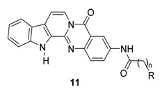
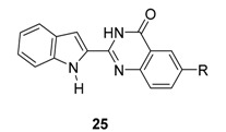
Abstract
Rutaecarpine is a pentacyclic indolopyridoquinazolinone alkaloid found in Evodia rutaecarpa and other related herbs. It has a variety of intriguing biological properties, which continue to attract the academic and industrial interest. Studies on rutaecarpine have included isolation from new natural sources, development of new synthetic methods for its total synthesis, the discovery of new biological activities, metabolism, toxicology, and establishment of analytical methods for determining rutaecarpine content. The present review focuses on the synthesis, biological activities, and structure-activity relationships of rutaecarpine derivatives, with respect to their antiplatelet, vasodilatory, cytotoxic, and anticholinesterase activities.
Keywords: alkaloid, rutaecarpine, antiplatelet activity, vasodilatory activity, anticancer activity, anti-cholinesterase activity, anti-obesity activity
1. Introduction
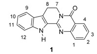
Rutaceous plants, especially Evodia rutaecarpa (its dried fruit is called ‘Wu-Chu-Yu’ in China), have long been used to treat gastrointestinal disorders, headache, amenorrhea, and postpartum hemorrhage in traditional oriental medicine [1,2]. The alkaloid, rutaecarpine (8,13-dihydroindolo-[2′,3′:3,4]pyrido[2,1-b]quinazolin-5(7H)-one, 1a, Figure 1) was first isolated in 1915 by Asahina and Kashiwaki from an acetone extract of base-treated Evodia rutaecarpa [3,4,5] and later from ‘Wu-Chu-Yu’ [6]. Interest in the molecule has since been growing, presumably due to its characteristic structure and intriguing biological properties (733 references were found in the SciFinder database provided by the American Chemical Society). In addition, 55 patents have been issued regarding its isolation, biological activity, synthesis, metabolism, and toxicology. Numbers of papers covering rutaecarpine are summarized in Table 1.
Figure 1.
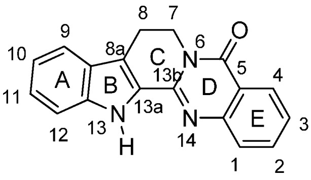
Structure of rutaecarpine (1a).
Table 1.
Numbers of references listed for recent years.
| Period | 1915–2007 | 2008 | 2009 | 2010 | 2011 | 2012 | 2013 | 2014 | 2015 | Total |
|---|---|---|---|---|---|---|---|---|---|---|
| Numbers | 339 | 42 | 46 | 43 | 59 | 69 | 64 | 52 | 19 | 733 |
Of the 17 review papers written to date, eight have focused on the synthesis of rutaecarpine [7,8,9,10,11,12,13,14], seven on pharmacology [15,16,17,18,19,20,21], one on the modulation of cytochrome P450 [22], and one on detection methods [23]. A review published in 1983 by Bergman [7] covers the nomenclature, structure, synthesis and pharmacological properties of rutaecarpine and of related quinazolinone alkaloids. The review of Wang et al., written in Chinese in 2006, provides details of the synthesis of rutaecarpine based on construction patterns of the five-ring system [10]. Shakhidoyatov and Elmurado’s review covered the most recent view on the general point of view for tricyclic quinazoline alkaloids [14]. A review written in 1999 by Sheu addressed the in vitro and in vivo pharmacology of rutaecarpine [15] and later described the cardiovascular pharmacological actions of rutaecarpine in his recent review [20]. More recently, in 2010, Jia and Hu reviewed its cardiovascular protective effects [19]. The present work focuses on the synthesis, biological activities, and structure-activity relationships, with respect to the antiplatelet, vasodilatory, cytotoxic, and anticholinesterase activities, of rutaecarpine derivatives, and complements our first review published in 2008 [24].
2. Synthesis of Rutaecarpine
A simple retrosynthetic analysis leads to tryptamine (2) and its equivalents for the indole moiety, and anthranilic acid (3a) and its equivalents for the quinazolinone moiety, which leaves an additional one-carbon unit needed for the C13b atom in rutaecarpine (Scheme 1).
Scheme 1.
Retrosynthetic analysis for rutaecarpine synthesis.
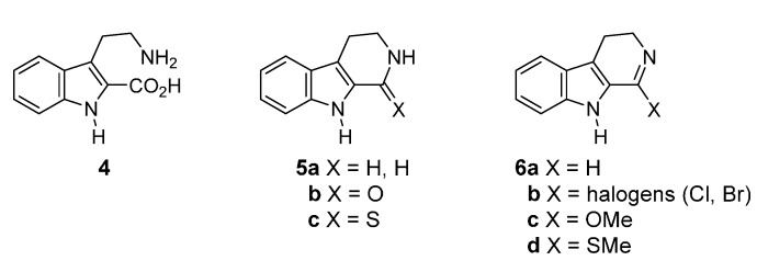
Tryptamine (2) has been one of most popular starting materials [1,11] and the compounds 4, 5, and 6 (Figure 2) have been used as alternative starting materials which provide the A,B,C-ring system and the one-carbon unit at C13b.
Figure 2.
Structures of tryptamine (2) equivalents for rutaecarpine synthesis.
On the other hand, a series of benzoic acid derivatives 3b–i with nitrogen at the ortho-position (Figure 3) were employed as an equivalent for 3a as the counterparts for tryptamine. In fact, in 1927 Asahina et al. reported two synthetic procedures for the synthesis of rutaecarpine using these equivalents as a starting materials—one procedure involved a three-step synthesis from 3-(2-aminoethyl)indole-2-carboxylic acid (4) and 2-nitrobenzoyl chloride (3e) (yield not given) [25] and the other a one-pot synthesis (in 24% yield) from 1,2,3,4-tetrahydro-1-oxo-β-carboline (5b) and methyl anthranilate (3d) in the presence of PCl3 [26] (Scheme 2).
Figure 3.
Structures of anthranilic acid (3) equivalents for rutaecarpine synthesis.
Scheme 2.
Since the classification of syntheses in our previous review [1] was based on the structures of starting materials, we kept the same classification in the present review, that is: (1) tryptamine-derived syntheses; (2) tetrahydro-β-carboline-derived syntheses; and (3) miscellaneous.
2.1. Synthesis Using Tryptamine
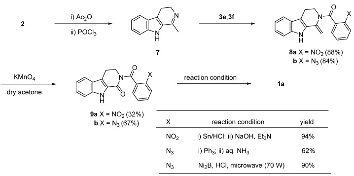
Lee et al. [27,28] and Kamal et al. [29] used a one-pot reductive-cyclization of nitro (9a) and azide compounds (9b), respectively, to construct the quinazolinone skeleton. Tryptamine was subjected to a Bischler-Napieralsky reaction to afford starting compound 7, which was then condensed with 3e and 2-azidobenzoyl chloride (3f) to afford 8a and 8b, respectively. Cleavage of the exocyclic double bond led to the corresponding ketone 9. It is worth noting that cleavage of the exocyclic double bond on 8 by ozonolysis failed, whereas oxidative cleavage with KMnO4 lead to ketones 9a and 9b in 32% and 67% yield, respectively (Scheme 3).
Scheme 3.
Synthesis of rutaecarpine by Lee et al. [27,28] and Kamal et al. [29].
The reduction of the nitro group in 9a by tin chloride resulted in subsequent cyclization giving 1a in 94% yield [27,28]. On the other hand, the related 2-azobenzamide (9b) undergoes an aza-Wittig reductive cyclization in the presence of Ph3P and NH4OH or Ni2B in HCl-MeOH under microwave irradiation [29].
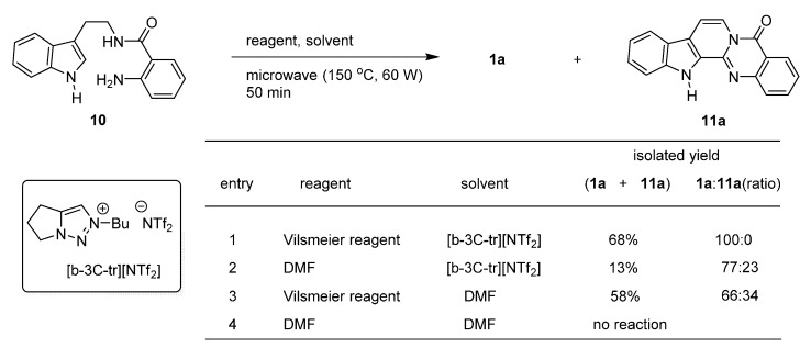
Tseng et al. studied the potential use of bicyclic 1,2,3-triazolium ionic liquids for the synthesis of rutaecarpine from a one-carbon unit reagent and 10 [30]. Microwave-assisted cyclization of 10 led to 1a and 7,8-dehydrorutaecarpine (11a) in ratios dependent on the reaction conditions (Scheme 4). The starting material 10 was prepared from tryptamine and isatoic anhydride (3h) in over 90% yield.
Scheme 4.
Synthesis of rutaecarpine by Tseng et al. [30].
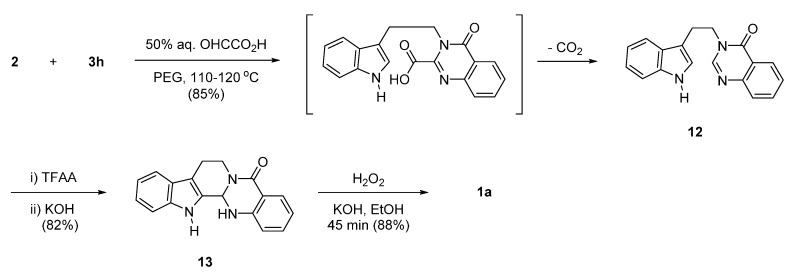
Rao et al. used 50% aqueous glyoxylic acid as a replacement for DMF or the Vilsmeier-Haack reagent in the above reaction. Reaction of 2 and isatoic anhydride (3h) in the presence of 50% aqueous glyoxylic acid led to 12, which was then subjected to acid-catalyzed cyclization followed by H2O2/KOH-catalyzed dehydrogenation to produce rutaecarpine (1a) [31] (Scheme 5). Although the authors did not mention a possible reaction mechanism, the high reaction temperature would lead to decarboxylation of the possible intermediate 3-[2-(1H-indol-3-yl)ethyl]-4-oxo-3,4-dihydroquinazoline-2-carboxylic acid, to produce 12.
Scheme 5.
Synthesis of rutaecarpine by Rao et al. [31].
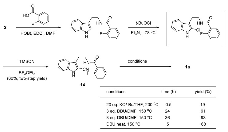
More recently, base-initiated intramolecular anionic cascade cyclization [32] of the 2-cyano compound 14 was applied to rutaecarpine synthesis by Liang, et al. [33]. The authors optimized the reaction conditions and found DBU was the reagent of choice for the conversion. The prerequisite 2-cyanoindole compound 14 was prepared in two-steps from tryptamine and 2-fluorobenzoic acid via a 2-chloroindolenine, generated by an electrophilic aromatic substitution reaction at the C2 position in the indole moiety by t-butyl hypochlorite, followed by nucleophilic substitution of the 2-chloro group by cyanide anion in the presence of BF3 (Scheme 6).
Scheme 6.
Synthesis of rutaecarpine by Liang et al. [33].
2.2. Synthesis Using Tetrahydro-β-Carboline
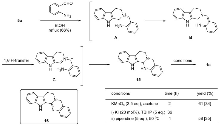
Zheng et al. reported the oxidation of s ring-fused aminal to rutaecarpine via an α-amination of an N-heterocycle as the key reaction [34,35,36] (Scheme 7). The α-position of 1,2,3,4-tetrahydro-β-carboline (5a) was activated by reacting with 2-aminobenzaldehyde to form an iminium ion (A), which was converted to the quinonoidal intermediate (B) by rearrangement of adjacent π-systems and a proton loss. 1,6-Hydrogen atom transfer in B led to the dipolar intermediate C, which ultimately afforded the cyclized aminal product (15). Oxidation of 15 by KMnO4 afforded rutaecarpine (1a) in 61% yield (Scheme 7). It should be noted that the MnO2 oxidation of dehydroaminal such as 16 yielded 1a and fully dehydrogenated product (11a) in an 8:1 ratio [37].
Scheme 7.
On the other hand, reaction between 3,4-dihydro-β-carboline (17) and o-azidobenzoyl chloride (3f) in the presence of Hünig’s base delivered 1a in 58% yield [38], while reaction with 3h afforded 1a in 93% yield [39] (Scheme 8).
Scheme 8.
Synthesis of rutaecarpine using 3,4-dihydro-β-carboline [38,39].
2.3. Miscellaneous
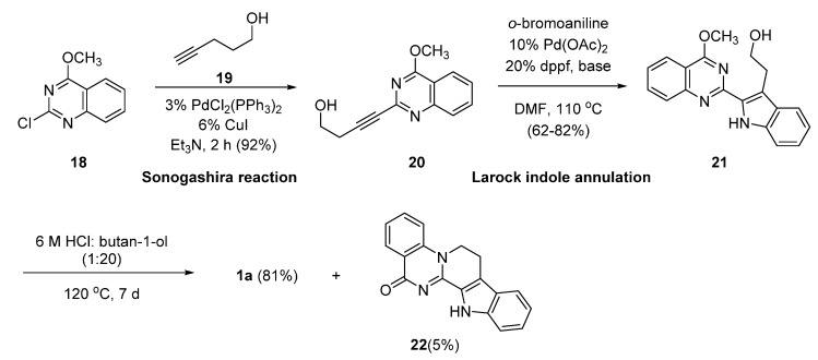
Synthetic methods not employing anthranilic acid, tryptamine, or their equivalents are very rare. Recently, Pan and Bannister employed a sequential Sonogashira reaction and Larock indole synthesis, whereby Sonogashira’s Pd(0)-catalyzed ethynylation of 19 to 18 led to 20, which subsequently underwent an intramolecular Pd(0)-catalyzed indole formation [40] to produce 21. The acid-catalyzed cyclization of 21 led to 1a in 81% yield with a trace of isomeric compound 22 [41] (Scheme 9). To the best of our knowledge, this procedure is the first example of construction of the C-ring to synthesize rutaecarpine via N6-C7 bond formation.
Scheme 9.
Synthesis of rutaecarpine by Pan and Bannister [41]
To synthesize rutaecarpine, C-ring construction can be pursued in three different ways via: (1) C13a-C13b bond formation; (2) N6-C7 bond formation; or (3) C8-C8a bond formation. Methods involving C13a-C13b bond formation at the final stage of synthetic sequences have been most commonly used [1,26,30,31,42,43,44,45,46,47]. However, a method employing C8-C8a bond formation has not been studied as yet.
2.4. Synthesis of Bioisosteres and Hybrid Rutaecarpine Systems
Bioisosteric replacement of the quinazolinone moiety of the pentacyclic system with benzothiadiazine 1,1-dioxide has been pursued [48] (Scheme 10). Bubenyák et al. reported a condensation of benzothiadiazine 1,1-dioxide analogue 23 with phenyldiazonium chloride led to a phenylhydrazone derivative, which was subjected to Fischer indole synthesis to afford the corresponding 5-sulfarutaecarpine (24). The starting 23 was prepared in two-steps from 2-aminobenzenesulfonic acid and 6-methoxy-2,3,4,5-tetrahydropyridine in 33% yield or 2-nitrobenzenesulfonyl chloride and piperidin-2-one in 66% yield.
Scheme 10.
Synthesis of 5-sulfarutaecarpine [48].
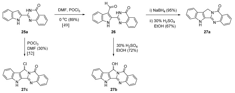
The same group [49] reported a hybrid between the alkaloids rutaecarpine and luotonin A [50,51]. Vilsmeier-Haack formylation of 2-(1H-indol-2-yl)quinazolin-4(3H)-one (25a) gave 26 which was subjected to either direct or indirect reduction to alcohol followed by acid catalyzed cyclization to produce 27a,b. On the other hand, a direct cyclization followed by chlorination under Vilsmeier-Haack conditions led to 27c [52] (Scheme 11). Such a chloro-compound represents a good substrate for introducing substituents by nucleophilic substitution [52] for the synthesis of related series of compounds.
Scheme 11.
Synthesis of a hybrid between rutaecarpine and luotonin A [50,52].
3. Biological Properties
A review written in 2003 by Hu and Li, comprehensively described the in vitro and in vivo pharmacology of rutaecarpine [18], in which pharmacological actions were classified as; cardiovascular effects, antiplatelet activity, antithrombotic activity, anticancer activity, anti-inflammatory and analgesic effects, effects on the endocrine system, anti-obesity and thermoregulatory effects, effects on smooth muscle (except cardiovascular), and others. In addition, rutaecarpine ameliorated body weight gain by inhibiting orexigenic neuropeptides NPY and AgRP in mice [53] and reducing lipid accumulation by AMPK (AMP activated protein kinase) activation and UPR (unfolded protein response) suppression [52]. Recently, Xu et al. reported the anti-atherosclerosis activity (EC50 = 0.27 μM) by up-regulating ATP-binding cassette transporter A1 (ABCA1) [54,55]. The present review addresses structure-activity relationships with respect to antiplatelet activity, vasodilator activity, cytotoxicity, and anticholinesterase activity.
3.1. Antiplatelet Activity
Early studies revealed that the antiplatelet activity of rutaecarpine was due to the inhibition of thromboxane formation and phosphoinositide breakdown [56]. Two different antiplatelet activities (85.2% aggregation at the concentration of 100 μg∙mL−1 [28] vs. 0% aggregation at the concentration of 20 μg∙mL−1 [57]) have been reported for rutaecarpine, whereas 2,3-methylenedioxyrutaecarpine (1b), 3-chlororutaecarpine (1c), and 3-hydroxyrutaecarpine (1d, IC50 = 1–2 μg∙mL−1) showed 100% inhibition towards arachidonic acid-induced aggregation at 5 μg∙mL−1. However, aggregations induced by ADP (0.22 μM), thrombin (0.1 unit∙mL−1), collagen (10 μM), and platelet activating factor (PAF, 2 μg∙mL−1, data not shown) were not affected by rutaecarpine or its derivatives except 3-methoxyrutaecarpine (1e, 19.8% aggregation at 100 μg∙mL−1 level) and butanoic acid derivative (1a, b, 19.8% aggregation at 100 μg∙mL−1). Results are summarized in Table 2.
Table 2.
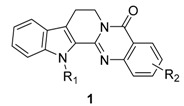 |
| Compound | R1 | R2 | Aggregation (%) | |||
|---|---|---|---|---|---|---|
| Inducer | ||||||
| ADP (0.22 μM) | Thrombin (0.1 unit∙mL−1) | A.A. a (100 μM) | Collagen (10 μg∙mL−1) | |||
| 1a | H | H | 99 b [56] | 85.7 c [28] | 85.2 c [28], 0 d [57] | 82.1 c [28], 22.1 c [57] |
| 1b [28] | H | 2,3-OCH2O- | 89.8 b | 0 e | ||
| 1c [28] | H | 3-Cl | 88.8 b | 0 e | 75.4 c | |
| 0.5% DMSO [28] | 88.2 | 90.1 | 88.5 | |||
| 1d [58] | H | 3-OH | 84.0 d | 0 e | 51.1 c | |
| 1e [58] | H | 3-OMe | 89.7 e | 0 f | 19.8 c | |
| aspirin (20 μg∙mL−1) [58] | 92.1 d | 0 e | 90.1 d | |||
| 1aa [57] | CH2CH2OH | H | 76 b | |||
| 1ab [57] | (CH2)3CO2H | H | 14 b | |||
| 1ac [57] | 3,4-(Me)2C6H3 | H | 88 b | |||
The values were given with standard error of the mean (SEM), but intentionally omitted SEM for clarity. Platelets were pre-incubated with 0.5% DMSO (control) or compounds for 3 min, then the inducer was added to trigger aggregation. a A.A. = arachidonic acid; b,d,e,f Values given were aggregation percentage in the presence of 100, 20, 5, and 50 μg∙mL−1 of rutaecarpines, respectively; c Aggregation percentage at the concentration of 10 μM.
3.2. Vasodilator Activity
An early study showed that phenylephrine-induced contraction of isolated rat mesenteric arterial segments with intact endothelium was relaxed by 90% by 0.1 mM rutaecarpine and that such relaxation was concentration-dependent in the 0.1 μM–0.1 mM range [59]. Further study revealed that NO-dependent vasodilation is primarily responsible for the vasodilatory activity of rutaecarpine [60]. Subsequently, the vasodilatory effect of rutaecarpine was also related to the stimulation of endogeneous calcitonin gene-related peptide (CGRP) release via the activation of transient receptor potential vanilloid subfamily, member 1 (TRPV1) [61,62]. Chen et al. synthesized 12 rutaecarpine derivatives and 11 analogues, and then evaluated their vasodilator activities (data not shown). These authors found two important trends regarding the vasodilator activities of rutaecarpine-related anti-hypertensives: (1) the N14 atom of rutaecarpine might be the key site, and (2) the 5-carbonyl probably makes a lower contribution, while simple substitution on the indole or quinazoline rings does not enhance vasodilatory effects [62]. Although the prepared compounds showed better activity than rutaecarpine (EC50 = 1.33 μM), such a finding would suggest a new direction for the discovery of valuable TRPV1 agonists as anti-hypertensive drugs if rutaecarpine had proper substituent (s) at the proper position (s).
3.3. Cytotoxicity
3.3.1. Rutaecarpine Derivatives
The findings from the studies on the cytotoxicity of rutaecarpine and its derivatives are summarized in Table 3. In general, ring substitution results in selectivity towards specific cell lines. Although the introduction of substitutions on ring A affected the cytotoxicity more significantly, the position is also important. 11-Methoxyrutaecarpine (1f) showed selective cytotoxicity against a human lung and renal cancer cells at concentrations of 0.75 and 0.31 μM, respectively, and the 10,11-methylenedioxy analogue 1b showed selective cytotoxicity for ovarian cancer cell lines, while 10-methoxyrutaecarpine (1e) showed no significant cytotoxicity at concentrations up to 25 μM [63]. 12-Fluororutaecarpine (1n) showed somewhat stronger cytotoxicity in the HT-29 human cell line compared to 2-chlororutaecarpine (1m) and those with fluorine on ring E (data not shown) [64]. 11,12-Dichlororutaecarpine (1o), a hybrid of bauerine (7,8-dichloro-9-methyl-2,9-dihydro-1H-pyrido[3,4-b]indol-1-one) [65] and rutaecarpine, showed the strongest inhibitory activity against HL-60 at the 0.15 μM level.
Table 3.
 |
| SF-295 | HT-29 | A549/ATCC | NCI-H460 | OVCAR-4 | 786-0 | CCRF-CEM/HL-60 | N87 | HS-578T | |
|---|---|---|---|---|---|---|---|---|---|
| 1a (rutaecarpine) | 14.1 [72] | 31.6 [73] | 14.5 [63] | 18.9 [63] | 19.8 [74] | 8.41 [74] | 22.6 [63] | ||
| 1b (10,11-OCH2O-) [63] | >25.0 | 1.55 | 1.50 | 1.08 | >25.0 | 5.05 | |||
| 1d (3-OH) [70] | 11.94 | ||||||||
| 1e (10-OCH3) [63] | >25.0 | >25.0 | >25.0 | >25.0 | >25.0 | ||||
| 1f (11-OCH3) [63] | 0.75 | 1.38 | >25.0 | 0.31 | 1.59 | ||||
| 1g (10-Br) [72] | 8.62 | 16.3 b | 6.43(11.1) c | ||||||
| 1h (1-OH) [73] | 7.39 | 10.43 | 10.1 [74], 8.34 [70] | 8.38 [74] | |||||
| 1i [7-OH(β)] [74] | 10.1 | 23.2 | |||||||
| 1j [7-OH(β), 8-OH(α)] [74] | 13.7 | 14.1 | |||||||
| 1k [7-OH(β), 8-OCH3(α) [74] | 7.82 | 22.3 | |||||||
| 1l [7-OH(β), 8-OEt(α)] [74] | 8.31 | 27.9 | |||||||
| 1m (2-Cl) [64] | 5.62 | 22.4 | 21.6 d | ||||||
| 1n (12-F) [64] | 1.26 | 8.4 | 3.18 d | ||||||
| 1o (11,12-diCl) [75] | 0.15 |
Tumor cell lines: CNS cancer (U251), human colon carcinoma (HT-29), human lung adenocarcinoma (A549/ATCC and NCI-11460), renal cancer (786-0, ACHN), ovarian cancer (OVCAR-4), leukemia (CCRF-CEM, HL-60, or P-388), human gastric carcinoma (N87), and breast cancer (HS-578T). a The cytotoxicity GI50 values are the concentrations corresponding to 50% growth inhibition, and they are the averages of at least two determinations; b for non-small cell lung cancer cell line; c Value in parenthesis was taken from ovarian OVCAR-8 cancer cell line; d for human breast cancer carcinoma MCF-7.
Regarding the mechanisms responsible for cytotoxicity, inhibitory activities against topoisomerase (topo) I and II have been studied. These inhibitory activities appeared to be affected by substitution on the E-ring but not by substitutions on rings A and/or C [64,66,67], except for 11-bromorutaecarpine. In fact, 10-bromorutaecarpine (1g) and 3-chlororutaecarpine (1c) showed strong inhibitory activities (79.54% and 84.35%, respectively) against topo I and were comparable to camptothecin (82.62%) at 100 μM against 0.2 unit topo I and similar to that against 0.2 units of topo II [67] (see Table 5). In addition, rutaecarpine inhibited tumor cell migration by approximately 30%–40% at 100 μg∙mL−1 [68], which would open a new study-window on the use of rutaecarpine as an antitumor agent.
Table 5.
 |
| Compound | Cell Lines (GI50, μM) | Topo I Inhibition (%) | Topo II Inhibition (%) | ||||
|---|---|---|---|---|---|---|---|
| HEK293 | MCF7 | DU145 | HCT116 | K562 | |||
| 1a | >50 | 19.57 | 31.65 | 33.89 | 25.77 | 1.20 | 1.25 |
| 1g | (see Table 3) | 79.54 | 35.43 | ||||
| 1c | NT | NT | NT | NT | NT | 84.36 | 69.51 |
| 30a | 36.57 | 11.42 | 27.54 | 9.94 | 25.38 | NA | 45.38 |
| 30b | >50 | 16.94 | 36.55 | 24.16 | 31.42 | 48.09 | 3.52 |
| 30c | >50 | 12.25 | 30.14 | 15.78 | 25.63 | 0.46 | 45.92 |
| 30d b | - | - | - | - | - | - | - |
| 30e | >50 | 12.06 | 29.64 | 25.36 | 28.39 | 28.11 | 52.74 |
| etoposide c | 4.09 | 3.02 | 3.75 | 1.88 | 2.23 | 52.52 | - |
| camptothecin c | 5.16 | 4.22 | 3.22 | 0.38 | 1.05 | - | 63.41 |
a The original data were from three different experiments performed in triplicate and given as mean +/− standard error of the mean (SEM), but intentionally omitted SEM for clarity. Cell lines used are embryonic kidney cell line (HEK293), human breast cancer (MCF7), human prostate tumor (DU145), human colorectal adenocarcinoma (HCT116), and human chronic myelogenous leukemia cell line (K562); b Not soluble enough to produce meaningful values; c Data was taken with 0.2 unit of topo I (or II), 100 μM of reference (camptothecin or etoposide) and benzorutaecarpines prepared.
3.3.2. Rutaecarpine-Isosteres and Hybrids
Annulation of aromatic rings, especially thiophene (27a–d), pyrrole (27e), and furan (27f) rings onto rutaecarpine ring E enhanced cytotoxicity toward selected human cancer cell lines [69]. All of these systems showed improved cytotoxicity against melanoma (UACC62), ovarian (SKVO3), prostate (DU145) and renal cancer (ACHN) cell lines but not against CNS or lung cancer cell lines.
On the other hand, dihydrorutaecarpines (29) showed increased activity and selectivity toward a CNS (U251) cancer cell line in the concentration range 0.02–7 μM and toward a renal cancer cell line at 0.08–20 μM (Table 4). The parent 30a showed strong activity (GI50 = 0.02 μM) and selectivity toward a CNS cancer cell line and the 10-methylthio compound (29b) showed strong activity (GI50 = 0.08 μM) and selectivity toward a renal (ACHN) cancer cell line.
Table 4.
 |
| Compound | X | R1 | R2 | R3 | U251 | H522 | UACC62 | SKOV3 | DU145 | ACHN |
|---|---|---|---|---|---|---|---|---|---|---|
| 28a | S | H | H | H | >100 | >100 | 7 | 10 | >100 | >100 |
| 28b | S | Cl | H | H | 53 | 74 | 2 | 12 | 12 | 2 |
| 28c | S | SCH3 | CH3 | CO2Et | >100 | >100 | 7 | 20 | 2 | 13 |
| 28d | S | F | H | H | >100 | >100 | 7 | 17 | 1 | 46 |
| 28e | N | Br | CH3 | H | 26 | 33 | 9 | 14 | 34 | 20 |
| 28f | O | Cl | t-Bu | H | 11 | - | 14 | 8 | 15 | 2.4 |
| 29a | - | H | H | H | 0.02 | 37 | >100 | 3 | 0.2 | 20 |
| 29b | - | SCH3 | H | H | 3 | 35 | 17 | 13 | 15 | 0.08 |
| 29c | - | SOCH3 | H | H | 7 | >100 | >100 | 27 | 0.1 | 2 |
| 29d | - | Br | H | H | 5 | 59 | >100 | >100 | 32 | 1 |
| 29e | - | Cl | H | H | 2.5 | 9 | >100 | 2 | 2 | 2 |
| 29f | - | Cl | H | CH3 | 6 | 10 | 7 | 11 | 2 | 1 |
| 29g | - | Br | H | CH3 | 0.3 | 18 | >100 | 3 | 3 | 0.4 |
| 29h | - | H | H | CH3 | 5 | 1 | 6 | 2 | 15 | 11 |
| 29i | - | H | NO2 | H | 3 | 3 | 2 | 12 | 3 | 3 |
Most of the values were given with standard error of the mean (SEM), but intentionally omitted SEM for clarity. Tumor cell lines: CNS cancer (U251), lung cancer (H522), melanoma cancer (UACC62), ovarian cancer (SKOV3), prostate cancer (DU145), and renal cancer (ACHN). a The cytotoxicity GI50 values are the concentrations corresponding to 50% growth inhibition, and are the averages of at least two determinations.
It is worth noting that evodiamine (29h) showed strong in vitro cytotoxicity against the following cell lines; human leukemia (HL-60, GI50 = 0.51 μM), human prostate cancer cell line (PC-3, GI50 = 14.35 μM) [70], murine fibrosarcoma (L929, GI50 = 20.3 μM), human breast adenocarcinoma (HeLa, GI50 = 15.4 μM), and human malignant melanoma (A375-S2, GI50 = 10.1 μM) [71].
Although the cytotoxicity of 13b,14-dihydrorutaecarpine (29a) is somewhat more potent than that of rutaecarpine (1a), it is not easy to establish any possible structure-activity relationship between 13b,14-dihydrorutaecarpine derivatives 29 and the corresponding rutaecarpine derivatives 1, not only because the substitution patterns are different but also because tested cancer cell lines are different.
The poor solubility of rutaecarpine and its derivatives in common organic solvents and in water results in poor bioavailability, which is the main obstacle that needs to be resolved for the further development of rutaecarpine and its derivatives as a drug. In fact, the excellent in vitro activities (GI50 = 1–8 μM) of compounds 29e, 29f, and 29i were not reflected by xenograft model results, presumably because of their poor bioavailability [69].
Generally, benzo-annulation increases electronic dispersion and planar dimensions thus may play an important role in interactions with receptor sites [76,77]. A series of benzo-annulated rutaecarpines were prepared using Fischer indole synthesis and their cytotoxicities against selected human cancer cell lines and their inhibitory effects on topo I and II were evaluated [78] (Table 5). However, currently available data are not sufficient to indicate any clear structure-activity relationships.
It should be noted that an isostere 24 with a sulfone moiety was not as active as rutaecarpine. The percentage of apoptotic cells corresponding to the sub-GI phase of 5-sulfarutaecarpine was found to be 29.2%, which compares with the 14.5% of rutaecarpine [48]. In addition, the hybrids 27a showed an increase of cytotoxic activities against HeLa cells and apoptosis inducing effects at a concentration comparable to that of etoposide. The percentages of apoptotic cells corresponding to the sub-G1 phase of 26 and 27a at the 10−6 mol∙L−1 were 38.6% and 24.1%, respectively, which are comparable to the 14.5% of rutaecarpine while a positive control (etoposide) gave 15.4%.
3.4. Inhibitory Activity on Acetylcholinesterase
The early studies on the strong inhibitory activity (64% inhibition at 100 μg∙mL−1) of fruits extract of Evodia officinalis against acetylcholinesterase [79] and its strong in vivo anti-amnesic activity (IC50 = 6.3 μM) led to the finding that dehydroevodiamine (31) was the origin of such biological activities [80] (Table 6 and Table 7). These results led to more systematic studies on the anti-cholinesterase activity of rutaecarpine [81].
Table 6.
 |
| Compound | R | X | AChE Inhibition (nM) a | BuChE Inhibition (nM) b | Selectivity Index c |
|---|---|---|---|---|---|
| 1a | H | O | >100,000 | >100,000 | |
| 1q | NHCOCH2NEt2 | O | 372.3 | 17,620 | 47.4 |
| 1r |  |
O | 131.2 | 696.1 | 5.3 |
| 1s |  |
O | 111.4 | 33,020 | 297.5 |
| 1t | NHCO(CH2)2NEt2 | O | 80.20 | 2848 | 35.5 |
| 1u |  |
O | 29.24 | 844.5 | 28.9 |
| 1v |  |
O | 21.40 | 2112 | 98.7 |
| 1w | H | H,H | 340 | 50 | 0.15 |
| tacrine | 222.7 | 29.98 | 0.1 | ||
| 31 [79] | 13,200 (37,800 [80], 6300 [83]) | 115,900 | 8.8 |
The original data were given as mean +/− standard error of the mean (SEM), but intentionally omitted SEM for clarity. a 50% inhibitory concentration (means of at least four independent experiments) of eeAChE from electric eel; b 50% inhibitory concentration (means of at least four independent experiments) of eqBuChE from equine serum; c Selectivity Index for AChE = IC50 (BuChE)/IC50 (AChE).
Table 7.
| Compound | R | N | AChE Inhibition (nM) a | BuChE Inhibition (nM) b | Selectivity Index c |
|---|---|---|---|---|---|
| 11b [81] | NHCOCH2NEt2 | 57.09 (70.4 [82]) | 11,360 | 198.9 | |
| 11c [81] |  |
1 | 23.56 (59.3 [82]) | 428.2 | 18.2 |
| 11d [81] |  |
1 | 10.07 (70.0 [82]) | 5429 | 539.1 |
| tacrine [81] | 222.7 | 29.98 | 0.1 | ||
| 11e [82] |  |
2 | 13.90 | 14,900 | 1072 |
| 11f [82] |  |
2 | 51.00 | 24,000 | 471 |
| 11g [82] |  |
1 | 0.80 | 2451 | 3225 |
| 11h [82] |  |
1 | 0.61 | 1855 | 3092 |
| 11i [82] |  |
1 | 3.09 | 7300 | 2362 |
| 11j [82] |  |
2 | 2.30 | 4291 | 1858 |
| 11k [82] |  |
2 | 2.10 | 3488 | 1638 |
| 11l [82] |  |
2 | 14.30 | 6340 | 450 |
| 11m [82] |  |
3 | 3.90 | 4160 | 1056 |
| tacrine [82] | 108.0 | 33.4 | 0.3 |
The original data were given as mean +/− standard error of the mean (SEM), but intentionally omitted SEM for clarity. a 50% inhibitory concentration (means of at least four independent experiments) of eeAChE from electric eel; b 50% inhibitory concentration (means of at least four independent experiments) of eqBuChE from equine serum; c Selectivity Index for AChE = IC50 (BuChE)/IC50 (AChE).
Wang, et al. prepared a series of rutaecarpine (compounds 1q–w) (Table 6) and 7,8-dehydrorutaecarpine (11, vide infra) derivatives (Table 7). Most of the rutaecarpine derivatives showed strong inhibitory activity against acetylcholinesterase (eeAChE) from electric eel and butylcholinesterase (eqBuChE) from equine serum with a selectivity on AChE over BuChE in the range 0.1–297.5 [81]. Additional structural modifications of 11 lead a dramatic increase in anticholinesterase activity up to 0.61 nM (11h) and selectivity on AChE up to over 3000 [81,82].
Interestingly, 5b, not only natural product but also one of the favored starting materials for rutaecarpine synthesis, showed promising inhibitory activity on AChE (IC50 = 83.38 μM) [84], implying a new vista towards designing new analogues of rutaecarpine for the treatment of Alzheimer’s disease. In addition to 5b, studies on truncated rutaecarpines have led to new promising lead compounds, such as 25 [85] (Table 8), 32 [86], and 33 [87] (Figure 4).
Table 8.
| Compound | R | AChE Inhibition (nM) a | BuChE Inhibition (nM) b | Selectivity Index c |
|---|---|---|---|---|
| 5b [84] | 83,380 | |||
| physostigmine [84] | 170 | 590 | 3.47 | |
| 25a [85] | H | >10,000 | >10,000 | |
| 25b [85] |  |
326.6 | 3103 | 9.50 |
| 25c [85] |  |
147.9 | 10,160 | 68.70 |
| 25d [85] |  |
20.98 | 7322 | 349.17 |
| tacrine [85] | 222.7 | 29.98 | 0.1 | |
The original data were given as mean +/− standard error of the mean (SEM), but intentionally omitted SEM for clarity. a 50% inhibitory concentration (means of at least 3 independent experiments) of eeAChE from electric eel; b 50% inhibitory concentration (means of at least 3 independent experiments) of eqBuChE from equine serum; c Selectivity Index for AChE = IC50 (BuChE)/IC50 (AChE).
Figure 4.
Selected examples of truncated rutaecarpines.
The anticholinesterase activity and the selectivity on AChE were somewhat related to the back-bone structure and the length of the side chain: The derivatives with a backbone with an aromatic C ring (11) showed better activity and selectivity than non-aromatic (1) and open-chain systems (25). The 7,8-dehydrogenated rutaecarpine derivative (11c, IC50 = 23.56 nM, SI = 18.20) is more active and more selective than the rutaecarpine derivative (1r, IC50 = 131.2 nM, SI = 5.3) and open C-ring derivative (25b, 326.6 nM, SI = 9.50) (Table 8). On the other hand, an increase of the length of the side chain would increase the activity (1q vs. 1t, 1r vs. 1u, and 1s vs. 1v; 11c vs. 11e; and 25c vs. 25d) and selectivity except in the case of 1s vs. 1v.
The selectivity indexes (SI, calculated by IC50 for BuChE/IC50 for AChE) on AChE of all the rutaecarpine derivatives ranged from 5.3–3225. Although the selectivity on AChE over BuChE is a concern for curing Alzheimer’s disease, clinically useful physostigmine (SI = 3.47) [84], galanthamine HBr (SI = 13.1 [86]) and donepezil (SI = 1252 [88]) show selectivity for AChE over BuChE while rivastigmine (SI ≤ 0.008 [86]) and neostigmine (SI = 0.58 [89]) show selectivity for BuChE over AChE. These results imply that it may not be an advantage for a cholinesterase inhibitor to be selective for AChE or BuChE, but instead suggest that higher efficacy requires a good balance between AChE and BuChE.
4. Conclusions
Rutaecarpine is one of the important alkaloids isolated from the Rutaceae and related plants, and it exhibits various interesting biological properties. Recent years have witnessed steady progress in understanding the chemistry and biology of rutaecarpine. Furthermore, it should be noted that reports have been issued on the beneficial effects of rutaecarpine analogues on controlling lipid accumulation [52], obesity [53], and atherosclerosis [54,55], and. The present review focuses on the synthesis of rutaecarpine derivatives and on their biological activities, especially on structure-activity relationships and their antiplatelet, vasodilatory, cytotoxic, and anticholinesterase activities. More efficient and/or practical methods are needed for the synthesis of rutaecarpine derivatives, not only to pursue structure-activity relationship studies but also to identify novel potent lead compounds for drug development.
Acknowledgments
Most of our results adopted herein were done by the financial support from the Korean Science Foundation.
Author Contributions
J.K.S. and Y.J. wrote the manuscript. H.W.C. critically revised the manuscript typically section for biological properties.
Conflicts of Interest
The authors declare no conflict of interest.
References
- 1.Chen A.L., Chen K.K. The constituents of Wuchuyin (Evodia rutaecarpa) J. Am. Pharm. Assoc. 1933;22:716–719. doi: 10.1002/jps.3080220804. [DOI] [Google Scholar]
- 2.Liao J.F., Chen C.F., Chow S.Y. Pharmacological studies of Chinese herbs (9). Pharmacological effects of Evodia fructus. J. Formos. Med. Assoc. 1981;80:30–38. [PubMed] [Google Scholar]
- 3.Asahina Y., Kashiwaki K. Chemical constituents of the fruits of Evodia rutaecarpa. J. Pharm. Soc. Jpn. 1915:1293. [Google Scholar]
- 4.Asahina Y., Mayeda S. Evodiamine and rutaecarpine, alkaloids of Evodia rutaecarpa. J. Pharm. Soc. Jpn. 1916:871. [Google Scholar]
- 5.Asahina Y., Fujita A. Constitution of rutaecarpine. J. Pharm. Soc. Jpn. 1921:863–869. [Google Scholar]
- 6.Chu J.H. Constituents of the Chinese drug Wu-Chu-Yu, Evodia rutaecarpa. Sci. Rec. (China) 1951;4:279–284. [Google Scholar]
- 7.Bergman J. The quinazolinocarboline alkaloids. In: Brossi A., editor. The Alkaloids: Chemistry and Pharmacology. Volume 21. Academic Press; New York, NY, USA: 1983. pp. 29–54. [Google Scholar]
- 8.Witt A., Bergman J. Recent developments in the field of quinazoline chemistry. Curr. Org. Chem. 2003;7:659–677. doi: 10.2174/1385272033486738. [DOI] [Google Scholar]
- 9.Kikelj D. Product Class 13: Quinazolines. Sci. Synth. 2004;16:573–749. [Google Scholar]
- 10.Wang C.L., Liu J.L., Ling Y.P. Progress in the synthesis of rutaecarpine. Chin. J. Org. Chem. 2006;26:1437–1443. [Google Scholar]
- 11.Kollenz G. Product Class 10: Imidoylketenes. Sci. Synth. 2006;23:351–380. [Google Scholar]
- 12.Mhaske S.B., Agrade N.P. The chemistry of recently isolated naturally occurring quinazolinone alkaloids. Tetrahedron. 2006;62:9787–9826. doi: 10.1016/j.tet.2006.07.098. [DOI] [Google Scholar]
- 13.Yu Q., Guo C., Cheng Z. Current advances in the study on rutaecarpine. Yaoxue Shijian Zazhi. 2007;25:353–357. [Google Scholar]
- 14.Shakhidoyatov K.M., Elmuradov B.Z. Tricyclic quinazoline alkaloids: Isolation, synthesis, chemical modification, and biological properties. Chem. Nat. Compd. 2014;50:781–800. doi: 10.1007/s10600-014-1086-6. [DOI] [Google Scholar]
- 15.Sheu J.-R. Pharmacological effects of rutaecarpine, an alkaloid isolated from Evodia rutaecarpa. Cardiovasc. Drug Rev. 1999;17:237–245. doi: 10.1111/j.1527-3466.1999.tb00017.x. [DOI] [Google Scholar]
- 16.Chen C.F., Chiou W.F., Chou C.J., Liao J.F., Lin L.C., Wang G.J., Ueng Y.F. Pharmacological effects of Evodia rutaecarpa and its bioactive components. Chin. Pharm. J. (Taiwan) 2002;54:419–435. [Google Scholar]
- 17.Hu C., Li Y. Research progress in pharmacological actions of evodiamine and rutaecarpine. Chin. Pharmacol. Bull. 2003;19:1084–1087. [Google Scholar]
- 18.Long M.I., Zhang Y.M., He L.P., Zeng Q. Pharmacology of rutaecarpine. Jiefangjun Yaoxue Xuebao. 2008;24:528–531. [Google Scholar]
- 19.Jia S., Hu C. Pharmacological effects of rutaecarpine as a cardiovascular protective agent. Molecules. 2010;15:1873–1881. doi: 10.3390/molecules15031873. [DOI] [PMC free article] [PubMed] [Google Scholar]
- 20.Jayakumar T., Sheu J.R. Cardiovascular pharmacological actions of rutaecarpine, a quinazolinocarboline alkaloid isolated from Evodia rutaecarpa. J. Exp. Clin. Med. 2011;3:63–69. doi: 10.1016/j.jecm.2011.02.004. [DOI] [Google Scholar]
- 21.Yang Z., Meng Y., Wang Q., Yang B., Kuang H. Study of the pharmaceutical effects and material basis of Fructus evodiae. Chin. Arch. Tradit. Chin. Med. 2011;29:2415–2417. [Google Scholar]
- 22.Wang Y., Gao Y. Advances in modulation of cytochrome P-450 by Chinese herbal medicine. Zhong Cao Yao. 2003;34:477–479. [Google Scholar]
- 23.Ueda J., Ohsawa K. Determination of main components in oriental pharmaceutical decoctions and extract preparations by ion-pair high-performance liquid chromatography. J. Tohoku Pharm. Univ. 2002;49:13–25. [Google Scholar]
- 24.Lee S.H., Son J.K., Jeong B.S., Jeong T.C., Chang H.W., Lee E.S., Jahng Y. Progress in the studies on rutaecarpine. Molecules. 2008;13:272–300. doi: 10.3390/molecules13020272. [DOI] [PMC free article] [PubMed] [Google Scholar]
- 25.Asahina Y., Irie T., Ohta T. Synthesis of rutaecarpine. II. J. Pharm. Soc. Jpn. 1927;543:51–52. [Google Scholar]
- 26.Asahina Y., Manske R.H.F., Robinson R. A synthesis of rutaecarpine. J. Chem. Soc. 1927;1708:1708–1710. doi: 10.1039/jr9270001708. [DOI] [Google Scholar]
- 27.Lee C.S., Liu C.K., Chiang Y.L., Cheng Y.Y. One-pot reductive-cyclization as key step for the synthesis of rutaecarpine alkaloids. Tetrahedron Lett. 2008;49:481–484. doi: 10.1016/j.tetlet.2007.11.094. [DOI] [Google Scholar]
- 28.Lee C.S., Liu C.K., Cheng Y.Y., Teng C.M. A new and facile synthesis of rutaecarpine alkaloids. Heterocycles. 2009;78:1047–1056. doi: 10.3987/COM-08-11606. [DOI] [Google Scholar]
- 29.Kamal A., Reddy M.K., Reddy T.S., Santos L.S., Shankaraiah N. Total synthesis of rutaecarpine and analogues by tandem azido reductive cyclization assisted by microwave irradiation. Synlett. 2011;1:61–64. doi: 10.1055/s-0030-1259095. [DOI] [Google Scholar]
- 30.Tseng M.C., Cheng H.T., Shen M.J., Chu Y.H. Bicyclic 1,2,3-triazolium ionic liquids: Synthesis, characterization, and application to rutaecarpine synthesis. Org. Lett. 2011;13:4434–4437. doi: 10.1021/ol201793v. [DOI] [PubMed] [Google Scholar]
- 31.Rao K.R., Raghunadh A., Mekala R., Meruva S.B., Pratap T.V., Krishna T., Kalita D., Laxminarayana E., Prasad B., Pal M. Glyoxylic acid in the reaction of isatoic anhydride with amines: A rapid synthesis of 3-(un)substituted quinazolin-4(3H)-ones leading to rutaecarpine and evodiamine. Tetrahedron Lett. 2014;55:6004–6006. doi: 10.1016/j.tetlet.2014.09.011. [DOI] [Google Scholar]
- 32.Peterson I.N., Crestey F., Kristensen J.L. Total synthesis of ascididemin via anionic cascade ring closure. Chem. Commun. 2012;48:9092–9094. doi: 10.1039/c2cc34725c. [DOI] [PubMed] [Google Scholar]
- 33.Liang L.N., An R., Huang T., Xu M., Hao X.J., Pan W.D., Liu S. A simple approach for the synthesis of rutaecarpine and its analogs. Tetrahedron Lett. 2015;56:2466–2468. doi: 10.1016/j.tetlet.2015.03.104. [DOI] [Google Scholar]
- 34.Zheng C., De C.K., Mal R., Seidel D. α-Amination of nitrogen heterocycles: Ring-fused aminals. J. Am. Chem. Soc. 2008;130:416–417. doi: 10.1021/ja077473r. [DOI] [PubMed] [Google Scholar]
- 35.Richers M.T., Zhao C., Seidel D. Selective copper(II) and potassium iodide catalyzed oxidation of aminals to dihydroquinazoline and quinazolinone alkaloids. Beilstein J. Org. Chem. 2013;9:1194–1201. doi: 10.3762/bjoc.9.135. [DOI] [PMC free article] [PubMed] [Google Scholar]
- 36.Richers M.T., Deb I., Platonova Y.A., Zhang C., Seidel D. Facile access to ring-fused aminals via direct a-amination of secondary amines with o-aminobenzaldehydes: Synthesis of vasicine, deoxyvasicine, deoxyvasicinone, mackinazolinone, and rutaecarpine. Synthesis. 2013;45:1730–1748. [PMC free article] [PubMed] [Google Scholar]
- 37.Möhrle H., Kamper C., Schmid R. Eine neue Synthese von Rutaecarin. Arch. Pharm. 1980;313:990–995. doi: 10.1002/ardp.19803131204. [DOI] [Google Scholar]
- 38.Granger B.A., Kaneda K., Martin S.F. Multicomponent assembly strategies for the synthesis of diverse tetrahydroisoquinoline scaffolds. Org. Lett. 2011;13:4542–4545. doi: 10.1021/ol201739u. [DOI] [PMC free article] [PubMed] [Google Scholar]
- 39.Huang G., Roos D., Patricia Stadtmüller P., Decker M. A simple heterocyclic fusion reaction and its application for expeditious syntheses of rutaecarpine and its analogs. Tetrahedron Lett. 2014;55:3607–3609. doi: 10.1016/j.tetlet.2014.04.120. [DOI] [Google Scholar]
- 40.Larock R.C., Yum E.K. Synthesis of indoles via palladium-catalyzed heteroannulation of internal alkynes. J. Am. Chem. Soc. 1991;113:6689–6690. doi: 10.1021/ja00017a059. [DOI] [Google Scholar]
- 41.Pan X., Bannister T.D. Sequential Sonogashira and Larock indole synthesis reactions in a general strategy to prepare biologically active β-carboline-containing alkaloids. Org. Lett. 2014;16:6124–6127. doi: 10.1021/ol5029783. [DOI] [PMC free article] [PubMed] [Google Scholar]
- 42.Kametani T., Loc C.V., Higa T., Koizumi M., Ihara M., Fukumoto K. Iminoketene cycloaddition. 2. Total syntheses of arborine, glycosminine, and rutecarpine by condensation of iminiketene with amides. J. Am. Chem. Soc. 1977;99:2306–2309. doi: 10.1021/ja00449a047. [DOI] [Google Scholar]
- 43.Bergman J., Bergman S. Studies of rutaecarpine and related quinazolinocarboline alkaloids. J. Org. Chem. 1985;50:1246–1255. doi: 10.1021/jo00208a018. [DOI] [Google Scholar]
- 44.Kaneko C., Chiba T., Kasai K., Miwa C. A short synthesis of rutecarpine and/or vasicolinone from 2-chloro-3-(indol-3-yl)ethylquinazolin-4(3H)-one: Evidence for the participation of the spiro intermediate. Heterocycles. 1985;23:1385–1390. doi: 10.3987/R-1985-06-1385. [DOI] [Google Scholar]
- 45.Mohanta P.K., Kim K. A Short Synthesis of quinazolinocarboline alkaloids rutaecarpine, hortiacine, euxylophoricine A and euxylophoricine D from methyl N-(4-chloro-5H-1,2,3-dithiazol-5-ylidene)anthranilates. Tetrahedron Lett. 2002;43:3993–3996. doi: 10.1016/S0040-4039(02)00711-6. [DOI] [Google Scholar]
- 46.Harayama T., Hori A., Serban G. Concise synthesis of quinazoline alkaloids, luotonins A and B, and rutaecarpine. Tetrahedron. 2004;60:10645–10649. doi: 10.1016/j.tet.2004.09.016. [DOI] [Google Scholar]
- 47.Bowman W.R., Elsegood M.R.J., Stein T., Weaver G.W. Radical reactions with 3H-quinazolinones: Synthesis of deoxyvasicinone, mackinazolinone, luotonin A, rutaecarpine, and tryptanthrin. Org. Biomol. Chem. 2007;5:103–110. doi: 10.1039/B614075K. [DOI] [PubMed] [Google Scholar]
- 48.Bubenyák M., Takács M., Blazics B., Rácz A., Noszál B., Püski L., Kökösi J., Hermecz I. Synthesis of bioisosteric 5-sulfa-rutaecarpine derivatives. ARKIVOC. 2010;11:291–302. doi: 10.3998/ark.5550190.0011.b23. [DOI] [Google Scholar]
- 49.Bubenyák M., Pálfi M., Takács M., Béni S., Szökö É., Noszál B., Kökösi J. Synthesis of hybrid between the alkaloids rutaecarpine and luotonins A, B. Tetrahedron Lett. 2008;49:4937–4940. doi: 10.1016/j.tetlet.2008.05.141. [DOI] [Google Scholar]
- 50.Ma Z.Z., Hano Y., Nomura T., Chen Y.J. Two new pyrroloquinzolinoquinoline alkaloids from Peganum nigellastrum. Heterocycles. 1997;46:541–546. [Google Scholar]
- 51.Liang J.U., Cha H.C., Jahng Y. Recent advances in the studies on luotonins. Molecules. 2011;16:4861–4833. doi: 10.3390/molecules16064861. [DOI] [PMC free article] [PubMed] [Google Scholar]
- 52.Chen Y.C., Zeng X.Y., He Y., Liu H., Wang B., Zhou H., Chen J.W., Liu P.Q., Gu L.Q., et al. Rutaecarpine analogues reduce lipid accumulation in adipocytes via inhibiting adipogenesis/lipogenesis with AMPK activation and UPR suppression. ACS Chem. Biol. 2013;8:2301–2311. doi: 10.1021/cb4003893. [DOI] [PubMed] [Google Scholar]
- 53.Kim S.J., Lee S.J., Lee S., Chae S., Han M.D., Mar W., Nam K.W. Rutecarpine ameliorates bodyweight gain through the inhibition of orexigenic neuropeptides NPY and AgRP in mice. Biochem. Biophys. Res. Commun. 2009;389:437–442. doi: 10.1016/j.bbrc.2009.08.161. [DOI] [PubMed] [Google Scholar]
- 54.Xu Y.N., Liu Q., Xu Y., Liu C., Wang X., He X.B., Zhu N.Y., Liu J.K., Wu Y.X., Li Y.Z., et al. Rutaecarpine suppresses atherosclerosis in ApoE−/−mice through up-regulating ABCA1 and SR-BI within RCT. J. Lipid Res. 2014;55:1634–1647. doi: 10.1194/jlr.M044198. [DOI] [PMC free article] [PubMed] [Google Scholar]
- 55.Li Y., Feng T., Liu P., Liu C., Wang X., Li D., Li N., Chen M., Xu Y., Si S. Optimization of rutaecarpine as ABCA1 up-regulator for treating atherosclerosis. ACS Med. Chem. Lett. 2014;5:884–888. doi: 10.1021/ml500131a. [DOI] [PMC free article] [PubMed] [Google Scholar]
- 56.Sheu J.R., Hung W.C., Lee Y.M., Yen M.H. Mechanism of inhibition of platelet aggregation by rutaecarpine, an alkaloid isolated from Evodia rutaecarpa. Eur. J. Phmacol. 1996;318:469–475. doi: 10.1016/S0014-2999(96)00789-3. [DOI] [PubMed] [Google Scholar]
- 57.Liu Q., Zheng W., Sang H., Chen J. Chin. Synthesis and aggregation activities in vitro of rutaecarpine derivatives. Zhongguo Yaowu Huaxue Zazhi. 2006;16:20–22. [Google Scholar]
- 58.Sheen W.S., Tsai I.L., Teng C.M., Ko F.N., Chen I.S. Indolopyridoquinazoline alkaloids with antiplatelet aggregation activity from Zanthoylum integrifoliolum. Planta Med. 1996;62:175–176. doi: 10.1055/s-2006-957846. [DOI] [PubMed] [Google Scholar]
- 59.Chiou W.F., Chou C.J., Liao J.F., Sham A.Y.C., Chen C.F. The mechanism of the vasodilator effect of rutaecarpine, and alkaloid isolated from Evodia rutaecarpa. Eur. J. Pharmacol. 1994;257:59–66. doi: 10.1016/0014-2999(94)90694-7. [DOI] [PubMed] [Google Scholar]
- 60.Chiou W.F., Liao J.F., Chen C.F. Comparative study on the vasodilatory effects of three quinazoline alkaloids isolated from Evodia rutaecarpa. J. Nat. Prod. 1996;59:374–378. doi: 10.1021/np960161+. [DOI] [PubMed] [Google Scholar]
- 61.Li Y.J., Deng H.W., Xiao L., Hu J.P. The depressor and vasodilator effects of rutaecarpine are mediated by calcitonin gene-related peptide. Planta Med. 2003;69:125–129. doi: 10.1055/s-2003-37703. [DOI] [PubMed] [Google Scholar]
- 62.Chen Z., Hua G., Li D., Chen J., Li Y., Zhou H., Xie Y. Synthesis and vasodilator effects of rutaecarpine analogues which might be involved transient receptor potential vanilloid subfamily, member 1 (TRPV1) Bioorg. Med. Chem. 2009;17:2351–2359. doi: 10.1016/j.bmc.2009.02.015. [DOI] [PubMed] [Google Scholar]
- 63.Yang L.M., Chen C.F., Lee K.H. Synthesis of rutaecarpine and cytotoxic analogs. Bioorg. Med. Chem. Lett. 1995;5:465–468. doi: 10.1016/0960-894X(95)00046-V. [DOI] [Google Scholar]
- 64.Jahng Y., Kim S.I., Lee E.S. Synthesis and cytotoxicities of rutaecarpine analogues; Abstracts of Papers of the American Chemical Society, Proceedings of the 227th ACS National Meeting; Anaheim, CA, USA. March 28–April 1 2004; Washington, DC, USA: American Chemical Society; MEDI 146. [Google Scholar]
- 65.Larsen L.K., Moore R.E., Patterson G.M.L. β-Carbolines from the blue-green alga Dichothrix baueriana. J. Nat. Prod. 1994;57:419–421. doi: 10.1021/np50105a018. [DOI] [PubMed] [Google Scholar]
- 66.Xu M.L., Moon D.C., Lee J.S., Woo M.H., Lee E.S., Jahng Y., Chang H.W., Lee S.H., Son J.K. Cytotoxicity and DNA topoisomerase inhibitory activity of constituents isolated from the fruits of Evodia officinalis. Arch. Pharm. Res. 2006;29:541–547. doi: 10.1007/BF02969262. [DOI] [PubMed] [Google Scholar]
- 67.Kim S.I., Lee S.H., Lee E.S., Lee C.S., Jahng Y. New topoisomerase inbihitors: Synthesis of rutaecarpine derivatives and their inhibitory activity against topoisomerases. Arch. Pharm. Res. 2012;35:785–789. doi: 10.1007/s12272-012-0504-1. [DOI] [PubMed] [Google Scholar]
- 68.Ogasawara M., Matsubara T., Suzuki H. Screening of natural compounds for inhibitory activity on colon cancer cell migration. Biol. Pharm. Bull. 2001;24:720–723. doi: 10.1248/bpb.24.720. [DOI] [PubMed] [Google Scholar]
- 69.Baruah B., Dasu K., Vaitilingam B., Mamnoor P., Venkata P.P., Rajagopal S., Yeleswarapu K.R. Synthesis and cytotoxic activity of novel quinazolino-β-carboline-5-one derivatives. Bioorg. Med. Chem. 2004;12:1991–1994. doi: 10.1016/j.bmc.2004.03.005. [DOI] [PubMed] [Google Scholar]
- 70.Zhao N., Li Z.L., Li D.H., Sun Y.T., Shan D.T., Bai J., Pei Y.H., Jing Y.K., Hua H.M. Quinolone and indole alkaloids from the fruits of Euodia rutaecarpa and their cytotoxicity against two human cancer cell lines. Phytochemistry. 2015;109:133–139. doi: 10.1016/j.phytochem.2014.10.020. [DOI] [PubMed] [Google Scholar]
- 71.Zhang Y., Zhang Q.H., Wu L.J., Tashiro S.I., Onodera S., Ikejima T. Atypical apoptosis in L929 cells induced by evodiamine isolated from Evodia rutaecarpa. J. Asian Nat. Prod. Res. 2004;6:19–27. doi: 10.1080/1028602031000119772. [DOI] [PubMed] [Google Scholar]
- 72.Yang L.M., Lin S.J., Lin L.C., Kuo Y.H. Antitumor agents. 2. Synthesis and cytotoxic evaluation of 10-bromorutaecarpine. Chin. Pharm. J. (Taiwan) 1999;51:219–225. [Google Scholar]
- 73.Chen J.J., Huang H.Y., Duh C.Y., Chen I.S. Cytotoxic constituents from the stem bark of Zanthoxylum pistaciiflorum. J. Chin. Chem. Soc. 2004;51:659–663. doi: 10.1002/jccs.200400099. [DOI] [Google Scholar]
- 74.Huang X., Zhang Y.B., Yang X.W. Indoloquinazoline alkaloids from Euodia rutaecarpa and their cytotoxic activities. J. Asian Nat. Prod. Res. 2011;13:977–983. doi: 10.1080/10286020.2011.602015. [DOI] [PubMed] [Google Scholar]
- 75.Huber K., Bracher F. Cytotoxic hybrids between the aromatic alkaloids bauerine C and rutaecarpine. Z. Naturforsch. B. 2007;62:1313–1316. doi: 10.1515/znb-2007-1013. [DOI] [Google Scholar]
- 76.Costes N., le Deit H., Michel S., Tillequin F., Koch M., Pfeiffer B., Renard P., Leonce S.S., Guilbaud N., Kraus-Berthier L., et al. Synthesis and cytotoxic and antitumor activity of benz[b]pyran[3,2-h]acridine-7-one analogues of acronycine. J. Med. Chem. 2000;43:2395–2402. doi: 10.1021/jm990972l. [DOI] [PubMed] [Google Scholar]
- 77.Michel S., Gaslonde T., Tillequin F. Benzo[b]acronycine derivatives: A novel class of antitumor agents. Eur. J. Med. Chem. 2004;39:649–655. doi: 10.1016/j.ejmech.2004.05.001. [DOI] [PubMed] [Google Scholar]
- 78.Hong Y.H., Lee W.J., Lee S.H., Son J.K., Kim H.L., Nam J.M., Kwon Y., Jahng Y. Synthesis and biological properties of benzo-annulated rutaecarpines. Biol. Pharm. Bull. 2010;33:1704–1709. doi: 10.1248/bpb.33.1704. [DOI] [PubMed] [Google Scholar]
- 79.Lee J.Y., Cha M.R., Choi C.W., Kim Y.S, Lee B.H., Ryu S.Y. Cholinesterase inhibitors isolated from the fruits extract of Evodia officinalis. Korean J. Pharmacogn. 2012;43:122–126. [Google Scholar]
- 80.Park C.H., Kim S.H., Choi W., Lee Y.L., Kim J.S., Kang S.S., Suh Y.H. Novel anticholinesterase and antiamnesic activity of dehydroevodiamine, a constituent of Evodia rutaecarpa. Planta Med. 1996;62:405–409. doi: 10.1055/s-2006-957926. [DOI] [PubMed] [Google Scholar]
- 81.Wang B., Mai Y.C., Li Y., Hou J.Q., Huang S.L., Ou T.M., Tan J.H., An L.K., Li D., Gu L.Q., et al. Synthesis and evaluation of novel rutaecarpine derivatives and related alkaloids derivatives as selective acetylcholinesterase inhibitors. Eur. J. Med. Chem. 2010;45:1415–1423. doi: 10.1016/j.ejmech.2009.12.044. [DOI] [PubMed] [Google Scholar]
- 82.He Y., Yao P.F., Chen S.B., Huang Z.H., Huang S.L., Tan J.H., Li D., Gu L.Q., Huang Z.S. Synthesis and evaluation of 7,8-dehydrorutaecarpine derivatives as potential multifunctional agents for the treatment of Alzheimer’s disease. Eur. J. Med. Chem. 2013;63:299–312. doi: 10.1016/j.ejmech.2013.02.014. [DOI] [PubMed] [Google Scholar]
- 83.Decker M. Novel inhibitors of acetyl- and butyrylcholinesterase derived from the alkaloids dehydroevodiamine and rutaecarpine. Eur. J. Med. Chem. 2005;40:305–313. doi: 10.1016/j.ejmech.2004.12.003. [DOI] [PubMed] [Google Scholar]
- 84.Fadaeinasab M., Hadi A.H.A., Kia Y., Basiri A., Murugaiyah V. Cholinesterase enzymes inhibitors from the leaves of Rauvolfia reflexa and their molecular docking study. Molecules. 2013;18:3779–3788. doi: 10.3390/molecules18043779. [DOI] [PMC free article] [PubMed] [Google Scholar]
- 85.Li Z., Wang B., Hou J.Q., Huang S.L., Ou T.M., Tan J.H., An L.K., Li D., Gu L.Q., Huang Z.S. 2-(2-Indolyl-)-4(3H)-quinazolines derivates as new inhibitors of AChE: Design, synthesis, biological evaluation and molecular modelling. J. Enzyme Inhib. Med. Chem. 2013;28:583–592. doi: 10.3109/14756366.2012.663363. [DOI] [PubMed] [Google Scholar]
- 86.Decker M., Krauth F., Lehmann J. Novel tricyclic quinazolinimines and related tetracyclic nitrogen bridged compounds as cholinesterase inhibitors with selectivity on butylcholinesterase. Bioorg. Med. Chem. 2006;14:1966–1977. doi: 10.1016/j.bmc.2005.10.044. [DOI] [PubMed] [Google Scholar]
- 87.Decker M. Homobivalent quinazolinimines as novel nanomolar inhibitors of cholinesterases with dirigible selectivity toward butyrylcholinesterase. J. Med. Chem. 2006;49:5411–5413. doi: 10.1021/jm060682m. [DOI] [PubMed] [Google Scholar]
- 88.Sugimoto H., Iimura Y., Yamaishi Y., Yamatsu K. Synthesis and structure-activity relationships of acetylcholinesterase inhibitors: 1-Benzyl-4-[(5,6-dimethoxy-1-oxoindan-2-yl)methylpiperidine hydrochloride and related compounds. J. Med. Chem. 1995;38:4821–4829. doi: 10.1021/jm00024a009. [DOI] [PubMed] [Google Scholar]
- 89.Khan I., Ibrar A., Zaib S., Ahmad S., Furtmann N., Hameed S., Simpson J., Bajorath J., Iqbal J. Active compounds from a diverse library of triazolothiadiazole and triazolothiadiazine scaffolds: Synthesis, crystal structure determination, cytotoxicity, cholinesterase inhibitory activity, and binding mode analysis. Bioorg. Med. Chem. 2014;22:6163–6173. doi: 10.1016/j.bmc.2014.08.026. [DOI] [PubMed] [Google Scholar]


