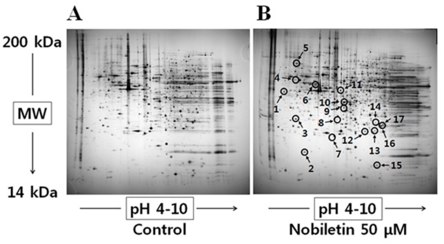
Figure 1.
Representative protein maps from human gastric cancer SNU-16 cells that were treated with (A) DMSO or (B) 50 μM nobiletin for 24 h. Spots with arrows represent proteins that changed their expression after nobiletin treatment; the PMF-based identification of these spots is summarized in Table 1.

