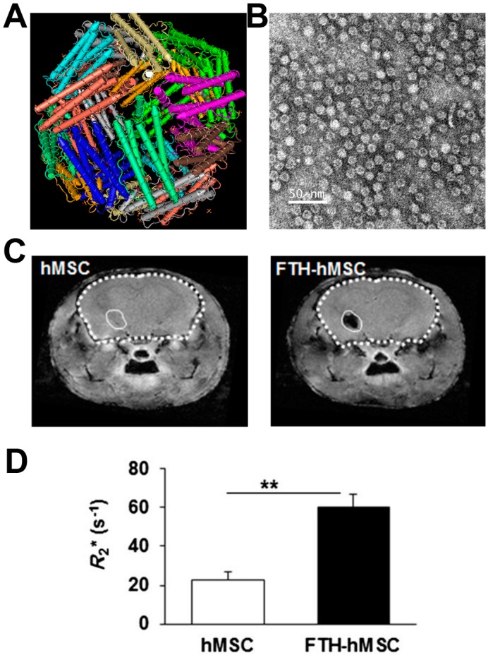
Figure 2.
Ferritin reporter gene was used to image human mesenchymal stem cells (hMSCs) by Hoe Suk Kim et al. (A) The structure of ferritin; (B) TEM of ferritin nanocages; (C) In vivo MRI R2* maps of mouse brain transplanted with hMSCs and ferritin reporter gene expressing hMSCs; (D) Bar chart showing the average R2* values measured from in vivo MR images at hMSCs-transplanted sites. Asterisks (**) indicate that the p value showed a statistically significant difference (p ≤ 0.01). Adapted with permission from [39].

