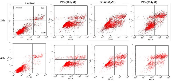
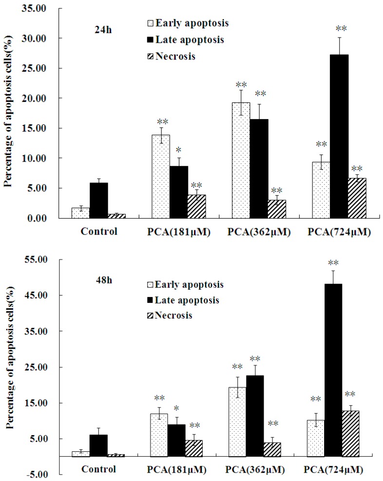
Figure 5.
Apoptosis induced by PCA on HT-29 cells. HT-29 cells were treated without (Control) and with PCA (181, 362, 724 μM) for 24 h and 48 h. Then, they were stained with FITC-conjugated Annexin V and PI for flow cytometry. All experiments were performed in trplicates. * p < 0.05, ** p < 0.01 vs. Control.


