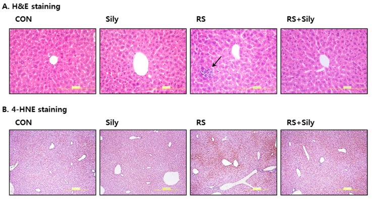
Figure 2.
Effect of silymarin on histological changes of the liver in mice subjected to restraint stress. (A) Hematoxylin and eosin (H & E) staining of representative liver sections shown at the same magnification (400×). Only mice subjected to restraint showed infilterated cells in the liver parenchyma. Black arrow represents inflammatory cell infiltration; (B) Representative pictures of 4-HNE protein adducts with brown color. 4-HNE positivity was observed mostly in hepatocytes around the central vein and it was most clear in the liver of restraint stressed mice. (CON; control mice, Sily; only silymarin-treated mice, RS; restraint stressed mice, RS + Sily; restraint-stressed mice with silymarin treatment).

