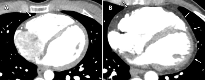
Figure 3:
Contrast-enhanced cardiac CT images in, A, 31-year-old healthy man with low epicardial adipose tissue (EAT) volume and, B, 39-year-old woman with arrhythmogenic right ventricular dysplasia/cardiomyopathy (ARVD/C) and high EAT volume. In the healthy control group participant, a small amount of epicardial fat is seen around right ventricle (arrow) but not around left ventricle. In patient with ARVD/C, there is extensive fat around both the left and right ventricles (arrows).

