ABSTRACT
Ocular features of Gaucher disease include gaze abnormalities, corneal clouding, ocular deposits and pigmentary changes in the macula. We report the presence of bilateral fovea sparing macular deposits in a 21-year-old woman with type 3 Gaucher disease. Macular deposits occur due to glucocerebroside accumulation within histiocytes and retinal deposits might correlate with the degree of systemic infiltration.
KEYWORDS: Gaucher disease, glycolipids, retinal Gaucher cells
Photo essay
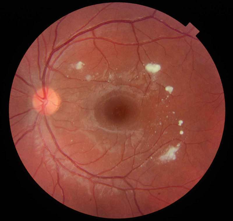
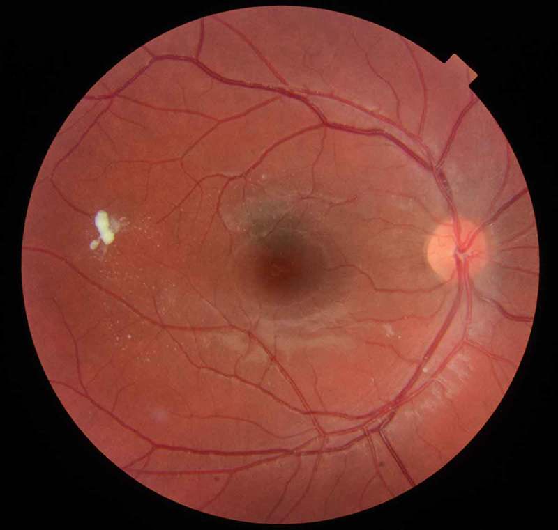
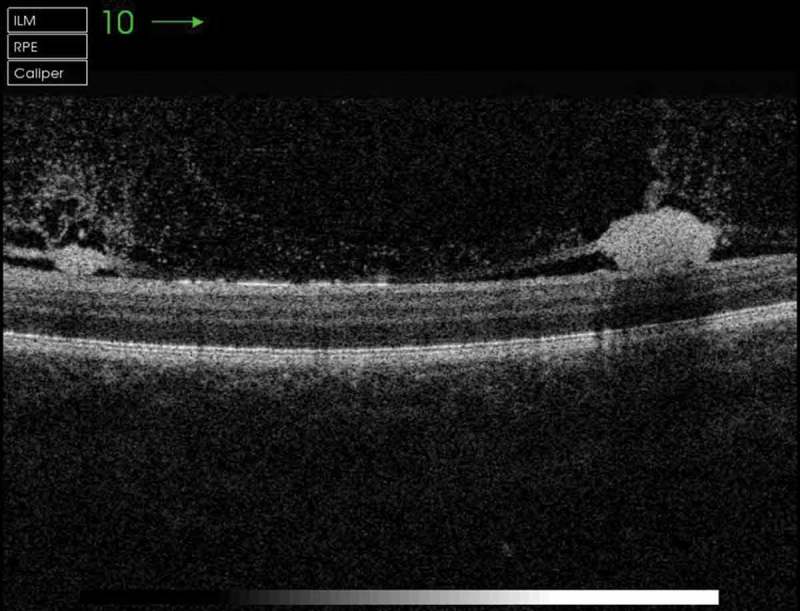
A 21-year-old woman with type 3 Gaucher disease (GD) on enzyme replacement therapy was reviewed in clinic. She had no visual symptoms and visual acuity was 6/6 bilaterally. She had clear corneas, quiet anterior chambers and no optic neuropathy. The macula showed white scattered deposits bilaterally, sparing the fovea (Figures 1 and 2). Optical Coherence Tomography examination showed these deposits to be preretinal (Figure 3).
Figure 1.

Retinal deposits in the left eye.
Figure 2.

Retinal deposits in the right eye.
Figure 3.

OCT image showing preretinal glycolopid deposits.
GD is an autosomal recessive disorder characterised by deficiency of glucocerebrosidase, resulting in abnormal accumulation of glycolipids within the reticuloendothelial system.1 There are three clinical types: Type 1 (GD1 – non-neuronopathic), which is distinguished from type 2 (GD2 – acute neuronopathic) and type 3 (GD3 – subacute neuronopathic) by the lack of characteristic involvement of the central nervous system.
Ocular features include oculomotor apraxia and supranuclear gaze abnormalities. Intraocular features include pingueculae, corneal clouding or white deposits on the corneal endothelium, pupillary margin, angle structures and in the ciliary body.2 There may be pigmentary changes3 in the macula and uveitis4 occurs infrequently. Cherry-red spots at the macula and pre retinal deposits5 have also been occasionally reported.
Macular deposits in GD occur due to accumulation of glucocerebroside within histiocytes6 and have been described as Gaucher cells.2,3 This can be confirmed histochemically by lectin staining methods.7 Gaucher cells are typically found in the liver, spleen and bone marrow. They are large, multinucleated cells with bluish grey cytoplasm with the appearance of ‘crumpled tissue paper’.3 It has been hypothesised that retinal Gaucher cell deposits may be linked to the degree of systemic infiltration.8
Acknowledgments
The authors thank Natalie Cook, Ophthalmic Photographer, Royal Free London Hospitals NHS Trust for retinal images.
Disclosure statement
The authors report no conflicts of interest. The authors alone are responsible for the content and writing of the article.
References
- 1.Beutler E. Gaucher disease: multiple lessons from a single gene disorder. Act Paediatr Suppl. 2006. April; 95(451):103–109. doi: 10.1080/08035320600619039. [DOI] [PubMed] [Google Scholar]
- 2.Petrohelos M, Tricoulis D, Kotsiras I, Vouzoukos A. Ocular manifestations of Gaucher disease. Am J Ophthalmol. 1975. December; 80(6):1006–1010. doi: 10.1016/0002-9394(75)90329-3. [DOI] [PubMed] [Google Scholar]
- 3.Rosenthal G, Wollstein G, Klemperer I, Lfghitz T.. Macular changes in type 1 Gaucher disease. Ophthalmic Surg Lasers. 2000. Jul-Aug; 31(4): 331–333. [PubMed] [Google Scholar]
- 4.Shrier EM, Barr CC, Grabowski GA. Vitreous opacities and retinal vascular abnormalities in Gaucher disease. Arch Ophthalmol. 2004;122(9):1395–1398. doi: 10.1001/archopht.122.9.1395. [DOI] [PubMed] [Google Scholar]
- 5.Sawicka-Gutaj N, Machaczka M, Kulińska-Niedziela I, et al. The appearance of newly identified intraocular lesions in Gaucher disease type 3 despite long-term glucocerebrosidase replacement therapy. Ups J Med Sci. 2016. August; 121(3):192–195. doi: 10.3109/03009734.2016.1158756. [DOI] [PMC free article] [PubMed] [Google Scholar]
- 6.Matsubara T, Yoshiya S, Maeda M, Shiba R, Hirohata K. Histologic and histochemical investigation of Gaucher cells. Clin Orthop Relat Res. 1982. June;(166):233–242. [PubMed] [Google Scholar]
- 7.Parkin JL, Brunning RD. Pathology of the Gaucher cell. Prog Clin Biol Res. 1982;95:151–175. [PubMed] [Google Scholar]
- 8.Gonzalez RMJ, Pintado CH, Lopez NC, Capdevila PA, torres CJP. Retinal Involvement In Gaucher’s Disease. J Fr Ophtalmol. 1992;15(3): 185–190. [PubMed] [Google Scholar]


