Abstract
Three-dimensional motion capture analysis is considered the gold standard for any movement research. Motion capture data were recorded for 7 healthy female participants with no prior throwing experience to investigate the learning process for overarm throwing during a selected period. Participants were monitored 3 times a week for 5 weeks. Each session consisted of 15 dominant and 15 nondominant hand side overarm throws. A total of 3,150 trials were recorded and preprocessed (labeling reflective markers) for further analysis. The presented dataset can provide valuable information about upper extremity kinematics of the learning process of overarm throwing without any kind of feedback. Furthermore, this dataset may be used for more advanced analysis techniques, which could lead to more insightful information.
Subject terms: Motor control, Physiology, Learning and memory
Background & Summary
Three-dimensional motion analysis systems, which use markers to track motion, are expensive, and they require long amounts of time. Therefore, due to the nature of the biomechanics research, most studies have had a highly limited number of trials, which makes it hard to use advanced data analysis methods, such as machine and deep learning. Most biomechanics studies with large numbers of data and researchers use educated guesses for data reduction before starting data acquisition, and they are limited to investigation of only several variables at specific events (e.g., shoulder flexion angle at ball release). With the advances of computers and computing methods, researchers have started to use more advanced analysis methods, such as principal component analysis, support vector machines, and regressions on larger datasets1–8. Studies that use more advanced analysis methods are mostly focused on cyclic movements such as gait and running. It has been shown that these methods may detect age-2 and fatigue-related5 changes, overall variability3,4,7, and the effects of different equipment6. However, limitations such as the sheer amount of rigorous work time and the financial support it takes to collect and process the data for the further calculations make it difficult for researchers to use more advanced analysis techniques.
Even though the use of reflective markers in 3D motion capture is very accurate (error < 1 mm), it is difficult to accurately track the shoulder and scapular joint movement. As the shoulder joint is connected to the scapular girdle, it has the ability to move in several planes simultaneously. This makes defining the shoulder joint biomechanics difficult and limited. Even with the work of the International Society of Biomechanics, a standard definition for shoulder biomechanics is not universally accepted9. Therefore, each research group uses its own model and marker set to define shoulder biomechanics according to the specific purpose of its study10–12. It was reported that rotation sequences have an effect on shoulder kinematics13,14. Some rotational sequences for the shoulder joint can result in gimbal lock occurrences more often than other combinations13,14. A modified marker set from other studies was used during data acquisition10,11. The aim of the modified marker set was for future studies to investigate differences of the upper extremity biomechanics’ definitions.
Another aspect of this study was to investigate the movement variability during the early skill acquisition for overarm throwing. In literature, extensive studies of upper extremity biomechanics during throwing activities15–18 and variability related to overarm throwing19–21 have been conducted. However, there has been no definitive conclusion about the role of variability, especially in sports biomechanics and performance22. Differences between the models and quantifications of variability are a source of discrepancy between results. It was reported that different variability quantifications showed different patterns, such as standard deviation increases while the coefficient in variation decreased23. In the traditional approach, less variability was concluded to be better because of the invariance of the performance, and the variability was regarded as the noise of the system24. However, recent studies showed that variability is not a noise of the system but can actually be useful24,25. Dynamical system theories applied to biomechanical datasets showed that variability is actually important for the human body as a system. It was reported that experts decrease their space-joint variability while increasing their trajectory variability19. Furthermore, the authors suggested that heavier balls can actually be used to achieve optimal variability for performance improvement of basketball free throws. Additionally, other studies showed that variability can be used to differentiate skill levels, injuries, and different populations12,26,27.
Therefore, healthy women participants with no prior overarm throwing experience or practice were recruited, and they performed several sessions for 5 weeks in a laboratory setting. They performed the throws with no feedback about their performance to make sure that they used only their sensory system for throwing the ball “as fast as possible”.
Methods
Participants
Eight female participants were recruited for this study. One participant was excluded because she injured herself during another activity during the experimental period, which left a total of seven participants for data acquisition. The study protocol was approved by the institutional review board of Kookmin University and was conducted according to the Helsinki declaration. The subjects gave their informed consent to participate in the study. All subjects were injury-free for both lower and upper extremities in the most recent six months, and they had no lower or upper extremity surgery in the last two years.
Procedures
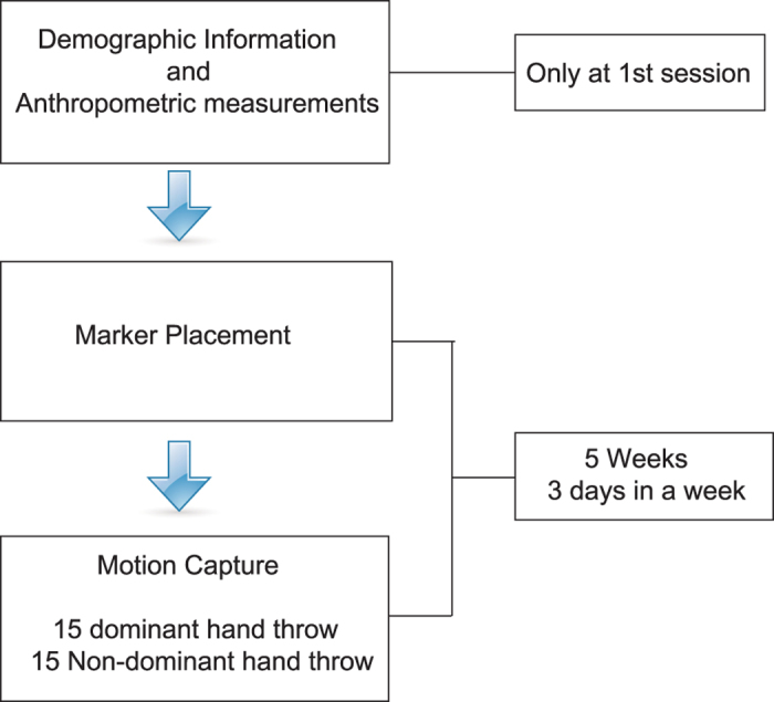
Participants visited the laboratory three times a week for five weeks. All participants’ demographic and anthropometric measurements needed for modeling were carried out during the first session (Table 1). Each participant performed 15 dominant and 15 nondominant hand throws with a baseball. Participants were asked to throw the ball “as fast as possible” into a foam cushion (approx. 3 m x 3 m), which was approximately 4 m away from the participant. The first 3 throws for each side have been classified as a warm-up. Ball speeds were measured with a speed gun for each trial. The experimental procedure is shown in Fig. 1.
Table 1. Demographic information of subjects.
| Subject | Age (Years) | Weight (kg) | Height (cm) | Wrist Width (mm) | Elbow Width (mm) | Knee Width (mm) | Ankle Width (mm) | Leg Length (cm) | Right Shoulder Offset (mm) | Left Shoulder Offset (mm) | Hand Thickness (mm) | Right Shoulder Circumference (cm) | Left Shoulder Circumference (cm) | Dominant Hand |
|---|---|---|---|---|---|---|---|---|---|---|---|---|---|---|
| S1 | 23 | 54.6 | 165 | 44 | 55 | 86 | 62 | 85 | 80 | 84 | 22 | 36.5 | 36.5 | Right |
| S2 | 26 | 54.8 | 160 | 43 | 50 | 85 | 54 | 82.5 | 83 | 83 | 16 | 35.3 | 35.3 | Left |
| S3 | 24 | 56.6 | 162 | 46 | 54 | 88 | 59 | 83.5 | 86 | 87 | 13 | 36.5 | 36.5 | Right |
| S4 | 30 | 57.1 | 159 | 47 | 53 | 87 | 59 | 82.5 | 73 | 73 | 18 | 37.5 | 37.5 | Right |
| S5 | 26 | 73.1 | 165 | 42 | 55 | 97 | 55 | 87.3 | 92 | 95 | 17 | 40.5 | 40.5 | Right |
| S6 | 23 | 51.1 | 160 | 40 | 50 | 85 | 55 | 82.2 | 61 | 63 | 27 | 34.3 | 34.3 | Right |
| S7 | 24 | 48.7 | 155 | 45 | 50 | 87 | 56 | 80.6 | 81 | 81 | 14 | 35.3 | 35.3 | Right |
| Mean±SD | 25.1±2.4 | 56.5±7.8 | 160.8±3.5 | 43.8±2.4 | 52.4±2.3 | 87.8±4.1 | 57.1±2.9 | 83.3±2.1 | 79.4±9.9 | 80.8±10.2 | 18.1±4.8 | 36.5±2.0 | 36.5±2.0 |
Figure 1. Experimental protocol.
Motion capture
Each session was performed in a laboratory fitted with 10 Vicon infrared cameras (T-10, T-40, Oxford Metrics Ltd., UK) and 2 force plates (BP-600900, AMTI, USA). Each camera connected to the Vicon MX Giganet and force plates were connected to the Giganet through a Vicon Datastation ADC patch panel. The giganet unit was connected to the main computer of the laboratory. Each of these cameras was equipped with an infrared strobe, which makes the reflective optical markers emit infrared light. Therefore, cameras recorded the trajectories of the markers via the emitted light from the markers. Marker trajectories were recorded through Vicon Nexus software (version 1.8.5, Oxford Metrics Ltd., UK). The marker trajectories and the force plate data were recorded synchronously at 200 and 1000 Hz sampling rates, respectively. A total of 450 trials were recorded for each participant.
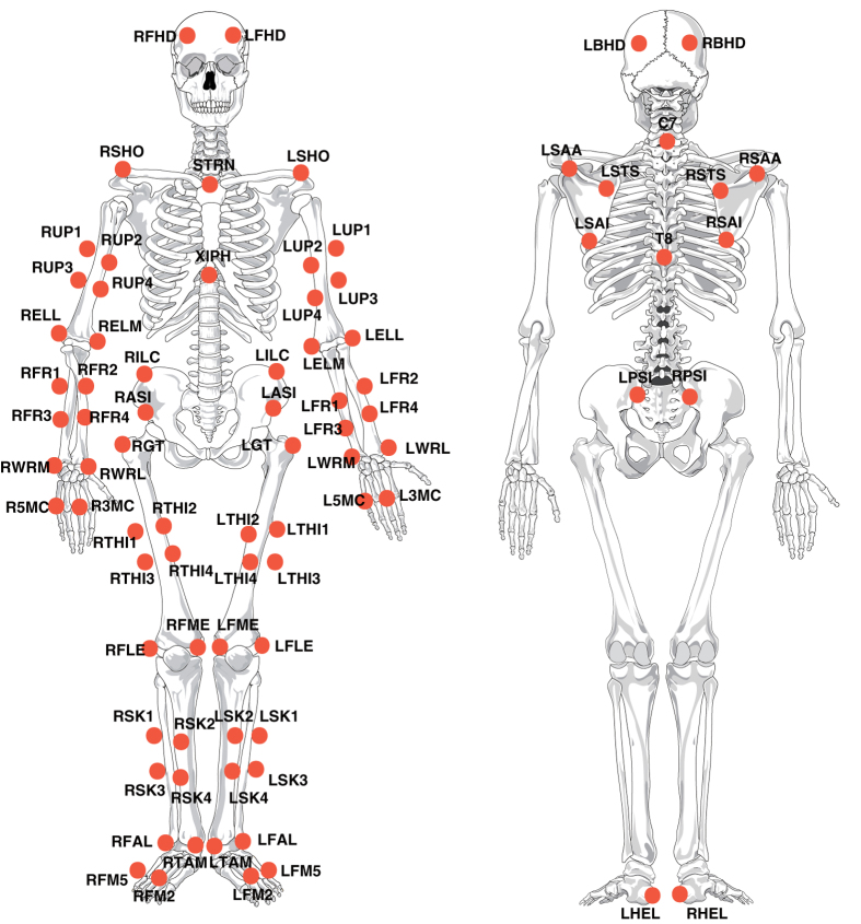
At each session, participants wore tight clothes before the reflective marker placement. Participants performed warm-up throws until they became comfortable with the markers and ready to start. Markers and cluster markers (Fig. 2) affixed to the anatomical landmarks and extremities are listed in Table 2 (available online only). Static trials needed for modeling purposes in the further analysis were captured in two different positions. Dynamic trials were captured with only the tracking markers, as listed in Table 2 (available online only). All throws were made while participants had each foot placed on one force plate. The marker trajectories were saved automatically by the system without any names attached to them. For further calculations, each marker had to be labeled with a specific name. Each trials’ marker trajectories were labeled (the naming process of the markers according to Table 2 (available online only)) using Vicon Nexus software (version 1.8.5, Oxford Metrics Ltd., UK).
Figure 2. Marker set used in motion capture.

Table 2. Marker set used in motion capture.
| Marker Name | Segment/Joint | Location | Tracking |
|---|---|---|---|
| LFHD | Head | Apprx. over the Left Temple | X |
| RFHD | Head | Apprx. over the Right Temple | X |
| LBHD | Head | Left Back of the Head | X |
| RBHD | Head | Right Back Of The Head | X |
| STRN | Trunk | Suprasternal Notch | X |
| XIPH | Trunk | Xiphoid Process | X |
| C7 | Trunk | 7thCervicalVertebra | X |
| T8 | Trunk | 8thThoracicVertebra | X |
| RASI | Pelvis | Right Ant Superior Iliac Crest | X |
| RPSI | Pelvis | Right Post Superior Iliac Crest | X |
| RILC | Pelvis | Right Iliac Crest | X |
| LASI | Pelvis | Left Ant Superior Iliac Crest | X |
| LPSI | Pelvis | Left Post Superior Iliac Crest | X |
| LILC | Pelvis | Left Iliac Crest | X |
| RSHO | Right Humerus | Acromion Process | X |
| RSTS | Right Humerus | Trigonum Scapulae | X |
| RSAI | Right Humerus | Angulus Inferior | X |
| RSAA | Right Humerus | Angulus Acromialis | X |
| RUP1 | Right Humerus | Upper Arm Cluster | X |
| RUP2 | Right Humerus | Upper Arm Cluster | X |
| RUP3 | Right Humerus | Upper Arm Cluster | X |
| RUP4 | Right Humerus | Upper Arm Cluster | X |
| RELL | Right Elbow Joint | Lateral Epicondyle | |
| RELM | Right Elbow Joint | Medial Epicondyle | |
| RFR1 | Right Forearm | Forearm Cluster | X |
| RFR2 | Right Forearm | Forearm Cluster | X |
| RFR3 | Right Forearm | Forearm Cluster | X |
| RFR4 | Right Forearm | Forearm Cluster | X |
| RWRL | Right Wrist | Lateral Point of Radial Styloid | |
| RWRM | Right Wrist | Medial Point of Ulnar Styloid | |
| R3MC | Right Hand | 3rdMetacarpalHead | X |
| R5MC | Right Hand | 5thMetacarpalHead1 | X |
| LSHO | Left Humerus | Acromion Process | X |
| LSTS | Left Humerus | Trigonum Scapulae | X |
| LSAI | Left Humerus | Angulus Inferior | X |
| LSAA | Left Humerus | Angulus Acromialis | X |
| LUP1 | Left Humerus | Upper Arm Cluster | X |
| LUP2 | Left Humerus | Upper Arm Cluster | X |
| LUP3 | Left Humerus | Upper Arm Cluster | X |
| LUP4 | Left Humerus | Upper Arm Cluster | X |
| LELL | Left Elbow Joint | Lateral Epicondyle | |
| LELM | Left Elbow Joint | Medial Epicondyle | |
| LFR1 | Left Forearm | Forearm Cluster | X |
| LFR2 | Left Forearm | Forearm Cluster | X |
| LFR3 | Left Forearm | Forearm Cluster | X |
| LFR4 | Left Forearm | Forearm Cluster | X |
| LWRL | Left Wrist | Lateral Point Of Radial Styloid | |
| LWRM | Left Wrist | Medial Point Of Ulnar Styloid | |
| L3MC | Left Hand | 3rdMetacarpalHead | X |
| L5MC | Left Hand | 5thMetacarpalHead1 | X |
| RGT | Right Femur | Femur Greater Trochanter | X |
| RTH1 | Right Femur | Thigh Cluster | X |
| RTH2 | Right Femur | Thigh Cluster | X |
| RTH3 | Right Femur | Thigh Cluster | X |
| RTH4 | Right Femur | Thigh Cluster | X |
| RFLE | Right Knee Joint | Femur Lateral Epicondyle | |
| RFME | Right Knee Joint | Femur Medial Epicondyle | |
| RSK1 | Right Shank | Shank Cluster | X |
| RSK2 | Right Shank | Shank Cluster | X |
| RSK3 | Right Shank | Shank Cluster | X |
| RSK4 | Right Shank | Shank Cluster | X |
| RFAL | Right Ankle Joint | Fibula Lateral Malleolus | |
| RTAM | Right Ankle Joint | Tibia Medial Malleolus | |
| RFM2 | Right Foot | 2ndMetatarsalHead | X |
| RFM5 | Right Foot | 5thMetatarsalHead | X |
| RHEL | Right Foot | Posterior of Calcaneus | X |
| LGT | Left Femur | Femur Greater Trochanter | |
| LTH1 | Left Femur | Thigh Cluster | X |
| LTH2 | Left Femur | Thigh Cluster | X |
| LTH3 | Left Femur | Thigh Cluster | X |
| LTH4 | Left Femur | Thigh Cluster | X |
| LFLE | Left Knee Joint | Femur Lateral Epicondyle | |
| LFME | Left Knee Joint | Femur Medial Epicondyle | |
| LSK1 | Left Shank | Shank Cluster | X |
| LSK2 | Left Shank | Shank Cluster | X |
| LSK3 | Left Shank | Shank Cluster | X |
| LSK4 | Left Shank | Shank Cluster | X |
| LFAL | Left Ankle Joint | Fibula Lateral Malleolus | |
| LTAM | Left Ankle Joint | Tibia Medial Malleolus | |
| LFM2 | Left Foot | 2ndMetatarsalHead | X |
| LFM5 | Left Foot | 5thMetatarsalHead | X |
| LHEL | Right Foot | Posterior of Calcaneus | X |
| Ball | Baseball | X |
Each trial has three events: the start of the movement, the ball release, and the end of the movement. The start of the movement was defined as the moment when the distance between the trunk and the hand marker increased 10% more than the start position. The ball release was defined as the moment when the distance between the ball marker and the hand marker increased 10%. The end of the movement was selected as the moment of the end of the follow through and the closest distance between hand and trunk. Vicon Nexus software lets users label events with only three names: ‘Foot Strike’, ‘Foot Off’ and ‘General’. For future usage and to prevent confusion, the start of the movement event was labeled ‘Foot Strike’; the ball release, ‘General’; and the end of the movement, ‘Foot Off’.
Data Records
c3d files are the standard file format that can be acquired from many motion capture systems and analyzed with different software. This file format is designed to include both three-dimensional point information and analog data. Therefore, each throw has its own c3d file that includes information about marker positions and all analog data. Additionally, comma-separated values (CSV) files, which include event times, positions of each marker, and force plate data, are included in the data records. The data records are available online from figshare (Data Citation 1), and they consist of c3d and CSV files and a detailed description file for each trial’s information (Trial Information, Data Citation 1).
Each subject has her own folder, and each subject folder has 15 folders which represent each session. Session folders were named ‘XwXd’, where X is the incremental integer for the week and day. For example, 1w1d represents the 1st week-1st day session.
Each session folder consists of c3d and CSV files for dominant and nondominant throw trials. Each trial was named systematically as ‘<S>_DP(or NDP)_<xx>_<m>’, where ‘S’ represents the subject and DP and NDP represent dominant and nondominant hand throws, respectively. ‘xx’ and ‘m’ are the incremental integer and information about each trial, respectively. For information about each trial, please refer to the Trial Information (Data Citation 1). Trial Information files (Data Citation 1) are provided due to the non-systematic incremental numbers of trials, ball speed, and other situations such as missing markers and errors. The key tab in a Trial Information file (Data Citation 1) consists of the meaning of each letter in ‘m’. For example, ‘S1_DP_01_O’ would represent Subject 1’s dominant hand first throw, and ‘O’ represents the trial having no problem.
Technical Validation
Markers were mostly affixed to the participants’ bodies by the first author. In some sessions, markers were affixed by other researchers but always checked by the first author before the recordings. Camera calibration was performed before each session, and the settings (such as strobe intensity and the threshold for centroid fitting) were chosen for optimal marker visibility and noise reduction.
Gap filling is a process used when there is a missing marker during the trial. It is used from the start to the end event for each trial. Gap filling via spline or pattern fill in Vicon Nexus software was used depending on which one concludes with the appropriate trajectory for movement. Experienced researchers execute gap filling during the labeling process. Some trials had missing markers from the start of the recording. Therefore, gap filling could not be performed in these trials. All trials were checked after labeling and the gap filling process by the first author. The Trial Information (Data Citation 1) includes information about each trial, whether or not it had any issues during acquisition or processing. The related description about the Trial Information (Data Citation 1) has been given in the Data Records.
Usage Notes
For more detailed information about c3d files and their possible uses with different software, refer to the c3d website (https://www.c3d.org). The modified marker set can be described as the combination of two previously published studies10,11. An upper extremity kinematics study based on this dataset has been published28. Furthermore, the needed anthropometric measurements for this modeling can be found in Table 1. There are two static trials for each session in case researchers want to use different definitions of upper extremity biomechanics. Additionally, researchers should be mindful of the data because the gap filling was made from the events of the start and end of the movement. Therefore, gap filling was not performed in frames other than those of the duration of the throwing. Researchers should be mindful of the trials with missing markers because they may affect the results, depending on the chosen model for further analysis.
Additional information
How to cite this article: Ozkaya, G. et al. Three-dimensional motion capture data during repetitive overarm throwing practice. Sci. Data. 5:180272 doi: 10.1038/sdata.2018.272 (2018).
Publisher’s note: Springer Nature remains neutral with regard to jurisdictional claims in published maps and institutional affiliations.
Supplementary Material
Acknowledgments
We greatly appreciate Associate Professor David O’Sullivan for his help in revising the manuscript. This study was financially supported by the Ministry of Education through the support of the National Research Foundation of Korea’s Basic Humanities and Social Research Support—General Joint Research Support Project (project number: NRF-2013S1A5A2A03045819). G.O. was supported by the Global Scholarship Program for Foreign Graduate Students at Kookmin University in Korea.
Footnotes
The authors declare no competing interests.
Data Citations
- Ozkaya G., et al. . 2018. figshare. https://doi.org/10.6084/m9.figshare.c.4017808
References
- Phinyomark A., Petri G., Ibáñez-Marcelo E., Osis S. T. & Ferber R. Analysis of Big Data in Gait Biomechanics: Current Trends and Future Directions. J. Med. Biol. Eng 38, 244–260 (2018). [DOI] [PMC free article] [PubMed] [Google Scholar]
- Fukuchi R. K., Eskofier B. M., Duarte M. & Ferber R. Support vector machines for detecting age-related changes in running kinematics. J. Biomech. 44, 540–542 (2011). [DOI] [PubMed] [Google Scholar]
- Federolf P., Tecante K. & Nigg B. A holistic approach to study the temporal variability in gait. J. Biomech. 45, 1127–1132 (2012). [DOI] [PubMed] [Google Scholar]
- Horst F. et al. Daily changes of individual gait patterns identified by means of support vector machines. Gait Posture 49, 309–314 (2016). [DOI] [PubMed] [Google Scholar]
- Janssen D. et al. Diagnosing fatigue in gait patterns by support vector machines and self-organizing maps. Hum. Mov. Sci 30, 966–975 (2011). [DOI] [PubMed] [Google Scholar]
- Onodera A. N., Gavião Neto W. P., Roveri M. I., Oliveira W. R. & Sacco I. C. Immediate effects of EVA midsole resilience and upper shoe structure on running biomechanics: a machine learning approach. PeerJ 5, e3026 (2017). [DOI] [PMC free article] [PubMed] [Google Scholar]
- Hoerzer S., von Tscharner V., Jacob C. & Nigg B. M. Defining functional groups based on running kinematics using Self-Organizing Maps and Support Vector Machines. J. Biomech. 48, 2072–2079 (2015). [DOI] [PubMed] [Google Scholar]
- Ferber R., Osis S. T., Hicks J. L. & Delp S. L. Gait biomechanics in the era of data science. J. Biomech. 49, 3759–3761 (2016). [DOI] [PMC free article] [PubMed] [Google Scholar]
- Wu G. et al. ISB recommendation on definitions of joint coordinate systems of various joints for the reporting of human joint motion - Part II: Shoulder, elbow, wrist and hand. J. Biomech. 38, 981–992 (2005). [DOI] [PubMed] [Google Scholar]
- Aguinaldo A. L., Buttermore J. & Chambers H. Effects of upper trunk rotation on shoulder joint torque among baseball pitchers of various levels. J. Appl. Biomech. 23, 42–51 (2007). [DOI] [PubMed] [Google Scholar]
- Gates D. H., Walters L. S., Cowley J., Wilken J. M. & Resnik L. Range of motion requirements for upper-limb activities of daily living. Am. J. Occup. Ther. 70, 7001350010p1–7001350010p10 (2016). [DOI] [PMC free article] [PubMed] [Google Scholar]
- Fleisig G. S., Chu Y., Weber A. & Andrews J. Variability in baseball pitching biomechanics among various levels of competition. Sport. Biomech. / Int. Soc. Biomech. Sport 8, 10–21 (2009). [DOI] [PubMed] [Google Scholar]
- Šenk M. & Chèze L. Rotation sequence as an important factor in shoulder kinematics. Clin. Biomech. 21, 3–8 (2006). [DOI] [PubMed] [Google Scholar]
- Bonnefoy-Mazure A. et al. Rotation sequence is an important factor in shoulder kinematics. Application to the elite players’ flat serves. J. Biomech 43, 2022–2025 (2010). [DOI] [PubMed] [Google Scholar]
- Chu Y., Fleisig G. S., Simpson K. J. & Andrews J. R. Biomechanical comparison between elite female and male baseball pitchers. J. Appl. Biomech. 25, 22–31 (2009). [DOI] [PubMed] [Google Scholar]
- Escamilla R. F., Fleisig G. S., Barrentine S. W., Zheng N. & Andrews J. R. Kinematic comparisons of throwing different types of baseball pitches. J. Appl. Biomech. 14, 1–23 (1998). [Google Scholar]
- Werner S. L., Suri M., Guido J. A., Meister K. & Jones D. G. Relationships between ball velocity and throwing mechanics in collegiate baseball pitchers. J. Shoulder Elbow Surg. 17, 905–908 (2008). [DOI] [PubMed] [Google Scholar]
- Wilk K., Meister K., Fleisig G. & Andrews J. R. Biomechanics of the overhead throwing motion. Sport. Med. Arthrosc. Rev 8, 124–134 (2000). [Google Scholar]
- Button C., MacLeod M., Sanders R. & Coleman S. Examining movement variability in the basketball free-throw action at different skill levels. Res. Q. Exerc. Sport 74, 257–269 (2003). [DOI] [PubMed] [Google Scholar]
- Urbin M. A., Stodden D. & Fleisig G. Overarm Throwing Variability as a Function of Trunk Action. J. Mot. Learn. Dev 1, 89–95 (2013). [Google Scholar]
- Wagner H., Pfusterschmied J., Klous M., von Duvillard S. P. & Müller E. Movement variability and skill level of various throwing techniques. Hum. Mov. Sci 31, 78–90 (2012). [DOI] [PubMed] [Google Scholar]
- Bartlett R., Wheat J. & Robins M. Is movement variability important for sports biomechanists? Sport. Biomech. 6, 224–243 (2007). [DOI] [PubMed] [Google Scholar]
- Cortes N., Onate J. & Morrison S. Differential effects of fatigue on movement variability. Gait posture 39, 888–893 (2014). [DOI] [PMC free article] [PubMed] [Google Scholar]
- Hamill J., Haddad J. M., Heiderscheit B. C., van Emmerik R. E. A. in Movement System Variability Davids K., Bennett S. & Newell K.eds. 153–181 (2006). [Google Scholar]
- Daffertshofer A., Lamoth C. J. C., Meijer O. G. & Beek P. J. PCA in studying coordination and variability: A tutorial. Clin. Biomech. 19, 415–428 (2004). [DOI] [PubMed] [Google Scholar]
- Heiderscheit B. C., Hamill J. & Van Emmerik R. E. A. Variability of stride characteristics and joint coordination among individuals with unilateral patellofemoral pain. J. Appl. Biomech. 18, 110–121 (2002). [Google Scholar]
- Van Wegen E. E. H., Van Emmerik R. E. A. & Riccio G. E. Postural orientation: Age-related changes in variability and time-to-boundary. Hum. Mov. Sci 21, 61–84 (2002). [DOI] [PubMed] [Google Scholar]
- Ozkaya G. et al. Relationship Between the Ball Velocity and Upper Extremity Kinematics During an Overarm Throwing Self-Practice Program. Korean J. Sport Biomech. 27, 19–23 (2017). [Google Scholar]
- skeleton-41548. Pixabay https://pixabay.com/en/skeleton-human-skeletal-anatomy-41548/ (2018).
- skeleton-41550. Pixabay https://pixabay.com/en/skeleton-human-skeletal-anatomy-41550/ (2018).
Associated Data
This section collects any data citations, data availability statements, or supplementary materials included in this article.
Data Citations
- Ozkaya G., et al. . 2018. figshare. https://doi.org/10.6084/m9.figshare.c.4017808


