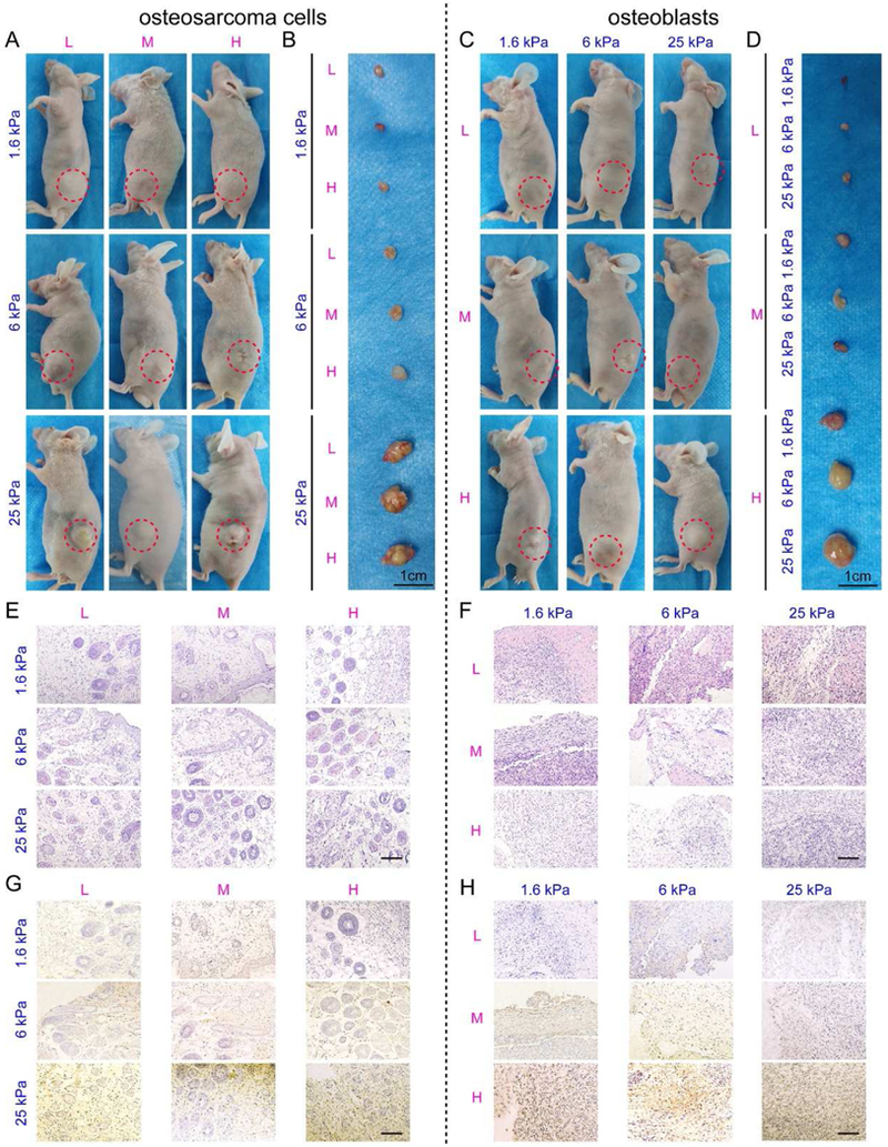
Fig. 5. The tumorigenesis of osteosarcoma cells and osteogenesis of osteoblasts cultured within the scaffolds in vivo.

(A-D) Representative images of nude mice and harvested samples constructed with (A, B) MG63 cells and (C, D) hFOB1.19 cells in 9 hydrogels. Red dotted circles indicate the tumor and the neoplasms. Scale bar: 1 cm. (E-F) HE staining of the (E) MG63 tumors and (F) hFOB1.19 neoplasms. Scale bar: 50 µm. (G) CD133 immunohistochemistry of the MG63 tumors. Scale bar: 50 µm. (H) OCN immunohistochemistry of the hFOB1.19 neoplasms. Scale bar: 50 µm.
