



Abstract
We describe the case of a 48-year-old patient who presented with an out-of-hospital cardiac arrest due to an acute ST-elevation myocardial infarction. Left ventriculography performed during the initial coronary angiography revealed the presence of structural abnormalities in the myocardial wall which subsequently, with the use of contrast echocardiography and cardiac magnetic resonance imaging, were confirmed to represent ventricular clefts. We reviewed the pathophysiological significance of these defects and compared them with similarly looking abnormalities such as aneurysms, pseudoaneurysms, and diverticula. In conclusion, recognition of ventricular clefts is needed to allow clinicians to differentiate them from other myocardial wall defects with different pathological profile and clinical significance.
<Learning objective: With emerging techniques in cardiac imaging some cardiac abnormalities such as left ventricular (LV) clefts are recognized more frequently. This case report describes an incidental finding of LV cleft and provides an extensive review of the literature regarding the clinical significance and differential from other similar findings of different pathology. That will help physicians to distinguish these overlapping findings and tailor patients’ management accordingly.>
Keywords: Ventricular, Cleft, Crypts, Fissure, Inferoseptal
Introduction
With increased availability of cardiac image modalities such as cardiac magnetic resonance imaging (cMRI), computed tomography coronary angiography (CTCA), and 3D echocardiography the incidental detection of left ventricular (LV) wall abnormalities is becoming more common. LV clefts were thought to be rare incidental findings, with the evidence derived from a small number of case reports [1], [2], [3] and few predominantly retrospective studies [4], [5], [6]. Despite studies suggesting that such structural variations of the myocardium might be more common than previously thought [4], [5], their pathophysiological significance remains unclear. We present a case of a patient found to have a myocardial cleft and discuss the possible significance of such findings based on current evidence.
Case report
A 48-year-old female with unremarkable personal and family history of cardiac disease, was brought directly to the catheterization laboratory, after suffering an out-of-hospital cardiac arrest. According to bystander information, the event was preceded by an episode of chest pain.
On arrival of the paramedic team, the patient was found to be in ventricular fibrillation and after receiving 3 cycles of cardiopulmonary resuscitation including 2 shocks, had return of spontaneous circulation.
Her initial electrocardiogram, following successful resuscitation, showed sinus rhythm at a heart rate of 54 bpm with ST elevation in leads I, aVL, V5, and V6. ST depression and T wave inversion were noted in leads III and aVF and poor R wave progression in precordial leads V1–V4. The patient underwent immediate coronary angiography which demonstrated total left anterior descending artery (LAD) occlusion at the level of D1. No other coronary disease was identified. Percutaneous coronary intervention of the LAD was performed, using a bare metal stent. Left ventriculography (Fig. 1) revealed a discrete protrusion of the LV cavity along the inferobasal wall, perpendicular to the long axis of the ventricle, that opacified with contrast.
Fig. 1.
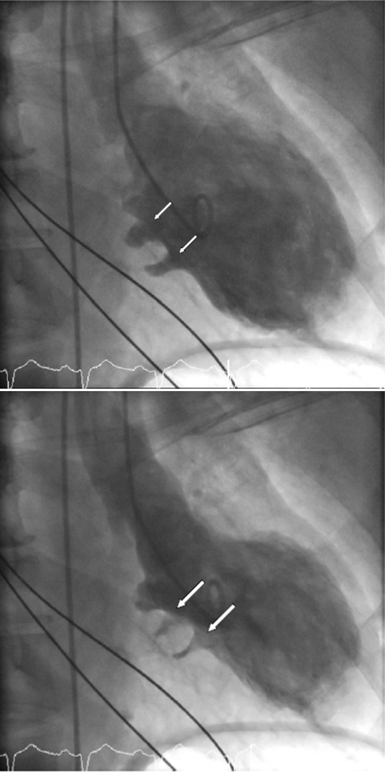
Upper: left ventriculography (RAO view) showing a pair of LV clefts (white arrows) at basal inferior wall in diastole. Lower: nearly complete obliteration of LV clefts (white arrows) during systole in left ventriculography (RAO view). LV, left ventricular; RAO, right anterior oblique.
A transthoracic echocardiogram (TTE) was also performed (Fig. 2) to assess myocardial and valve function but also to further characterize the LV wall abnormality. The test showed mildly impaired LV function with ejection fraction of approximately 45%. A fissure-like protrusion was identified, which was confined in the compacted myocardium and was penetrating more than 50% of wall thickness on the basal inferior wall. It showed near-total obliteration during systole. Repeat TTE with SonoVue contrast agent (Bracco, Milan, Italy) (Fig. 2) showed paired myocardial clefts in the basal inferoseptal/basal inferior region. No intracardiac thrombus was identified.
Fig. 2.

Transthoracic echocardiogram. Upper left: slightly angulated apical 2-chamber view. LV cleft (white arrow) in basal inferior wall in diastole. Upper right: 2-chamber view (slightly angulated). Obliteration of LV cleft (white arrow) in systole. Lower left: more prominent demonstration of LV clefts (white arrows) with contrast echocardiography in diastole (slightly rotated and angulated apical 2-chamber view). LV, left ventricular.
The patient subsequently underwent cMRI for further characterization of fissure-like protrusions of the inferior LV wall. That showed LV of normal cavity size with low normal overall systolic function. There was a chronic transmural mid to apical anterior wall infarct with associated hypokinesis. Two clefts in basal inferior wall were confirmed (Fig. 3).
Fig. 3.
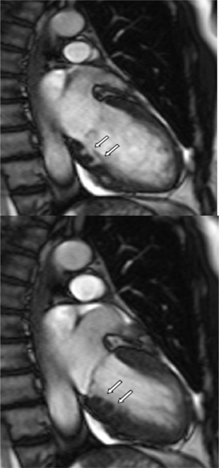
Cardiac magnetic resonance imaging. Upper: clear demonstration of paired LV clefts (white arrows) in diastole. Lower: pair of LV clefts (white arrows) nearly obliterated in systole. LV, left ventricular.
The patient made a remarkable recovery, with no residual neurological deficit and complete return to her previous functional status. No arrhythmias were noted whilst in hospital.
Discussion
On reviewing the literature, there is lack of a clear definition of ventricular clefts, with terminology such as ventricular “clefts,” “crypts,” “crevices,” or “fissures” often used interchangeably. In the majority of cases the authors describe the presence of cleft-like fissures, predominantly in the inferoseptal basal/mid wall which do not extend beyond the epicardial contour [1].
A more detailed definition of a ventricular cleft was given by Johansson et al. [4], who described these clefts as discrete V-shaped extensions of blood signal, penetrating >50% of the thickness of adjoining compact myocardium in long-axis views on cMRI. More importantly, in their study they described narrowing or complete occlusion of the cleft during systole, without evidence of local hypokinesia or dyskinesia.
Left ventricular clefts are congenital defects of the myocardial wall which were initially described in post-mortem hearts in patients with hypertrophic cardiomyopathy (HCM) [6]. A further correlation between these ventricular wall abnormalities and HCM was proposed by Germans et al. [7]. Using cMRI, their group reported the presence of “crypts” in the basal and mid-inferoseptal walls of 13 out of 16 patients with HCM mutations who had not yet developed echocardiographic evidence of hypertrophy. Interestingly, none of the 16 age- and gender-matched healthy volunteers had evidence of these structural anomalies, suggesting that these clefts represented early pathological alterations of the myocardium.
Subsequent work by Johansson et al. [4] argued against such hypothesis, as they retrospectively analyzed cMRI images of 399 individuals, including 120 healthy volunteers and 91 patients with HCM. They categorized clefts according to location in LV wall. Basal inferior myocardial clefts were visible in 7 out of 120 volunteers (7%) and 5 out of 91 HCM patients (5.5%), 5 out of 44 hypertensives (11.4%), 1 out of 104 repaired Tetralogy of Fallot patients (1%), and 9 out of 40 patients with surgically relieved congenital pulmonary valve stenosis (22.5%). Septal clefts were also seen in the mid to apical parts of the interventricular septum in 24 (6%) of the 399 individuals including 6 of the 120 volunteers (5%).
More recent data published by Maron et al. [8] revealed a strong association between myocardial clefts and HCM. Indeed, their group suggested that the presence of clefts could represent a distinctive pathological expression of HCM, with different frequency between patients with and without LV hypertrophy. In their study, a total of 292 consecutive patients with HCM were analyzed, including 31 genotype-positive/phenotype-negative patients without LV hypertrophy. These patients were compared against 98 controls. The results showed that the clefts were much more prevalent in the phenotypically negative HCM patients (without LV hypertrophy) with 19 out of 31 patients identified as having myocardial clefts. Interestingly, none of the controls was found to have clefts and only 10 out of the 261 patients with HCM and confirmed LV hypertrophy were found to have clefts. Similar findings were observed by Brouwer et al. [9] which also demonstrated that the presence of multiple crypts in the absence of LV hypertrophy were highly specific for HCM mutation carriership. Indeed this group suggested that detection of 2 or more crypts had a sensitivity of 51% and specificity of 94% for carriership. The argument that the presence of clefts can assist in the identification of gene carriage before the development of hypertrophy, could mean that these defects can be used as phenotypic markers of HCM allowing closer monitoring of these patients and possibly the detection of LV hypertrophy at an earlier stage.
The largest retrospective study was done by Erol et al. [5] who used CTCA images of 2093 patients and evaluated various congenital LV wall abnormalities. In their study, they identified 141 patients (6.7%) with LV clefts. There was no significant difference between the sexes and the majority of clefts were observed at the basal to mid inferoseptal segment of the LV (55.4%). In a mean follow-up of 36 months (143 patients with LV defects of which 141 with LV clefts) and 37 months (143 control patients) there was no significant difference in the incidence of major adverse cardiac events or stroke. Besides, no thrombus was identified within the cleft in any of the 141 patients. However, their study was limited by the lack of clinical background of patients, and the retrospective design of their study.
The etiology and pathogenesis of these myocardial structural defects remains unclear. The process of cardiac embryogenesis is an interesting area of research, with only some understanding of the underlying molecular and cellular mechanisms involved in ventricular morphogenesis and development of ventricular trabeculation and compaction [10]. Interestingly, one hypothesis regarding the pathophysiological mechanism of formation of myocardial clefts, is drawn by the initial work of Kuribayashi and Roberts [6], who identified deep tissue clefts at the interventricular junction in hearts of patients with HCM, at the same areas where extensive myocardial disarray was observed. Moon and McKenna [11] consider that LV clefts may represent a sign of abnormal fetal cardiomorphogenesis triggered by the in utero consequences of the underlying sarcomeric mutation. They consider that the results published by Maron et al. [8] support an extended role of sarcomeric proteins beyond simply contraction and relaxation that may include embryonic cardiomorphogenesis. However, it remains unknown how such mutations could cause clefts and they challenge this theory with a possible consideration that LV clefts may be analogous with LV non-compaction (LVNC). Based on previous experimental findings in murine models [10], they consider that HCM may demonstrate features similar to LVNC in an overlap appearance. That could lead to the hypothesis that it may not be the mutations creating these clefts, but the pre-LVH HCM heart is architecturally neonatal and arrested in a premature developmental phase, where such clefts are persistent and visible to our current technology.
Certain reports [1], [2] highlighted the need for a more clear differentiation between other anatomical anomalies of the LV such as pseudoaneurysms, true aneurysms, and diverticula, since these structural variations of the myocardium have distinct anatomical and histological characteristics as well as different prognostic significance [12], [13].
LV pseudoaneurysms form when cardiac rupture is contained by adherent pericardium or scar tissue. Most common causes are myocardial infarction (MI) and surgery [14]. The site of pseudoaneurysm varies with etiology. In a review of 52 patients [15], pseudoaneurysms were primarily seen in the inferior or posterolateral wall after MI, in the right ventricular outflow tract after congenital heart surgery, in the posterior subannular region of the mitral valve after mitral valve replacement, and in the subaortic region after aortic valve replacement. They are differentiated by LV clefts as they are not contained by contracting myocardium. They need to be recognized promptly as they are prone to rupture and carry a high risk of mortality.
A true LV aneurysm is defined as a well delineated, thin, scarred or fibrotic wall, devoid of muscle or containing necrotic muscle, that is a result of a healed transmural MI. The involved wall segment is either akinetic (without movement) or dyskinetic (with paradoxical ballooning) during systole. Aneurysms of the apex and anterior wall are more than four times as common as those of the inferior or inferoposterior walls. There are a number of serious complications that can occur as a consequence of LV aneurysm, particularly heart failure, ventricular arrhythmias, and thromboembolism.
Finally, LV diverticulum is defined as an outpouching structure that contains endocardium, myocardium, and pericardium and displays normal contraction [16]. Epidemiology of LV diverticulum is not clear, although earlier studies have been cited as reporting a prevalence of 0.4%, or 3 of 750 cardiac necropsy cases [17], [18]. The natural history of LV diverticulum has not been systematically studied. Major concerns are thrombosis, embolism, rupture, congestive heart failure, ventricular arrhythmias, and valvular abnormalities [12], [16]. The true incidence of these complications is not known because of the rarity of LV diverticulum. Midline thoracoabdominal defects and other congenital cardiac malformations are found in a substantial proportion of patients who have LV diverticula. Most LV diverticula are found in the apex [19]. Diverticula can be further subdivided into fibrous or muscular, based on their location and structure. Fibrous divertucla are subannular in location (submitral or subaortic). Their significance is that they are associated with annular weakness that is considered to be congenital [2] but are not related to cardiac and somatic anomalies. On the other hand, muscular diverticula are usually apical and involve all myocardial layers [1], [2]. They may be related with cardiac and/or extracardiac defects such as Cantrell's syndrome, atrial or ventricular septal defect, pulmonary stenosis, or Tetralogy of Fallot [1], [2]. Interestingly, although both clefts and diverticula contract with systole, their differentiation lies in the fact that diverticula are thought to be narrow mouthed with a wide outpouching and extend beyond the anatomic boundaries of the left ventricle [20].
Conclusion
Following review of the literature, there is some evidence to suggest an association between ventricular clefts and the presence of HCM gene carriership without the presence of LV hypertrophy. Indeed, such an association might be clinically beneficial, as identification of such myocardial wall abnormalities could serve as a screening tool for familial cases of HCM. Nevertheless, up to now there is no evidence to link the presence of clefts with any pathological consequence. In our case, there is no evidence to suggest that the presence of the ventricular cleft contributed to the presentation of our patient, as an alternative cause was identified.
In summary, the availability of high resolution cardiac imaging encourages the identification of structural variations of the myocardium. LV clefts are cleft-like fissures, predominantly located in the basal to mid-inferoseptal segment of the left ventricle, which become narrowed or completely occluded during systole. Such areas need to be carefully described and distinguished from similarly looking abnormalities such as aneurysms, pseudoaneurysms, and diverticula. There is certainly a need for the use of a consistent definition when describing these findings which would in turn facilitate the accumulation of data and the demonstration of a possible clinical significance. Further research should be warranted in HCM gene carriers to establish long-term follow-up and investigate their clinical significance.
Conflict of interest
None declared.
Acknowledgments
Dr. Andreas Seraphim reviewed the literature and wrote the first draft.
Dr. Alexandros Papachristidis revised the article, deepened the discussion, collected and commented on images, and submitted the article acting as corresponding author.
Dr. Naomi Bratt collected clinical details and all relevant information from patient's notes and wrote the case report section.
Dr. Man Fai Shiu supervised the whole project and provided expert opinion throughout.
References
- 1.Wein M., Wolf-Puetz A., Niehues R., Klein T., Kilner P.J., Klein R.M. Multiple left ventricular inferoseptal clefts. Multimodality imaging appearance and differential diagnosis. Herz. 2011;36:438–443. doi: 10.1007/s00059-011-3424-9. [DOI] [PubMed] [Google Scholar]
- 2.Afonso L., Kottam A., Khetarpal V. Myocardial cleft, crypt, diverticulum, or aneurysm? Does it really matter? Clin Cardiol. 2009;32(8):E48–E51. doi: 10.1002/clc.20466. [DOI] [PMC free article] [PubMed] [Google Scholar]
- 3.Maron B.J., Lindberg J., Lesser J.R. Ventricular septal crypt in hypertrophic cardiomyopathy. Eur Heart J. 2010;31:1923. doi: 10.1093/eurheartj/ehq140. [DOI] [PubMed] [Google Scholar]
- 4.Johansson B., Maceira A.M., Babu-Narayan S.V., Moon J.C., Pennell D.J., Kilner P.J. Clefts can be seen in the basal inferior wall of the left ventricle and the interventricular septum in healthy volunteers as well as patients by cardiovascular magnetic resonance. J Am Coll Cardiol. 2007;50:1294–1295. doi: 10.1016/j.jacc.2007.06.026. [DOI] [PubMed] [Google Scholar]
- 5.Erol C., Koplay M., Olcay A., Kivrak A.S., Ozbek S., Seker M., Paksoy Y. Congenital left ventricular wall abnormalities in adults detected by gated cardiac multidetector computed tomography: clefts, aneurysms, diverticula and terminology problems. Eur J Radiol. 2012;81:3276–3281. doi: 10.1016/j.ejrad.2012.03.025. [DOI] [PubMed] [Google Scholar]
- 6.Kuribayashi T., Roberts W.C. Myocardial disarray at junction of ventricular septum and left and right ventricular free walls in hypertrophic cardiomyopathy. Am J Cardiol. 1992;70:1333–1340. doi: 10.1016/0002-9149(92)90771-p. [DOI] [PubMed] [Google Scholar]
- 7.Germans T., Wilde A.A., Dijkmans P.A., Chai W., Kamp O., Pinto Y.M., van Rossum A.C. Structural abnormalities of the inferoseptal left ventricular wall detected by cardiac magnetic resonance imaging in carriers of hypertrophic cardiomyopathy mutations. J Am Coll Cardiol. 2006;48:2518–2523. doi: 10.1016/j.jacc.2006.08.036. [DOI] [PubMed] [Google Scholar]
- 8.Maron M.S., Rowin E.J., Lin D., Appelbaum E., Chan R.H., Gibson C.M., Lesser J.R., Lindberg J., Haas T.S., Udelson J.E., Manning W.J., Maron B.J. Prevalence and clinical profile of myocardial crypts in hypertrophic cardiomyopathy. Circ Cardiovasc Imaging. 2012;5:441–447. doi: 10.1161/CIRCIMAGING.112.972760. [DOI] [PubMed] [Google Scholar]
- 9.Brouwer W.P., Germans T., Head M.C., van der Velden J., Heymans M.W., Christiaans I., Houweling A.C., Wilde A.A., van Rossum A.C. Multiple myocardial crypts on modified long-axis view are a specific finding in pre-hypertrophic HCM mutation carriers. Eur Heart J Cardiovasc Imaging. 2012;13:292–297. doi: 10.1093/ehjci/jes005. [DOI] [PubMed] [Google Scholar]
- 10.Chen H., Zhang W., Li D., Cordes T.M., Payne R.M., Shou W. Analysis of ventricular hypertrabeculation and noncompaction using genetically engineered mouse models. Pediatr Cardiol. 2009;30:626–634. doi: 10.1007/s00246-009-9406-5. [DOI] [PMC free article] [PubMed] [Google Scholar]
- 11.Moon J., McKenna W. Myocardial crypts: a prephenotypic marker of hypertrophic cardiomyopathy? Circ Cardiovasc Imaging. 2012;5:431–432. doi: 10.1161/CIRCIMAGING.112.975888. [DOI] [PubMed] [Google Scholar]
- 12.Marijon E., Ou P., Fermont L., Concordet S., Le Bidois J., Sidi D., Bonnet D. Diagnosis and outcome in congenital ventricular diverticulum and aneurysm. J Thorac Cardiovasc Surg. 2006;131:433–437. doi: 10.1016/j.jtcvs.2005.09.046. [DOI] [PubMed] [Google Scholar]
- 13.Ohlow M.A. Congenital left ventricular aneurysms and diverticula: definition, pathophysiology, clinical relevance and treatment. Cardiology. 2006;106:63–72. doi: 10.1159/000092634. [DOI] [PubMed] [Google Scholar]
- 14.Frances C., Romero A., Grady D. Left ventricular pseudoaneurysm. J Am Coll Cardiol. 1998;32:557–561. doi: 10.1016/s0735-1097(98)00290-3. [DOI] [PubMed] [Google Scholar]
- 15.Yeo T.C., Malouf J.F., Oh J.K., Seward J.B. Clinical profile and outcome in 52 patients with cardiac pseudoaneurysm. Ann Intern Med. 1998;128:299–305. doi: 10.7326/0003-4819-128-4-199802150-00010. [DOI] [PubMed] [Google Scholar]
- 16.Makkuni P., Kotler M.N., Figueredo V.M. Diverticular and aneurysmal structures of the left ventricle in adults. Tex Heart Inst J. 2010;37:699–705. [PMC free article] [PubMed] [Google Scholar]
- 17.Pressoir R., Downing J.W. Congenital diverticula of the right ventricle of the heart: a case report. J Natl Med Assoc. 1980;72:262–264. [PMC free article] [PubMed] [Google Scholar]
- 18.Skapinker S. Diverticulum of the left ventricle of the heart; review of the literature and report of a successful removal of the diverticulum. AMA Arch Surg. 1951;63:629–634. doi: 10.1001/archsurg.1951.01250040643009. [DOI] [PubMed] [Google Scholar]
- 19.Walton-Shirley M., Smith S.M., Talley J.D. Left ventricular diverticulum: case report and review of the literature. Cathet Cardiovasc Diagn. 1992;26:31–33. doi: 10.1002/ccd.1810260108. [DOI] [PubMed] [Google Scholar]
- 20.Aquaro G.D., Di Bella G., Strata E., Deiana M., De Marchi D., Pingitore A., Lombardi M. Cardiac magnetic resonance findings in isolated congenital left ventricular diverticuli. Int J Cardiovasc Imaging. 2007;23:43–47. doi: 10.1007/s10554-006-9120-9. [DOI] [PubMed] [Google Scholar]