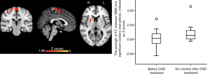
Figure 4:
Images show serial changes in functional connectivity (FC) mapping of nucleus basalis of Meynert (NBM) cholinergic network (hereafter, NBM FC) between baseline and 6 months after undergoing cholinesterase inhibitor (ChEI) treatment in 18 patients with mild cognitive impairment (MCI). NBM FC significantly increased from baseline to 6 months after ChEI in right anterior striatum and prefrontal cortex (red-yellow). Box plot shows distribution of strength of FC between NBM and significant clusters (red-yellow) before and 6 months after ChEI, indexed as Z score. All results were masked by gray matter masks obtained from Montreal Neurological Institute 152 standard-space T1-weighted average structural template image. Significance level was at familywise error–corrected P ˂ .05 (corrected for multiple comparisons) and corrected for age and mean displacement.

