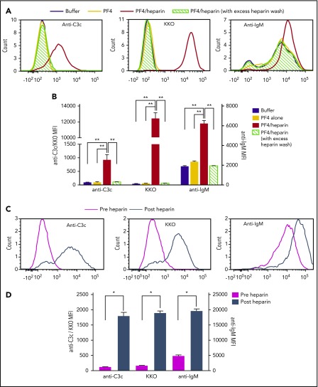
Figure 6.
Plasma IgM colocalizes with PF4/heparin and C3 fragments on B cells in healthy donors and patients on heparin therapy. (A-B) Whole blood from a representative healthy donor was incubated with buffer or PF4 (25 μg/mL) ± heparin (0.25 U/mL) and binding of C3c, PF4/heparin, and IgM on B cells was determined by flow cytometry. Binding of C3c/anti-PF4/heparin (KKO)/IgM to B cells is shown with and without PF4 ± heparin by histogram overlays in normal and excess heparin wash conditions (A) and as mean fluorescent intensity (B). Results are shown from a representative experiment involving 3 donors tested on 3 different occasions. (C-D) Binding of C3c, PF4/heparin, and IgM on B cells in the circulation of heparinized patients was determined by flow cytometry. Binding of C3c/anti-PF4/heparin (KKO)/IgM to B cells is shown pre- and postheparin exposure in the patient by overlay histograms (C) and as mean fluorescence intensity (D). Results are shown from 1 representative patient out of 3 patients studied. *P < .005; **P < .0001.

