

QUESTION
A 58-year-old male patient with melena and severe anemia presented to our attention. His medical history included hypercholesterolemia and arterial hypertension; nine months before admission, the patient underwent coronary stent implantation because of acute myocardial infarction and was started on dual anti-platelet therapy with aspirin and clopidogrel. He underwent urgent splenectomy because of trauma 30 years earlier. Melena was reported in the last two weeks; therefore, a gastroscopy and colonoscopy were performed on the patient, the results of which were negative. Small-bowel investigation with capsule endoscopy revealed an ulcerated lesion of the ileum protruding into the lumen causing a delayed passage of the capsule (Figure 1). A subsequent small-bowel CT-enteroclysis demonstrated the presence of four nodules through the ileal wall, the larger measuring 30×20 mm, with a strong contrast enhancement in the arterial phase (Figure 2). An oral double-balloon enteroscopy (DBE) was performed and the endoscope was advanced 3 m beyond the ligament of Treitz but the described lesion was not found. DBE through anal route was planned; however, the patient presented an acute bowel obstruction and underwent urgent surgery. Laparotomy revealed a small-bowel obstruction and an ileal resection was performed; on macroscopic evaluation, 27 cm of ileum presented multiple parietal nodules.
Figure 1.

Capsule endoscopy revealed an ulcerated lesion of the ileum
What is the patient’s most likely diagnosis?
Figure 2.

Small-bowel CT-enteroclysis demonstrated the presence of four nodules through the ileal wall, the larger measuring 30×20 mm, with a strong contrast enhancement in the arterial phase
ANSWER
Pathology examination revealed the presence of typical splenic parenchyma consisting of cords of red pulp alternating with white pulp (Figure 3–4). The patient was discharged after 5 days and subsequent follow-up was unremarkable. Splenosis is a frequent consequence/complication of traumatic splenic rupture because of the “transplantation” of splenic tissue through local mechanical seeding or hematogenous dissemination. The exact mechanism through which splenosis occurs in not clearly understood; the most plausible hypothesis is that splenic cells reach adjacent organs after capsule rupture; the preferential sites of splenosis are stomach, small bowel, peritoneum, and colon (1,2).
Figure 3.
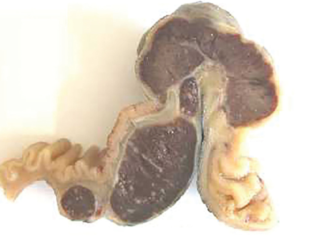
Macroscopic evaluation of ileum presented multiple parietal nodules
Figure 4.

Pathology examination revealed the presence of typical splenic parenchyma consisting of cords of red pulp alternating with white
The diagnosis of splenosis could be done a few months after splenectomy or even after many decades. Those patients are usually asymptomatic and the diagnosis is accidental as a result of radiological examinations or surgery. However, in some cases, splenosis could present as an acute abdominal pain, bowel obstruction, or bleeding (3).
From a diagnostic point of view, splenosis can mimic several intra-abdominal neoplasia. Differential diagnoses of splenosis include endometriosis, peritoneal mesothelioma, renal neoplasms, abdominal lymphomas, GIST, small-bowel tumors, or peritoneal metastatic implants (4). In conclusion, splenosis must be considered in the differential diagnosis of patients with a history of splenectomy and suspected masses of unknown etiology.
Footnotes
Informed Consent: Informed consent was obtained from patient who participated in this study.
Peer-review: Externally peer-reviewed.
Author Contributions: Endoscopic Examinations - A.M., C.A.A.P., M.L.B.; Writing - R.A., S.G.; Critical Reviews - A.L., O.T.
Conflict of Interest: The authors have no conflict of interest to declare.
Financial Disclosure: The authors declared that this study has received no financial support.
REFERENCES
- 1.Akay S, Ilica AT, Battal B, Karaman B, Guvenc I. Pararectal mass: An atypical location of splenosis. J Clin Ultrasound. 2012;40:443–7. doi: 10.1002/jcu.20843. https://doi.org/10.1002/jcu.20843 [DOI] [PubMed] [Google Scholar]
- 2.Brewster DC. Splenosis. Am J Surg. 1973;126:14–9. doi: 10.1016/s0002-9610(73)80086-8. https://doi.org/10.1016/S0002-9610(73)80086-8 [DOI] [PubMed] [Google Scholar]
- 3.Younan G, Wills E, Hafner G. Splenosis: A Rare Etiology for Bowel Obstruction-A Case Report and Review of the Literature. Case Rep Surg. 2015;2015:890602. doi: 10.1155/2015/890602. https://doi.org/10.1155/2015/890602 [DOI] [PMC free article] [PubMed] [Google Scholar]
- 4.Imbriaco M, Camera L, Manciuria A, Salvatore M. A case of multiple intra-abdominal splenosis with computed tomography and magnetic resonance imaging correlative findings. World J Gastroenterol. 2008;14:1453–5. doi: 10.3748/wjg.14.1453. https://doi.org/10.3748/wjg.14.1453 [DOI] [PMC free article] [PubMed] [Google Scholar]