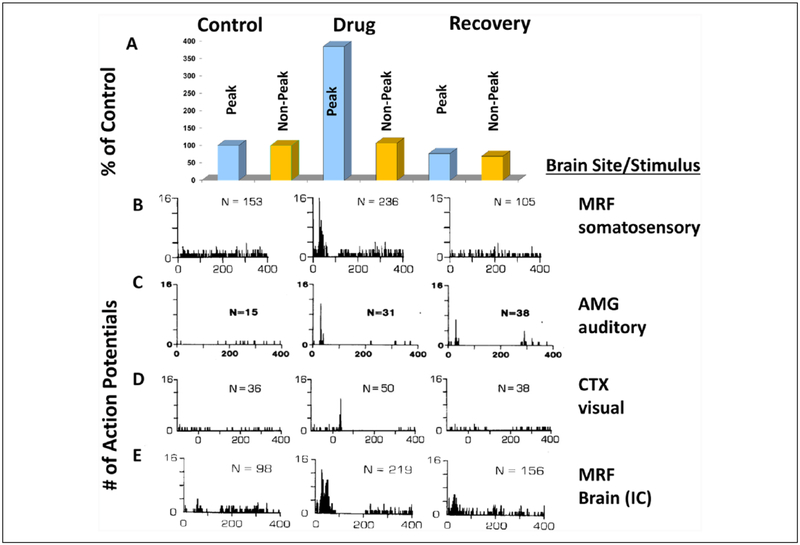
Figure 5.
Changes in extracellular action potential firing of conditional multireceptive (CMR) neurons in several brain regionsin response to sensory stimuli induced by administration of GABAA receptor antagonists (in subconvulsant doses). In predrug conditions (control column of the poststimulus histograms or PSTHs) presentations of electrical stimuli to the sciatic nervein line B to a mesencephalic reticular formation (MRF) neuron, line C amygdala (AMG) neuron response to auditory stimuli, line D response of a pericruciate cortex (CTX) neuron to visual stimuli and response of a MRF neuron to electrical stimulus within the brain to the inferior colliculus (IC) in (line E). Although spontaneous firing was observed, very little evidence of responsiveness to the stimuli is detectable, as shown by the absence of clear time-locked response peaks to the stimuli in the PSTHs (control column). After administration of a GABAA receptor antagonist these CMR neurons exhibit major responsiveness increases, which includes the induction of a very striking time-locked peak of responsiveness to each stimulus in each PSTH (drug column). These peaks indicate that the drug had induced extensive responsiveness of the CMR neurons to these stimuli. The bar graphs in A compare changes in the PSTH peak period and the rest of the recorded period for the PSTHs in line B. The peak (20–55 ms period after stimulus onset) shows a much greater percent increase as compared to the rest of the 400 ms sampling period as shown by the bar graphs for the nonpeak versus peak time points shown above each PSTH. Several of these CMR neurons showed response enhancement to more than one stimulus modality (not shown), indicating the multimodality responsiveness common in CMR neurons. This neuroplastic effect is short-term, since the firing patterns of these neurons recover to unresponsiveness with time (right column). Specific GABAA receptor antagonists and doses used are as follows: line B, pentylenetetrazol, 15 mg/kg; line C, bemegride, 3.6 mg/kg; line D, bicuculline, 0.03 mg/kg; line E, pentylenetetrazol, 5 mg/kg. Recovery times ~20 minutes after i.v. drug administration. Stimulus parameters: 0.5 Hz stimulus rate, 50 stimulus presentations (100 μs single bipolar pulses in B and E, 95 dB SPL clicks in C, and 18.5 lux visual stimulus [strobe] in D). PSTHs: bin width = 1 ms, scan length= 400 ms. Note the histograms in line D are peristimulus time histograms. N = number of action potentials per PSTH. This effect was seen in 88% of more than 700 bulbar and midbrain reticular formation neurons, 57% of more than 100 AMG, and 67% of more than 100 CTX neurons in unanesthetized cat and rat and was also seen with direct (iontophoretic) application of drug. (A, B, E: modified from Faingold and Feng 2014; C: modified from Faingold 2014b; D: modified from Faingold and others 1983.)

