



Abstract
Inherited arrhythmia syndromes comprise an increasingly complex group of diseases involving mutations in multiple genes encoding ion channels, ion channel accessory subunits and channel interacting proteins, and various regulatory elements. These mutations serve to disrupt normal electrophysiology in the heart, leading to increased arrhythmogenic risk and death. These diseases have added impact as they often affect young people, sometimes without warning. Although originally thought to alter ion channel function, it is now increasingly recognized that mutations may alter ion channel protein and messenger RNA processing, to reduce the number of channels reaching the surface membrane. For many of these mutations, it is also known that several interventions may restore protein processing of mutant channels to increase their surface membrane expression toward normal. In this article, we reviewed inherited arrhythmia syndromes, focusing on long QT syndrome type 2, and discuss the complex biology of ion channel trafficking and pharmacological rescue of disease-causing mutant channels. Pharmacological rescue of misprocessed mutant channel proteins, or their transcripts providing appropriate small molecule drugs can be developed, has the potential for novel clinical therapies in some patients with inherited arrhythmia syndromes.
Keywords: cardiac ion channel, inherited arrhythmia, protein trafficking, hERG, pharmacological rescue
INTRODUCTION
Cardiac arrhythmias leading to symptoms and sudden death were first linked to specific arrhythmia mechanisms (eg, reentry) approximately 100 years ago. With the ability to record cellular electrophysiology with microelectrodes, additional arrhythmia mechanisms (eg, triggered activity and enhanced automaticity) were identified approximately 50 years ago, and over ensuing decades, the cellular machinery for these arrhythmogenic mechanisms (eg, specific ion channels, cell Ca2+, regulatory pathways, cell coupling, and so on) continues to be investigated. At the same time, the first genetically based1–3 and drug-induced arrhythmia syndromes4 emerged, both linked to prolongation of action potentials and the QT interval on the electrocardiogram. With the first successful cloning of Na+ channel genes from electric eel and rat brain5 and the subsequent cloning of the first human arrhythmia disease-related gene (human ether-a-go-go–related gene, hERG or KCNH26,7), rapid progress has been achieved in understanding the genes and functional regulation for many ion channels. In this article, we review the field of inherited arrhythmia disorders, focusing on the mechanisms underlying KCNH2-related long QT syndrome (LQT2) and the potential for new therapeutic approaches.
Our present understanding of human inherited arrhythmia diseases has become increasingly complex. At least 12 clinical syndromes have been identified as human inherited arrhythmia diseases and these are summarized in Table 1A. At least 26 genes are known to cause these diseases, and these genes, their encoded proteins or currents, and the associated syndromes are given in Table 1B. These are distinguished from inherited cardiac abnormalities that cause structural heart disease (mutations in structural proteins and gene regulators). Congenital LQT was the first inherited arrhythmia disorder for which gene mutations were defined, and these occur predominantly in KCNQ1 (LQT1), KCNH2 (LQT2), less frequently in SCN5A (LQT3), with additional very rare forms (LQT4–12) known. Mutations associated with inherited arrhythmia syndromes occur in ion channel pore-forming proteins, associating subunit proteins and channel interacting proteins (CHIPS), Ca2+ handling proteins, components of the ion channel macromolecular complex, and regulatory pathways. Although most inherited arrhythmia syndromes are rare clinical findings, sometimes with just a single family described, they demonstrate the underlying (patho)physiology of gene expression, protein processing, regulatory processes, and surface membrane expression of ion channel complexes. It seems certain that more genes for inherited arrhythmia syndromes and new roles for known genes await discovery (Table 1).
TABLE 1A.
Inherited Arrhythmia Syndromes (Structurally Normal Hearts)
| LQTS (LQT 1–12): Romano-Ward, Andersen, Timothy Syndromes |
| LQTS (JLN 1–2): Jervell & Lange-Nielsen Syndrome |
| Early repolarization syndrome (ERS 1) |
| Brugada Syndrome (BrS 1–7) |
| Sudden Infant Death Syndrome (SIDS) |
| Idiopathic Ventricular Fibrillation (IVF 1) |
| Catecholaminergic Ventricular Tachycardia (CPVT 1–3) |
| Cardiac Conduction Disease (CCD 1–2) |
| Sick Sinus Syndrome (SSS 1–2) |
| Atrial Fibrillation (FAF 1–9) |
| Short QT Syndrome (SQT 1–3) |
| Drug-induced LQT (diLQT) |
TABLE 1B.
Affected Genes, Protein or Current, and Clinical Syndromes
| Gene | Protein or Current | Clinical Syndromes |
|---|---|---|
| ABCC9 | sur2 | FAF |
| AKAP9 | mAKAP or yotiao | LQT11 |
| ANKB | IK?, INa? | LQT4, FAF |
| CACNA1C | ICa | LQT8, BrS3 |
| CACNAB2 | ICa | BrS4 |
| CASQ2 | Ca release | CPVT2 |
| CAV3 | INa | LQT9, SIDS |
| DPP6 | Ito | IVF1 |
| GINS3 | ? | diLQT |
| GJA5 | connexin 40 | FAF |
| GPD1L | INa | BrS2, SIDS |
| HCN4 | If | SSS2 |
| KCNE1 | IKs | LQT5, JLN2, diLQT |
| KCNE2 | IKr | LQT6, FAF, diLQT |
| KCNE3 | Ito | BrS7 |
| KCNH2 | IKr | LQT2, SQT1, diLQT, SIDS |
| KCNJ2 | KIR2.1, IK1 | LQT7, FAF, SQT3, CPVT3 |
| KCNJ8 | IKATP | ERS1 |
| KCNQ1 | IKs | LQT1, JLN1, FAF, SQT2, diLQT, SIDS |
| RYR2 | Ca release | CPVT1 |
| SCN5A | INa | LQT3, BrS1, IVF, CCD, SSS1, FAF, diLQT, SIDS |
| SCN1B | INa | BrS5, CCD, FAF |
| SCN2B | INa | FAF |
| SCN3B | INa | BrS6 |
| SCN4B | INa | LQT10 |
| SNTA1 | INa | LQT12 |
CARDIAC CELLULAR ELECTROPHYSIOLOGY
The trigger for each heartbeat is the cardiac action potential. Its shape results from the orchestrated movement of inward and outward charge over the cell membrane through voltage-gated ion channels and electrogenic exchangers. Although action potential depolarization and conduction are mainly dependent on Na+ and Ca2+ channels along with gap junctions for cell-to-cell coupling, the plateau and repolarization phases depend on a delicate balance of multiple currents. The interplay of several currents to control the action potential shape and the electrocardiogram is illustrated in Figure 1. In different mammalian species, ion channel types vary in their relative density, membrane distribution, and gene sequence, and in nonmammalian cardiac myocytes, entire families of currents may be lacking (eg, IKr and IKs are minimally present in rodent cardiomyocytes).
FIGURE 1.
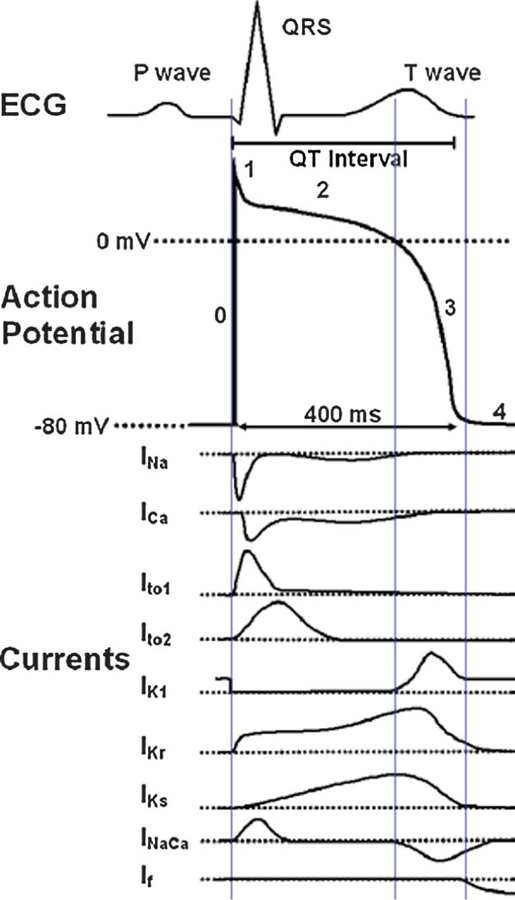
Major currents underlying the cardiac ventricular action potential. A simulated electrocardiogram is shown above the action potential waveform and major individual currents. INa, sodium current; ICa, L-type calcium current; Ito1, transient outward current 1; Ito2, transient outward current 2; IK1, inward rectifier potassium current; IKr, rapidly activating delayed rectifier potassium current; IKs, slowly activating delayed rectifier potassium current; INaCa, sodium–calcium exchanger current; If, pacemaker current. For current traces, dotted line is zero current. Modified from He et al.92
Mutations Alter Ion Channel Current: Complex Underlying Mechanisms
Disease-causing mutations in inherited arrhythmia syndromes usually alter a specific current to cause a ‘‘loss of function’’ or ‘‘gain of function’’ phenotype. For example, in congenital LQTS, mutations in delayed rectifier K+ channels (altered IKr or IKs) diminish outward current amplitude, which results in a loss of repolarizing K+ current and lengthening of the action potential duration. In contrast, mutations in Na+ channels (LQT3) cause increased late or slowly decaying INa, which represents a gain in depolarizing current, again to prolong action potential duration. In other inherited arrhythmia syndromes, opposite effects on these channel currents occur. For example, in Brugada syndrome and cardiac conduction disease, Na+ channel mutations result in decreased INa, whereas in short QT syndrome, a gain of function at most voltages has been shown for several K+ channel currents. In some forms of inherited arrhythmia syndromes, the effects are less direct through altered cell Ca2+, disruption of ion channel macromolecular complex signaling, or altered membrane localization of the channel complex, and possibly these may affect multiple channels. Thus, the molecular mechanisms underlying these loss or gain of function cellular phenotypes have become increasingly multifaceted and include (1) dominant negative and haploinsufficiency effects in channels that undergo subunit coassembly to form the channel pore, (2) altered biophysical properties of channel gating or ion permeation, (3) altered protein processing (trafficking) of channel proteins to the cell membrane, (4) altered regulation of channel function, (5) altered localization of channels in the cell membrane, (6) altered processing of mutant messenger RNA (mRNA), and (7) altered rates of exocytosis and endocytosis. Added to this is the ion channel background structure in which mutations are expressed; at least for Na+ channels, the isoform may dictate the expression profile of a mutation.8,9
Ether a go-go Gene and Human Ether a-go-go–related Gene
In 1969, Kaplan and Trout10 identified a mutation in Drosophila based on a rhythmic leg shaking phenotype after their exposure to ether. The mutation was named as the ether a-go-go gene (eag) and electrophysiological analysis of eag mutations in larval Drosophila neuromuscular junctions revealed larger and more prolonged synaptic potentials.11,12 Molecular studies and subsequent expression of eag in Xenopus oocytes confirmed that eag polypeptide assembled into voltage activated K+ (KV) channels.13,14
In 1994, Warmke and Ganetzky6 identified a human eag-related gene by screening a human hippocampus complementary DNA library with mouse eag derived from Drosophila eag. The newly identified gene, with 49% amino acid homology compared with Drosophila eag, represented a new branch of the eag family. It was located on chromosome 7 and named human-eag-related gene (h-erg or hERG or KCNH2). When expressed in Xenopus oocytes, the hERG (KV11.1) channels were shown to have kinetic properties and pharmacological sensitivity appropriate for the native IKr, thus it was concluded that KCNH2 encoded the α subunits for the IKr channel in human heart.15,16 Subsequently, 2 isoforms (ERG2 and ERG3) were found in rat brain,17 and an α subunit alternate transcript was identified that modifies gating (hERG1b18). Based on homology to Shaker KV channels and other KV channels, hERG channels are composed of 4 α subunit proteins. Each α subunit consists of a central hydrophobic core with 6 transmembrane α-helical domains (S1–S6) with the S1–S4 transmembrane regions forming a voltage sensing region, whereas S5–S6 form the K+ selective pore and binding domain for most drugs that block the channel.19 The selectivity filter in the pore of KV channels consists of a highly conserved sequence of amino acids. In hERG, the amino acid sequence is serine–valine–glycine–phenylalanine–glycine (S-V-G-F-G20) with G-F-G considered as the signature sequence.21 The hydrophobic core is flanked by cytoplasmic C and N termini. The consensus site for N-glycosylation is located just upstream of pore region (N598) and consensus site for protein kinase C phosphorylation located in the proximal C terminus. Even though hERG has many similar structural features with other KV channels, it also has unique features. hERG has longer N terminal and shorter C terminal compared with other eag channels. The extracellular S5-pore linker in hERG is longer than most other KV channels and contains a site for proteolytic digestion.22,23 hERG also lacks some H bonds between amino acid residues around the outer pore region. Lack of these H-bonds increases the flexibility of the outer pore of the channel.24 The proline residues found in the S6 region of most KV channels form a PXP motif that is absent in the pore region of hERG channels making the inner vestibule larger and more flexible, and the S6 region also contains aromatic residues (Y652 and F656) that are important for drug binding. These structural features may contribute to the unique gating and drug block properties of the channel.
During action potential depolarization, hERG channels open (activate) slowly from closed states and then rapidly inactivate; hence, the probability of channels being open initially is relatively low and IKr is small. During action potential repolarization, hERG channels rapidly recover from inactivation to reopen and then very slowly close (deactivate); hence, the probability of channels being open during repolarization is greater to give a larger IKr. These unique gating characteristics explain the channel’s ability to conduct maximum current during cardiac action potential repolarization.15,16,25,26
hERG Mutations and the Trafficking Pathway
Curran et al7 showed that KCNH2 is localized to chromosome 7q35–q36 and it is strongly expressed in the heart. They also identified mutations in KCNH2 in patients with LQT2. In LQT2, as with most inherited diseases, the majority of mutations are nonsynonymous single nucleotide changes causing single amino acid substitutions (missense mutations) in the channel protein.27 LQT2-associated mutations occur in hERG1a, except for a recent report of a hERG1b-specific A8V missense mutation.28 Although earlier studies in Xenopus oocytes suggested prominent roles for dominant negative versus haploinsufficient assembly and for gating abnormalities in regulating current amplitude, later studies in mammalian expression systems cultured at physiological temperatures suggested a central role for abnormal trafficking of mutant channel protein.29
A cartoon showing a trafficking pathway for hERG channel proteins is shown in Figure 2A. After DNA transcription in the nucleus, mRNA is transported into the cytosol and translated in rough endoplasmic reticulum (ER). hERG channel protein subunits are synthesized as monomeric proteins that then coassemble into multimeric channel proteins. In the ER, the mechanisms regulating protein folding are vital to ensuring correct tertiary structure and function. Many chaperone molecules that are present on luminal side of ER (eg, calnexin, and so on) and cytosolic side of ER such as heat shock proteins (Hsp70/Hsc70 and Hsp 90) and FK506-binding protein (FKBP38) interact to facilitate proper folding.30–33 hERG protein also undergoes N-linked core glycosylation in the ER. Stringent quality control mechanisms exist in ER where misfolded or unprocessed proteins are retained and eventually degraded. One mechanism is the RXR motif.34 Exposure of this motif caused by misfolding of a protein promotes its ER-associated retention. The forward trafficking of proteins from ER to Golgi depends on its proper packaging into transport vesicles, which is a complex process involving coat-associated proteins (COP II). Studies by Delisle et al35 using dominant negative strategies showed that hERG undergoes ER export in COP II vesicles and it may undergo endosomal recycling before being processed in the Golgi. Further processing occurs in Golgi including complex N-glycosylation, sorting and targeting of channels36; specific events taking place in this compartment remain poorly understood. Complex N-linked glycosylation is not mandatory for surface membrane expression of hERG protein but promotes channel stability.37 From the surface membrane, hERG channels are retrieved into endosomes where they not only can be ubiquitinated and degraded in lysosomes32 but also may be recycled back to the surface membrane, possibly via Golgi recycling.
FIGURE 2.
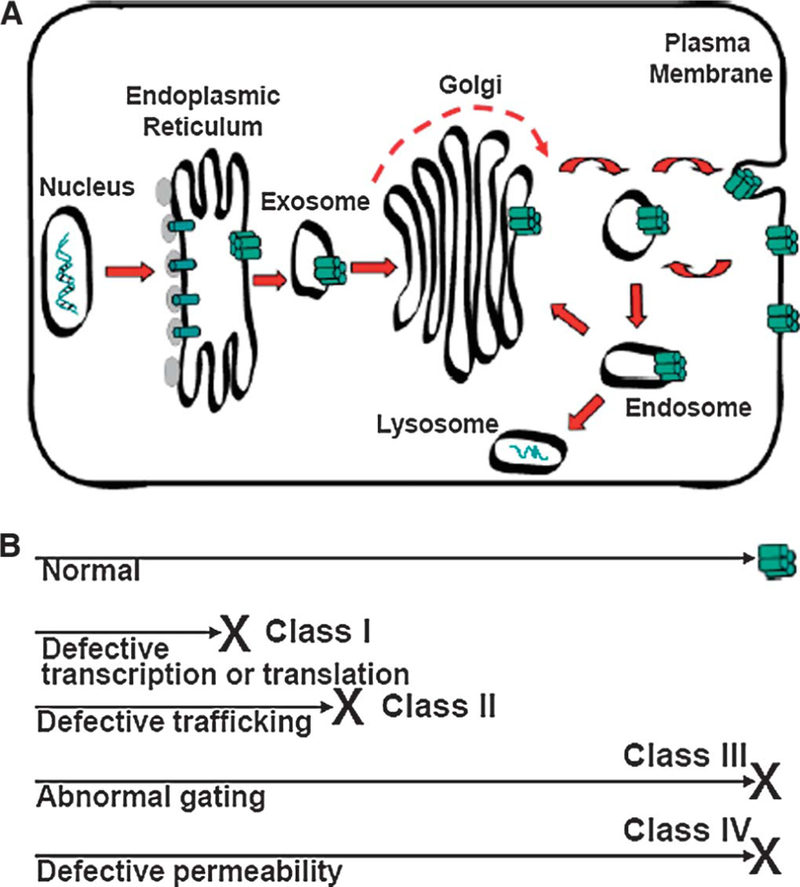
A proposed trafficking model for hERG K+ channels. A, Protein pathway for synthesis, recycling, and degradation. Dashed arrow indicates a nonconventional pathway. B, Disease mechanisms in LQT2.
The current through ion channels in the surface membrane (I) is the product of the number of functional channels on cell surface (N), their probability of opening (Po), and the ion conductance of each channel (i), and this equation (I = N × Po × i) is one approach to understanding the effect mutations have on hERG channels. These mechanisms for regulating hERG current (IhERG) are illustrated in Figure 2B.38 Normal [wild type (WT)] channels reach the surface membrane to function. Class 1 mutations undergo altered transcription and/or translation to reduce N. Recent evidence suggests that some LQT2 nonsense mutations that generate premature termination codons (W1001X, R1014X, and Q1070X) decrease mutant mRNA by nonsense-mediated decay (NMD), thereby reducing subsequent mutant hERG protein generation on the cell surface.39,40 Because the mutant allele mRNA selectively undergoes NMD, the WT allele mRNA can generate WT hERG channels that traffic to the surface membrane causing haploinsufficiency. Class 2 mutations generate protein that is trafficking-deficient with immature channels retained in the ER, ultimately leading to loss of functional channel density (N) on the cell surface. Providing WT and mutant hERG proteins coassembled in the ER before being retained, this mechanism potentially creates a more severe loss in N. Class 3 mutations alter Po and arise through mutations that affect channel gating. Class 4 mutations alter i and arise through mutations that affect channel permeability/selectivity (pore mutations). In channels that form as multimeric proteins (ie, K+ channels), dominant-negative interactions between coassembled mutant and WT channel subunits can potentially affect N, Po, or i. Further-more, a mutation can reduce IhERG by combinations of these cellular mechanisms.29,38,41
Anderson et al42 studied the mechanisms underlying the loss of IhERG by expressing 34 missense LQT2 mutations in HEK293 cells and characterizing their biophysical and biochemical properties. Their findings demonstrated that 28 channels had a trafficking-deficient phenotype (class 2 mechanism), one channel (T421M) trafficked normally but with altered gating (class 3 mechanism), one channel (G628S) trafficked normally but with disrupted permeation (class 4 mechanism), and 4 channels seemed to traffic and function normally. Thus, the dominant LQT2 disease mechanism was defective protein trafficking to reduce N resulting in little or no detectable IhERG. On Western blot analysis, immature (135 kD) core-glycosylated protein was present, whereas the mature (155 kD) complexly glycosylated protein was absent. Using immunocytochemistry, several investigators showed directly that trafficking-deficient LQT2 channel protein (T65P, A561V, G601S, Y611H, R752W, V822M, and N861I) was located predominantly intracellularly with a perinuclear pattern,43,44 and 3 mutations (T65P, N470D, and Y611H) directly colocalized with resident ER proteins.31,45 Recently, Amin et al46 described fever-induced QT interval prolongation in patients with the missense mutation A558P hERG. A558P is trafficking deficient and coassembles with WT subunits to cause dominant-negative behavior, and its current density fails to increase with increasing temperature similar to the increase found with WT channels.
It was evident from early genotyping and expression studies that LQT2 mutations are not restricted to a single region of the hERG channel protein; rather, trafficking-deficient mutations were identified in several regions of the protein, including the N terminus (T65P), transmembrane regions (N470D and A561V), pore region (G601S, Y611H, V612L, T613M, and L615V), and the C terminus including the cyclic nucleotide-binding domain (R752W, F805C, V822M, R823W, and N861I).45,47–52 Recently, Anderson showed that LQT2-asociated missense mutations are found in regions of highly ordered structures, particularly α-helices, to disrupt protein folding and trafficking. Figure 3 shows this structural model for one hERG α-subunit protein using data from one laboratory.42,46,53 Trafficking-deficient channels are shown in red (class 2 mechanism) and normally trafficking mutations in blue (class 3 or 4 mechanism). Interestingly, single amino acid substitutions in channels that function normally (yellow), most of which represent putative polymorphisms, tend to localize intracellularly in regions of the hERG protein that are thought not to contain α-helical or β-sheet structures. Thus, mutation location may influence the electrophysiological and clinical phenotype.54
FIGURE 3.
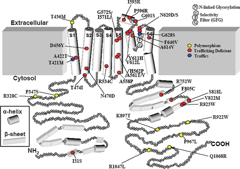
A structural model of the a-subunit of hERG protein showing localization of 39 missense mutations or putative polymorphisms. See text for description and discussion. Modified from Anderson et al.42
LQT2 mutant subunits differ in their ability to coassemble with WT channel subunit proteins.41 Coexpression of the trafficking-deficient N470D, A561V, V612L, T613M, and L615M mutations with WT hERG subunits resulted in a variable reduction of IhERG because of dominant-negative effects.50,55 Ficker et al47 showed that A561V coassembled with WT subunits, trapping most of the hERG protein in the ER. In contrast, the trafficking-deficient LQT2 mutants G601S and R752W did not cause dominant-negative suppression of IhERG.49,56 Gong et al57 studied the N470D mutant in mammalian cells and showed coassembly of mutant with WT hERG subunits to cause a dominant-negative effect. Thus, at least some trafficking-deficient LQT2 mutant subunits have dominant-negative interactions that severely suppress the surface expression of WT subunits.
Therapeutic Strategies for Reversing the Trafficking-deficient Phenotype: Correctors and Activators
Correction of the processing of trafficking-deficient proteins, as a therapeutic approach in patients, was recognized first in cystic fibrosis for the cystic fibrosis transmembrane conductance regulator (CFTR) transporter. Reducing the culture temperature (temperature-dependent correction) of cells expressing the ΔF508 deletion was shown to promote temperature-dependent export of mutant but functional CFTR proteins that had been retained in the ER.58 The concept that specific small molecule drugs (pharmacological rescue) can restore cell processing of mutated trafficking-deficient proteins was first shown using a P-glycoprotein–engineered mutagenesis model where cells expressing mutated P-glycoprotein multidrug transporter were cultured in specific drug substrates.59 The drug substrates caused the mutant protein to reach the surface membrane as a functional drug transporter. For LQTS, temperature-dependent correction of a trafficking-deficient channel protein was first shown in a mammalian expression system for the LQT2 missense mutation N470D cultured at 27°C (versus 37°C), and pharmacological rescue was shown by culturing the cells in the drug E-4031, a high-affinity blocker of hERG channels; these approaches restored protein trafficking to improve surface membrane expression of functional K+ channels.48 Multiple approaches have now been identified for increasing IhERG in missense containing LQT2 channels, including incubating cells, (1) at reduced culture temperature, (2) in drugs that cause high-affinity hERG channel block or their analogues, (3) in molar concentrations of glycerol, (4) in the sarcoplasmic/ER Ca-adenosine triphosphatase inhibitor thapsigargin, and (5) with the addition of specific second mutations (intragenic suppression, a form of ‘‘genetic rescue’’). Small molecule drugs that rescue loss of function mutations are also sometimes referred to as ‘‘correctors.’’ For hERG nonsense (premature termination codon) mutations, partial correction of the loss of function phenotype was shown for 2 of 4 mutations (R1014X and W927X but not R863X and E698X) with gentamicin, which reads through the premature termination codon to generate a full-length channel protein and increased IhERG to represent a different mechanism of pharmacological rescue.60 Whether some frameshift mutations, such as in the distal C terminus, similarly can undergo this to produce a functional channel is not known. Another approach to pharmacological rescue is altering gating of existing surface membrane channels to increase Po, and several drugs (activators) have been shown to increase IhERG by this mechanism (for review, see Grunnet et al61). Experimentally, hERG trafficking also can be affected by altering the cell culture K+ concentration62 or by over-expression of specific small guanosine triphosphatases involved in its protein processing.35 These approaches are summarized in Table 2. In addition, for the R56Q LQT2 mutation in the N terminus, when studied in Xenopus oocytes, deactivation gating was restored by the addition of recombinant N-terminal residues.72
TABLE 2.
Small Molecule Drugs and Interventions in LQT2
| Mode of Intervention | Compound Name | Reference | |
|---|---|---|---|
| Small molecule interventions (pharmacological rescue) |
hERG correctors |
E4031 | Zhou et al48 |
| Glycerol | Zhou et al48 | ||
| Astemizole | Ficker et al63 | ||
| Norastemizole | Ficker et al63 | ||
| Quaternary ammonium |
Ficker et al63 | ||
| Cisapride | Ficker et al63 | ||
| Quinidine | Ficker et al63 | ||
| Thapsigargin | Delisle et al43 | ||
| Fexofenadine | Rajamani et al64 | ||
| VRT-325 | Van Goor et al65 | ||
| Gentamicin | Yao et al60 | ||
| hERG activators |
RPR260243 | Kang et al66 | |
| NS1643 | Hansen et al67 | ||
| NS3623 | Hansen et al68 | ||
| PD-118057 | Zhou et al69 | ||
| PD-307243 | Zhou et al69 | ||
| Mallotoxin | Zeng et al70 | ||
| Biological interventions |
Low temperature |
Incubation at 27°C |
Zhou et al48 |
| Intragenic suppression |
Y652C/G601S Y652S/G601S |
Delisle et al71 Delisle et al71 |
|
| Protein chaperones |
Small GTPases |
Delisle et al35 |
|
| FKBP38 | Walker et al33 | ||
| Internalization | Extracellular K+ | Guo et al62 | |
GTPases, guanosine triphosphatases.
Pharmacological rescue, however, has added complexity. Several laboratories have shown that not all missense mutations can be corrected by reduced temperature or rescued with E-4031 or thapsigargin. Anderson et al42 showed that 19 of 28 trafficking-deficient LQT2 mutations could have their trafficking improved. The most common patterns were pharmacological rescue with E-4031 and temperature-dependent correction; however, a total of 8 patterns were present. These authors speculated that the different patterns of correction and rescue represent distinct steps at which mutations disrupt hERG channel biogenesis and correction improves channel biogenesis by promoting protein folding to stabilize intermediate steps in the protein-folding pathway. A second complexity is that the molecular mechanisms for defective trafficking and pharmacological rescue are poorly understood, and few studies have addressed this. Gong et al studied the LQT2 N470D mutation and showed that it had a prolonged association (compared with WT) with the ER chaperone protein calnexin. In the presence of E-4031, this association time was shortened that these authors interpreted as E-4031 inducing proper folding of the mutant channel protein to facilitate its exit from the ER.31 Thus, one mechanism proposed for drugs causing pharmacological rescue is that they act as chemical ligands to facilitate protein stability in a native folding state.
Many drugs that cause pharmacological rescue also block the channel. Amino acid residues on the inner portion of the pore-S6 segment of the 4 α-subunit proteins that form the hERG channel (F656, Y652, G648, V625, and T623) constitute a drug-binding domain for hERG channel–blocking drugs.73–75 Drugs bind to different combinations of these residues within this domain and the spatial association also may change with channel gating. The relationship between this drug-binding domain for channel block and pharmacological rescue remains controversial. Ficker et al63 studied the LQT2 pore mutation G601S and showed that the potency of hERG channel–blocking drugs and pharmacological rescue varied in parallel, although for most drugs higher concentrations were required for pharmacological rescue than for block, and mutating the drug-binding domain (F656C) minimized both drug block and pharmacological rescue. WT channels, as well as other LQT2 mutations (R752W, F805C, and R823W) in the C terminus, did not undergo pharmacological rescue,56,63 although F805C can be partially rescued by other approaches (FKBP38 overexpression33 and thapsigargin43). Ficker et al interpreted their findings to support a cotranslational folding model in which the different functional domains of hERG proteins fold sequentially and independently during translation. They suggested that the inner pore drug-binding domain may mediate both pharmacological rescue and drug block of IhERG and identified the need to target sites on the hERG channel protein other than the inner vestibule of the pore for pharmacological rescue.63 Other studies, however, have identified drugs causing pharmacological rescue at concentrations that do not cause hERG channel block. Fexofenadine is the primary metabolite of terfenadine (withdrawn from the marketplace for drug-induced LQTS). Terfenadine rescued the N470D mutation but blocked the channels, whereas fexofenadine caused an increase in IhERG amplitude with a half-maximal rescue concentration of 177 nmol/L, which was approximately 350-fold lower than the half-maximal channel block concentration.64 The G601S mutation was also rescued without channel block but not V822M. Thapsigargin, an inhibitor of sarcoplasmic/ER Ca-adenosine triphosphatase channels, can also rescue the trafficking-deficient LQT2 mutations G601S and F805C but not N470D. The mechanism of thapsigargin-mediated pharmacological rescue is distinct from rescue by hERG channel blockers for 2 reasons. Thapsigargin does not block IhERG at concentrations that cause pharmacological rescue, and it rescues LQT2 mutations that some hERG channel-blocking drugs do not.42,43 The mechanism underlying thapsigargin rescue is speculative, although it seems to involve alterations in luminal ER Ca2+ that directly affect protein-folding or Ca2+-dependent molecular chaperones. These different methods of pharmacological rescue did not increase the surface expression of WT channels.
In the studies of Anderson et al,42 mutations in the proximal N terminus and C-terminal nucleotide-binding domain seemed to not be correctable; however, other mutations including in the pore also showed this. For example, although G601S responded to reduced temperature, E-4031, and thapsigargin, other closely located missense mutations both upstream (H562P, and so on) and downstream (Y611H, and so on) from G601S did not, suggesting that pharmacological rescue is not simply a domain-restricted process as previously suggested,63 rather it seems to be drug and mutation specific. Thus, although studying single mutations is important, generalized conclusions need to be done with caution, and these findings indicate the need for more understanding of the molecular processes controlling normal and mutant channel generation. Taken together, the data suggest that temperature-dependent correction and pharmacological rescue selectively increase the probability that a mutant protein will reach its native conformation and the surface membrane. The observation that different approaches do not affect similarly all LQT2 trafficking-deficient channels suggests that these mutations, and approaches to rescue, differentially affect the protein-folding and oligomerization steps.
Drugs also can disrupt trafficking of WT hERG channels, and this has emerged as a potential concern to regulatory agencies as a new mechanism for causing drug-induced LQTS (for review, see Heyden et al76). hERG channel trafficking disruption and block are not tightly linked because some drugs disrupt trafficking at concentrations that do not cause channel block76 and some potent hERG channel blockers do not disrupt trafficking even at very high drug concentrations.77 The antihypertensive agent doxazosin has also been reported to cause apoptosis in hERG-expressing cell lines as yet another potential mechanism for drug toxicity.78
Other Cardiac Channels and Other Diseases
Defective protein trafficking of inherited arrhythmia mutation ion channel subunits has also been reported for KCNQ1 (LQT1), KCNE1 (LQT5), and SCN5A (Brugada syndrome). The Brugada syndrome SCN5A mutations, T351I, R367H, R1232W, R1232W/T1620M, R1432G, and G1743R are trafficking deficient to have small INa.79–81 Culture with antiarrhythmic drugs (flecainide, mexiletine, or quinidine) increased INa for most but not all mutations, the efficiency of pharmacological rescue varied with the mutation and with the drug, and INa was increased even for WT SCN5A channels. The R282H SCN5A Brugada syndrome mutation, when coexpressed with the H558R SCN5A polymorphism, rescues the defective INa,82 suggesting that the allele products interact to facilitate expression of the mutant channels, and this may be a mechanism for variable penetrance. For KATP (Kir6.2) channels coexpressed with mutant sulfonylurea receptor 1, reduced surface expression can be rescued by culture in sulfonylureas to reverse trafficking defects.83 In contrast, several mutant KCNQ1 channels found in LQT1 have been shown to be trafficking deficient yet failed to respond to temperature-dependent correction or pharmacological rescue.84
The greatest progress in developing new small molecule drugs as potential clinical therapies has been achieved in cystic fibrosis, a disease that follows a recessive inheritance pattern. Although more than 1200 mutations are known to occur in the CFTR transporter, one mutation (ΔF508) accounts for approximately 80% of the disease, and most mutations are thought to result in a trafficking-deficient CFTR protein including ΔF508. At least 3 small molecule drugs that improve trafficking and surface membrane function of mutant CFTR protein are in clinical trials [VX-770, VX-809, and PTC-124 (Ataluren)], and these act as correctors, activators, and readthrough premature termination codons, respectively. In addition to pharmacological rescue to salvage defective proteins, other approaches such as genetic modification of mutant proteins and use of proteosome inhibitors have been developed in other disease models.85,86
Therapeutic Implications and Challenges
The finding that IhERG of some LQT2 mutant channels can be increased has obvious therapeutic implications. Missense mutations are the dominant gene abnormality found in patients with LQT2, followed by premature termination codon (nonsense and frameshift) mutations, and together these account for approximately 93% of KCNH2 gene abnormalities identified in a patient cohort.27 For many trafficking-deficient missense mutations, as well as some premature termination codon nonsense mutations, the delivery of functional channels to the surface membrane can be improved. Before clinically significant approaches for pharmacological rescue of LQT2 channels can be achieved, several key issues must be considered.
-
(1)
Specificity: Although several compounds have been shown to cause pharmacological rescue of missense LQT2 mutations, there is little information about their specificity. At least some drugs exert effects in more than 1 disease, possibly suggesting that they may act in a nonspecific way on protein processing. Thapsigargin causes pharmacological rescue of mutant proteins in experimental models of both CFTR and LQT2. VRT-325 causes pharmacological rescue in 3 different experimental models including CFTR,65 LQT2 (G601S),65 and G 268V P-glycoprotein mutant.87 Cisapride is a potent hERG channel blocker that causes pharmacological rescue of LQT2-mutant channels. It was also reported to cause partial rescue of trafficking-deficient L1825P SCN5A mutant channels to increase late INa to initiate LQTS and torsades de pointes.88 Although drugs may act to fool the quality control mechanisms for mutant hERG or other channels, they may also do the same for other proteins that should be retained because of misfolding. Even less is known for the pharmacological rescue of mutations creating premature termination codons.60 PTC124 is a small molecule developmental compound that induces ribosomal readthrough of single premature, but not normal, termination codons in models of CFTR and muscular dystrophy and has been suggested to have broad therapeutic potential in multiple genetic disorders.89,90
-
(2)
Sensitivity: How much rescue is needed? In the ΔF508 CFTR mutation, estimates suggest that restoring mutant CFTR Cl− transport to 5%–30% of WT level may ameliorate clinical symptoms of the disease. Similar information is lacking in LQT2 or other inherited arrhythmia syndromes. In LQT2, undercorrection potentially will leave patients still vulnerable to increased risk of torsades de pointes. Overcorrection potentially would create short QT syndrome.
-
(3)
Uncoupling hERG channel block from pharmacological rescue: Most small molecule drugs that cause efficient pharmacological rescue of missense mutations are hERG channel blockers. A few drugs cause this without blocking hERG channels. Channel block is thought to be mediated by specific amino acid residues lining the inner pore of the channel. A key question to answer is how tightly the drug-binding domains mediating pharmacological rescue are linked to the drug-binding domain mediating channel block.
-
(4)
Impact of mutated channel properties: Is it wise to force surface membrane expression of mutant homomeric channels or mutant subunit containing heteromeric channels? Pharmacologically, rescued LQT2 mutant channels express IhERG with a range of biophysical properties, from having WT-like IhERG (eg, G601S) to having marked biophysical abnormalities such as altered gating (eg, N470D) or permeability (eg, G628S). Gong et al57 studied the impact of pharmacological rescue of N470D hERG coexpressed with WT hERG. Without pharmacological rescue, the IhERG was small in amplitude but with WT-like activation properties. After pharmacological rescue, IhERG amplitude was increased but activation was shifted negatively, presumably reflecting the increased presence of N470D subunits in the channel complex. Thus, for some missense mutations, pharmacological rescue may result in the generation of hERG channels that gate differently from WT channels. The antiarrhythmic benefit of increased IhERG versus potential proarrhythmic risk of an abnormal IhERG is not known, and the benefit versus risk of pharmacological rescue may be mutation specific.
-
(5)
Complexity of the diseases: Inherited arrhythmia syndromes, including LQTS, are complex diseases involving hundreds of mutations, dozens of genes, and multiple cellular mechanisms. Unlike CFTR, where a dominant mutation (eg, ΔF508) is present in a single gene, inherited arrhythmia syndromes have no dominant mutations to target. Effective therapy, if this is to be achieved, will require multiple approaches and drugs directed to specific cellular defects. In LQT2, pharmacological rescue of mutant trafficking-deficient channels with ‘‘correctors’’ to increase channel density (N) in the surface membrane seems to be a reasonable approach. For premature termination codon mutations where the mutant allele mRNA may undergo NMD leaving a reduced surface membrane density of mainly WT channels (haploinsufficiency), ‘‘activators’’ may have greater importance, as would drugs with the ability to readthrough a single premature termination codon to produce more full-length mRNA and functional channels. Combinations of drugs sharing these properties, or one drug with multiple properties, may have added benefit.
-
(6)
Expression models: A trafficking deficiency may result from disturbed interactions with other proteins essential for normal protein maturation and subcellular localization. In the Brugada syndrome E1053K mutation in the putative ankyrin-binding motif of SCN5A, channels were minimally expressed on the surface membrane of rabbit cardiomyocytes, whereas expression in HEK293 cells efficiently targeted mutant channels to the surface membrane to generate INa.91 The authors concluded that for some mutations, cell-specific trafficking mechanisms exist in HEK293 cells and cardiomyocytes that may affect a genotype–phenotype correlation. Noncardiac expression systems also may lack key subunits or elements of ion channel macromolecular complexes, thus the expression and study of mutations in cardiomyocyte models, including human derived, seems prudent.
In conclusion, a great deal has been learned in the last several years about the inherited arrhythmia syndromes and the individual diseases that comprise it. LQT2 is now understood to be a disease dominated by missense mutations and trafficking-deficient channels. Many of these trafficking-deficient channels can undergo pharmacological rescue to generate functional channels in the surface membrane. Further work is required including (1) mechanistic studies for WT and mutated ion channel biogenesis, (2) screening of small molecule compound libraries for different modalities of pharmacological rescue, (3) expression studies of mutant channels in native cardiac cells, and (4) potentially the study of human cardiac myocytes derived from patients with inherited arrhythmia diseases. These new approaches should permit the development of therapeutic interventions targeting protein synthesis and trafficking to treat human disease.
ACKNOWLEDGMENT
We thank Mrs Thankful Sanftleben for assistance in editing the article.
Supported by National Institutes of Health NHLBI R01 HL60723 (C. T. January), Cardiovascular Research Center T32 HL 007936 (C. L. Anderson), and American Heart Association 0815624G (E. C. Lin).
Footnotes
The authors have no conflicts of interest to disclose.
REFERENCES
- 1.Jervell A, Lange-Nielsen F. Congenital deaf-mutism, functional heart disease with prolongation of the Q-T interval and sudden death. Am Heart J 1957;54:59–68. [DOI] [PubMed] [Google Scholar]
- 2.Romano C, Gemme G, Pongiglione R. [Rare cardiac arrhythmias of the pediatric age. Syncopal attacks due to paroxysmal ventricular fibrillation.]. Clin Pediatr (Bologna) 1963;45:656–683. [PubMed] [Google Scholar]
- 3.Ward OC. A new familial cardiac syndrome in children. J Ir Med Assoc 1964;54:103–106. [PubMed] [Google Scholar]
- 4.Selzer A, Wray HW. Quinidine syncope. Paroxysmal ventricular fibrillation occurring during treatment of chronic atrial arrhythmias. Circulation 1964;30:17–26. [DOI] [PubMed] [Google Scholar]
- 5.Noda M, Ikeda T, Suzuki H, et al. Expression of functional sodium channels from cloned cDNA. Nature 1986;322:826–828. [DOI] [PubMed] [Google Scholar]
- 6.Warmke JW, Ganetzky B. A family of potassium channel genes related to eag in Drosophila and mammals. Proc Natl Acad Sci U S A 1994;91: 3438–3442. [DOI] [PMC free article] [PubMed] [Google Scholar]
- 7.Curran ME, Splawski I, Timothy KW, et al. A molecular basis for cardiac arrhythmia: HERG mutations cause long QT syndrome. Cell 1995;80: 795–803. [DOI] [PubMed] [Google Scholar]
- 8.Tan BH, Valdivia CR, Rok BA, et al. Common human SCN5A polymorphisms have altered electrophysiology when expressed in Q1077 splice variants. Heart Rhythm 2005;2:741–747. [DOI] [PubMed] [Google Scholar]
- 9.Tan BH, Valdivia CR, Song C, et al. Partial expression defect for the SCN5A missense mutation G1406R depends on splice variant background Q1077 and rescue by mexiletine. Am J Physiol Heart Circ Physiol 2006;291:H1822–H1828. [DOI] [PubMed] [Google Scholar]
- 10.Kaplan WD, Trout WE III. The behavior of four neurological mutants of Drosophila. Genetics 1969;61:399–409. [DOI] [PMC free article] [PubMed] [Google Scholar]
- 11.Ganetzky B, Wu CF. Neurogenetic analysis of potassium currents in Drosophila: Synergistic effects on neuromuscular transmission in double mutants. J Neurogenet 1983;1:17–28. [DOI] [PubMed] [Google Scholar]
- 12.Ganetzky B, Wu CF. Neurogenetics of membrane excitability in Drosophila. Annu Rev Genet 1986;20:13–44. [DOI] [PubMed] [Google Scholar]
- 13.Guy HR, Durell SR, Warmke J, et al. Similarities in amino acid sequences of Drosophila eag and cyclic nucleotide-gated channels. Science 1991; 254:730. [DOI] [PubMed] [Google Scholar]
- 14.Bruggemann A, Pardo LA, Stuhmer W, et al. Ether-a-go-go encodes a voltage-gated channel permeable to K+ and Ca2+ and modulated by cAMP. Nature 1993;365:445–448. [DOI] [PubMed] [Google Scholar]
- 15.Sanguinetti MC, Jiang C, Curran ME, et al. A mechanistic link between an inherited and an acquired cardiac arrhythmia: HERG encodes the IKr potassium channel. Cell 1995;81:299–307. [DOI] [PubMed] [Google Scholar]
- 16.Trudeau MC, Warmke JW, Ganetzky B, et al. HERG, a human inward rectifier in the voltage-gated potassium channel family. Science 1995; 269:92–95. [DOI] [PubMed] [Google Scholar]
- 17.Shi W, Wymore RS, Wang HS, et al. Identification of two nervous system-specific members of the erg potassium channel gene family. J Neurosci 1997;17:9423–9432. [DOI] [PMC free article] [PubMed] [Google Scholar]
- 18.Jones EM, Roti Roti EC, Wang J, et al. Cardiac IKr channels minimally comprise hERG 1a and 1b subunits. J Biol Chem 2004;279: 44690–44694. [DOI] [PubMed] [Google Scholar]
- 19.Torres AM, Bansal PS, Sunde M, et al. Structure of the HERG K+ channel S5P extracellular linker: role of an amphipathic alpha-helix in C-type inactivation. J Biol Chem 2003;278:42136–42148. [DOI] [PubMed] [Google Scholar]
- 20.Sanguinetti MC, Tristani-Firouzi M. HERG potassium channels and cardiac arrhythmia. Nature 2006;440:463–469. [DOI] [PubMed] [Google Scholar]
- 21.Tseng GN. I(Kr): the hERG channel. J Mol Cell Cardiol 2001;33: 835–849. [DOI] [PubMed] [Google Scholar]
- 22.Pardo-Lopez L, Zhang M, Liu J, et al. Mapping the binding site of a human ether-a-go-go-related gene-specific peptide toxin (ErgTx) to the channel’s outer vestibule. J Biol Chem 2002;277:16403–16411. [DOI] [PubMed] [Google Scholar]
- 23.Rajamani S, Anderson CL, Valdivia CR, et al. Specific serine proteases selectively damage KCNH2 (hERG1) potassium channels and I(Kr). Am J Physiol Heart Circ Physiol 2006;290:H1278–H1288. [DOI] [PubMed] [Google Scholar]
- 24.Liu J, Zhang M, Jiang M, et al. Structural and functional role of the extracellular s5-p linker in the HERG potassium channel. J Gen Physiol 2002;120:723–737. [DOI] [PMC free article] [PubMed] [Google Scholar]
- 25.Spector PS, Curran ME, Zou A, et al. Fast inactivation causes rectification of the IKr channel. J Gen Physiol 1996;107:611–619. [DOI] [PMC free article] [PubMed] [Google Scholar]
- 26.Zhou Z, Gong Q, Ye B, et al. Properties of HERG channels stably expressed in HEK 293 cells studied at physiological temperature. Biophys J 1998;74:230–241. [DOI] [PMC free article] [PubMed] [Google Scholar]
- 27.Kapplinger JD, Tester DJ, Salisbury BA, et al. Spectrum and prevalence of mutations from the first 2,500 consecutive unrelated patients referred for the FAMILION long QT syndrome genetic test. Heart Rhythm 2009;6: 1297–303. [DOI] [PMC free article] [PubMed] [Google Scholar]
- 28.Sale H, Wang J, O’Hara TJ, et al. Physiological properties of hERG 1a/1b heteromeric currents and a hERG 1b-specific mutation associated with long-QT syndrome. Circ Res 2008;103:e81–e95. [DOI] [PMC free article] [PubMed] [Google Scholar]
- 29.Zhou Z, Gong Q, Epstein ML, et al. HERG channel dysfunction in human long QT syndrome. Intracellular transport and functional defects. J Biol Chem 1998;273:21061–21066. [DOI] [PubMed] [Google Scholar]
- 30.Ficker E, Dennis AT, Wang L, et al. Role of the cytosolic chaperones Hsp70 and Hsp90 in maturation of the cardiac potassium channel HERG. Circ Res 2003;92:e87–e100. [DOI] [PubMed] [Google Scholar]
- 31.Gong Q, Jones MA, Zhou Z. Mechanisms of pharmacological rescue of trafficking-defective hERG mutant channels in human long QT syndrome. J Biol Chem 2006;281:4069–4074. [DOI] [PMC free article] [PubMed] [Google Scholar]
- 32.Gong Q, Keeney DR, Molinari M, et al. Degradation of trafficking-defective long QT syndrome type II mutant channels by the ubiquitin-proteasome pathway. J Biol Chem 2005;280:19419–19425. [DOI] [PubMed] [Google Scholar]
- 33.Walker VE, Atanasiu R, Lam H, et al. Co-chaperone FKBP38 promotes HERG trafficking. J Biol Chem 2007;282:23509–23516. [DOI] [PubMed] [Google Scholar]
- 34.Kupershmidt S, Yang T, Chanthaphaychith S, et al. Defective human ether-a-go-go-related gene trafficking linked to an endoplasmic reticulum retention signal in the C terminus. J Biol Chem 2002;277:27442–27448. [DOI] [PubMed] [Google Scholar]
- 35.Delisle BP, Underkofler HA, Moungey BM, et al. Small GTPase determinants for the golgi processing and plasmalemmal expression of human ether-a-go-go related (hERG) K+ channels. J Biol Chem 2009;284:2844–2853. [DOI] [PMC free article] [PubMed] [Google Scholar]
- 36.Gu F, Crump CM, Thomas G. Trans-Golgi network sorting. Cell Mol Life Sci 2001;58:1067–1084. [DOI] [PMC free article] [PubMed] [Google Scholar]
- 37.Gong Q, Anderson CL, January CT, et al. Role of glycosylation in cell surface expression and stability of HERG potassium channels. Am J Physiol Heart Circ Physiol 2002;283:H77–H84. [DOI] [PubMed] [Google Scholar]
- 38.Delisle BP, Anson BD, Rajamani S, et al. Biology of cardiac arrhythmias: ion channel protein trafficking. Circ Res 2004;94:1418–1428. [DOI] [PubMed] [Google Scholar]
- 39.Gong Q, Zhang L, Vincent GM, et al. Nonsense mutations in hERG cause a decrease in mutant mRNA transcripts by nonsense-mediated mRNA decay in human long-QT syndrome. Circulation 2007;116:17–24. [DOI] [PMC free article] [PubMed] [Google Scholar]
- 40.Bhuiyan ZA, Momenah TS, Gong Q, et al. Recurrent intrauterine fetal loss due to near absence of HERG: clinical and functional characterization of a homozygous nonsense HERG Q1070X mutation. Heart Rhythm 2008;5:553–561. [DOI] [PMC free article] [PubMed] [Google Scholar]
- 41.Sanguinetti MC, Curran ME, Spector PS, et al. Spectrum of HERG K+-channel dysfunction in an inherited cardiac arrhythmia. Proc Natl Acad Sci U S A 1996;93:2208–2212. [DOI] [PMC free article] [PubMed] [Google Scholar]
- 42.Anderson CL, Delisle BP, Anson BD, et al. Most LQT2 mutations reduce Kv11.1 (hERG) current by a class 2 (trafficking-deficient) mechanism. Circulation 2006. 113:365–373. [DOI] [PubMed] [Google Scholar]
- 43.Delisle BP, Anderson CL, Balijepalli RC, et al. Thapsigargin selectively rescues the trafficking defective LQT2 channels G601S and F805C. J Biol Chem 2003;278:35749–35754. [DOI] [PubMed] [Google Scholar]
- 44.Cui J, Kagan A, Qin D, et al. Analysis of the cyclic nucleotide binding domain of the HERG potassium channel and interactions with KCNE2. J Biol Chem 2001;276:17244–17251. [DOI] [PubMed] [Google Scholar]
- 45.Paulussen A, Raes A, Matthijs G, et al. A novel mutation (T65P) in the PAS domain of the human potassium channel HERG results in the long QT syndrome by trafficking deficiency. J Biol Chem 2002;277:48610–48616. [DOI] [PubMed] [Google Scholar]
- 46.Amin AS, Herfst LJ, Delisle BP, et al. Fever-induced QTc prolongation and ventricular arrhythmias in individuals with type 2 congenital long QT syndrome. J Clin Invest 2008;118:2552–2561. [DOI] [PMC free article] [PubMed] [Google Scholar]
- 47.Ficker E, Dennis AT, Obejero-Paz CA, et al. Retention in the endoplasmic reticulum as a mechanism of dominant-negative current suppression in human long QT syndrome. J Mol Cell Cardiol 2000;32:2327–2337. [DOI] [PubMed] [Google Scholar]
- 48.Zhou Z, Gong Q, January CT. Correction of defective protein trafficking of a mutant HERG potassium channel in human long QT syndrome. Pharmacological and temperature effects. J Biol Chem 1999;274:31123– 31126. [DOI] [PubMed] [Google Scholar]
- 49.Furutani M, Trudeau MC, Hagiwara N, et al. Novel mechanism associated with an inherited cardiac arrhythmia: defective protein trafficking by the mutant HERG (G601S) potassium channel. Circulation 1999;99:2290–2294. [DOI] [PubMed] [Google Scholar]
- 50.Huang FD, Chen J, Lin M, et al. Long-QT syndrome-associated missense mutations in the pore helix of the HERG potassium channel. Circulation 2001;104:1071–1075. [DOI] [PubMed] [Google Scholar]
- 51.Moss AJ, Zareba W, Kaufman ES, et al. Increased risk of arrhythmic events in long-QT syndrome with mutations in the pore region of the human ether-a-go-go-related gene potassium channel. Circulation 2002; 105:794–799. [DOI] [PubMed] [Google Scholar]
- 52.Akhavan A, Atanasiu R, Shrier A. Identification of a COOH-terminal segment involved in maturation and stability of human ether-a-go-go-related gene potassium channels. J Biol Chem 2003. 278:40105–40112. [DOI] [PubMed] [Google Scholar]
- 53.Anson BD, Ackerman MJ, Tester DJ, et al. Molecular and functional characterization of common polymorphisms in HERG (KCNH2) potassium channels. Am J Physiol Heart Circ Physiol 2004;286:H2434–H2441. [DOI] [PubMed] [Google Scholar]
- 54.Shimizu W, Moss AJ, Wilde AA, et al. Genotype-phenotype aspects of type-2 long-QT syndrome. J Am Coll Cardiol 2009;54:2052–2062. [DOI] [PMC free article] [PubMed] [Google Scholar]
- 55.Kagan A, Yu Z, Fishman GI, et al. The dominant negative LQT2 mutation A561V reduces wild-type HERG expression. J Biol Chem 2000;275: 11241–11248. [DOI] [PubMed] [Google Scholar]
- 56.Ficker E, Thomas D, Viswanathan PC, et al. Novel characteristics of a misprocessed mutant HERG channel linked to hereditary long QT syndrome. Am J Physiol Heart Circ Physiol 2000;279:H1748–H1756. [DOI] [PubMed] [Google Scholar]
- 57.Gong Q, Anderson CL, January CT, et al. Pharmacological rescue of trafficking defective HERG channels formed by coassembly of wild-type and long QT mutant N470D subunits. Am J Physiol Heart Circ Physiol 2004;287:H652–H658. [DOI] [PubMed] [Google Scholar]
- 58.Denning GM, Anderson MP, Amara JF, et al. Processing of mutant cystic fibrosis transmembrane conductance regulator is temperature-sensitive. Nature 1992;358:761–764. [DOI] [PubMed] [Google Scholar]
- 59.Loo TW, Clarke DM. Identification of residues in the drug-binding site of human P-glycoprotein using a thiol-reactive substrate. J Biol Chem 1997; 272:31945–31948. [DOI] [PubMed] [Google Scholar]
- 60.Yao Y, Teng S, Li N, et al. Aminoglycoside antibiotics restore functional expression of truncated HERG channels produced by nonsense mutations. Heart Rhythm 2009;6:553–560. [DOI] [PubMed] [Google Scholar]
- 61.Grunnet M, Hansen RS, Olesen SP. hERG1 channel activators: a new anti-arrhythmic principle. Prog Biophys Mol Biol 2008;98:347–362. [DOI] [PubMed] [Google Scholar]
- 62.Guo J, Massaeli H, Xu J, et al. Extracellular K+ concentration controls cell surface density of IKr in rabbit hearts and of the HERG channel in human cell lines. J Clin Invest 2009;119:2745–2757. [DOI] [PMC free article] [PubMed] [Google Scholar]
- 63.Ficker E, Obejero-Paz CA, Zhao S, et al. The binding site for channel blockers that rescue misprocessed human long QT syndrome type 2 ether-a-go-go-related gene (HERG) mutations. J Biol Chem 2002;277:4989–4998. [DOI] [PubMed] [Google Scholar]
- 64.Rajamani S, Anderson CL, Anson BD, et al. Pharmacological rescue of human K(+) channel long-QT2 mutations: human ether-a-go-go-related gene rescue without block. Circulation 2002;105:2830–2835. [DOI] [PubMed] [Google Scholar]
- 65.Van Goor F, Straley KS, Cao D, et al. Rescue of DF508-CFTR trafficking and gating in human cystic fibrosis airway primary cultures by small molecules. Am J Physiol Lung Cell Mol Physiol 2006;290: L1117–L1130. [DOI] [PubMed] [Google Scholar]
- 66.Kang J, Chen XL, Wang H, et al. Discovery of a small molecule activator of the human ether-a-go-go-related gene (HERG) cardiac K+ channel. Mol Pharmacol 2005;67:827–836. [DOI] [PubMed] [Google Scholar]
- 67.Hansen RS, Diness TG, Christ T, et al. Activation of human ether-a-go-go-related gene potassium channels by the diphenylurea 1,3-bis-(2-hydroxy-5-trifluoromethyl-phenyl)-urea (NS1643). Mol Pharmacol 2006;69:266–277. [DOI] [PubMed] [Google Scholar]
- 68.Hansen RS, Diness TG, Christ T, et al. Biophysical characterization of the new human ether-a-go-go-related gene channel opener NS3623 [N-(4-bromo-2-(1H-tetrazol-5-yl)-phenyl)-N’-(3’-trifluoromethylphenyl)urea]. Mol Pharmacol 2006;70:1319–1329. [DOI] [PubMed] [Google Scholar]
- 69.Zhou J, Augelli-Szafran CE, Bradley JA, et al. Novel potent human ether-a-go-go-related gene (hERG) potassium channel enhancers and their in vitro antiarrhythmic activity. Mol Pharmacol 2005;68: 876–884. [DOI] [PubMed] [Google Scholar]
- 70.Zeng H, Lozinskaya IM, Lin Z, et al. Mallotoxin is a novel human ether-a-go-go-related gene (hERG) potassium channel activator. J Pharmacol Exp Ther 2006;319:957–962. [DOI] [PubMed] [Google Scholar]
- 71.Delisle BP, Slind JK, Kilby JA, et al. Intragenic suppression of trafficking-defective KCNH2 channels associated with long QT syndrome. Mol Pharmacol 2005;68:233–240. [DOI] [PubMed] [Google Scholar]
- 72.Gustina AS, Trudeau MC. A recombinant N-terminal domain fully restores deactivation gating in N-truncated and long QT syndrome mutant hERG potassium channels. Proc Natl Acad Sci U S A 2009;106:13082–13087. [DOI] [PMC free article] [PubMed] [Google Scholar]
- 73.Perrin MJ, Subbiah RN, Vandenberg JI, et al. Human ether-a-go-go related gene (hERG) K+ channels: function and dysfunction. Prog Biophys Mol Biol 2008;98:137–148. [DOI] [PubMed] [Google Scholar]
- 74.Lees-Miller JP, Duan Y, Teng GQ, et al. Molecular determinant of high-affinity dofetilide binding to HERG1 expressed in Xenopus oocytes: involvement of S6 sites. Mol Pharmacol 2000;57:367–374. [PubMed] [Google Scholar]
- 75.Mitcheson JS, Chen J, Lin M, et al. A structural basis for drug-induced long QT syndrome. Proc Natl Acad Sci U S A 2000;97:12329–12333. [DOI] [PMC free article] [PubMed] [Google Scholar]
- 76.van der Heyden MA, Smits ME, Vos MA. Drugs and trafficking of ion channels: a new pro-arrhythmic threat on the horizon? Br J Pharmacol 2008;153:406–409. [DOI] [PMC free article] [PubMed] [Google Scholar]
- 77.Rajamani S, Eckhardt LL, Valdivia CR, et al. Drug-induced long QT syndrome: hERG K+ channel block and disruption of protein trafficking by fluoxetine and norfluoxetine. Br J Pharmacol 2006;149:481–489. [DOI] [PMC free article] [PubMed] [Google Scholar]
- 78.Thomas D, Bloehs R, Koschny R, et al. Doxazosin induces apoptosis of cells expressing hERG K+ channels. Eur J Pharmacol 2008;579:98–103. [DOI] [PMC free article] [PubMed] [Google Scholar]
- 79.Valdivia CR, Runwaldt KM, Makielski JC. Trafficking defective SCN5A Brugada syndrome mutations rescue by different class I antiarrhythmic drugs [abstract]. Heart Rhythm 2005;2:S136. [Google Scholar]
- 80.Valdivia CR, Tester DJ, Rok BA, et al. A trafficking defective, Brugada syndrome-causing SCN5A mutation rescued by drugs. Cardiovasc Res 2004;62:53–62. [DOI] [PubMed] [Google Scholar]
- 81.Pfahnl AE, Viswanathan PC, Weiss R, et al. A sodium channel pore mutation causing Brugada syndrome. Heart Rhythm 2007;4:46–53. [DOI] [PMC free article] [PubMed] [Google Scholar]
- 82.Poelzing S, Forleo C, Samodell M, et al. SCN5A polymorphism restores trafficking of a Brugada syndrome mutation on a separate gene. Circulation 2006;114:368–376. [DOI] [PubMed] [Google Scholar]
- 83.Yan FF, Casey J, Shyng SL. Sulfonylureas correct trafficking defects of disease-causing ATP-sensitive potassium channels by binding to the channel complex. J Biol Chem 2006;281:33403–33413. [DOI] [PubMed] [Google Scholar]
- 84.Wilson AJ, Quinn KV, Graves FM, et al. Abnormal KCNQ1 trafficking influences disease pathogenesis in hereditary long QT syndromes (LQT1). Cardiovasc Res 2005;67:476–486. [DOI] [PubMed] [Google Scholar]
- 85.Ulloa-Aguirre A, Janovick JA, Brothers SP, et al. Pharmacologic rescue of conformationally-defective proteins: implications for the treatment of human disease. Traffic 2004;5:821–837. [DOI] [PubMed] [Google Scholar]
- 86.Bonuccelli G, Sotgia F, Capozza F, et al. Localized treatment with a novel FDA-approved proteasome inhibitor blocks the degradation of dystrophin and dystrophin-associated proteins in mdx mice. Cell Cycle 2007;6:1242–1248. [DOI] [PubMed] [Google Scholar]
- 87.Loo TW, Bartlett MC, Clarke DM. Rescue of DeltaF508 and other misprocessed CFTR mutants by a novel quinazoline compound. Mol Pharm 2005;2:407–413. [DOI] [PubMed] [Google Scholar]
- 88.Liu K, Yang T, Viswanathan PC, et al. New mechanism contributing to drug-induced arrhythmia: rescue of a misprocessed LQT3 mutant. Circulation 2005;112:3239–3246. [DOI] [PubMed] [Google Scholar]
- 89.Welch EM, Barton ER, Zhuo J, et al. PTC124 targets genetic disorders caused by nonsense mutations. Nature 2007;447:87–91. [DOI] [PubMed] [Google Scholar]
- 90.Rowe SM, Clancy JP. Pharmaceuticals targeting nonsense mutations in genetic diseases: progress in development. BioDrugs 2009;23:165–174. [DOI] [PubMed] [Google Scholar]
- 91.Mohler PJ, Rivolta I, Napolitano C, et al. Nav1.5 E1053K mutation causing Brugada syndrome blocks binding to ankyrin-G and expression of Nav1.5 on the surface of cardiomyocytes. Proc Natl Acad Sci U S A 2004; 101:17533–17538. [DOI] [PMC free article] [PubMed] [Google Scholar]
- 92.He JQ, January CT, Thomson JA, et al. Human embryonic stem cell-derived cardiomyocytes: drug discovery and safety pharmacology. Expert Opin Drug Discov 2007;2:1–16. [DOI] [PubMed] [Google Scholar]