Abstract
AIM
To assess the effects of probiotic Medilac-S® as adjunctive therapy for the induction of remission of ulcerative colitis (UC) in a Chinese population through a systematic review and meta-analysis.
METHODS
A systematic literature search was conducted to find randomized, controlled trials in a Chinese population with at least two study arms - a control arm which receives a conventional, oral aminosalicylate drug, and a treatment arm, which administers the same conventional drug in conjunction with the probiotic Medilac-S® per os. Both English and Chinese databases were searched, including PubMed, EMBASE, Google Scholar, Chinese National Knowledge Infrastructure, Wanfang Data, and VIP Search, and study data was extracted onto standardized abstraction sheets. Meta-analyses were conducted for primary and secondary outcomes of interest using a fixed or random effects model. The primary outcome was the induction of clinical remission and the secondary outcomes included changes in Sutherland index, endoscopic and histological scores, proportion of reported clinical symptoms and adverse events (AEs). For outcomes with sufficient data, the type of conventional drug therapy was also assessed to determine if the effects of combination therapy with Medilac-S® was influenced by drug type. All tests were conducted using a type I error rate of 0.05 and all confidence intervals (CI) were based on a 95% confidence level. Review protocol was uploaded to PROSPERO (CRD42018085658 upon completion).
RESULTS
Fifty-three clinical trials with a total of 3984 participants were identified and included in the review. Medilac-S® adjunctive therapy significantly improved induction of clinical remission (RR = 1.21; 95%CI: 1.18-1.24; P < 0.0001) with the estimated likelihood of effective treatment, on average, 21% higher for those consuming the probiotic. Sutherland index scores showed the control mean was on average 3.10 (CI: 2.41-3.78; P = 0.0428) units greater than the treatment mean, thereby demonstrating significant improvement in participants taking the probiotic. Similarly, a significant difference was seen between the overall reduction of endoscopic and histological scores of control and treatment arm participants, with score decreases in the control groups 0.71 (CI: 0.3537-1.0742) and 1.1 (CI: 0.9189-1.2300) units smaller than treatment group score decreases. The proportion of participants reporting clinical symptoms, (abdominal pain, tenesmus, blood and mucous in stool, and diarrhea) was significantly reduced after combination therapy with Medilac-S® (P < 0.0001) and estimated to be on average 44% (RR = 0.44, CI: 0.32-0.59), 53% (RR = 0.53, CI: 0.38-74), 40% (RR = 0.40, CI: 0.28-0.58) and 47% (RR = 0.47 CI: 0.36-0.42) respectively, of the proportion of individuals reporting the aforementioned symptoms after conventional therapy alone. The risk of AEs was also significantly reduced with adjunctive Medilac-S® therapy. The proportion of individuals in the treatment groups reporting AEs was an estimated 72% of the proportion of individuals in the control groups reporting AEs (RR = 0.72, CI: 0.55-0.94, P = 0.0175). Upon comparing effect means for different drug types in conjunction with Medilac-S®, evidence of significant variability (P < 0.0001) was observed, and sulfasalazine was found to be the most effective drug in both primary and secondary outcomes.
CONCLUSION
Evidence suggests Medilac-S® adjunctive therapy should be considered standard care for UC in a Chinese population because it aids in the induction of clinical remission, improves symptoms of the gastrointestinal tract and reduces risk of AEs.
Keywords: Clinical remission, Systematic review, Meta-analysis, Mesalazine, Sulfasalazine, Ulcerative colitis, Medilac-S®
Core tip: Growing evidence demonstrates the important role of probiotics in the treatment of ulcerative colitis (UC), however past reviews evaluating the efficacy of probiotics as UC treatment often demonstrate significant heterogeneity, making it difficult to interpret results accurately. In this systematic review and meta-analysis, only one disease state, one probiotic and one population are reviewed and a focused analysis is conducted on the effects of the probiotic Medilac-S® in conjunction with conventional drug therapy to improve symptoms of UC and induce clinical remission within a Chinese population.
INTRODUCTION
Ulcerative colitis (UC) is an inflammatory bowel disease (IBD) of the colonic gastrointestinal (GI) tract, characterized by chronic, recurring inflammation, irritation and the formation of ulcers on the inner lining of the large intestine[1]. While the etiology of UC remains unknown, growing evidence suggests a connection between UC pathogenesis and host-specific microbial composition changes within the colonic environment[2].
The average human GI tract contains an estimated 1000 bacterial species[3], which form the microbial communities involved in regulating various aspects of normal host physiology, including host nutrition and metabolism, protection against pathogens and immunomodulation[4]. Recent studies show the enteric microbiota play a fundamental role in the onset of GI disorders, including IBD, as a result of overly aggressive immune responses to the natural microflora in genetically predisposed individuals[1,2]. The immune response may result in loss of the natural balance of intestinal microbiota, commonly known as gut dysbiosis[5].
Traditionally, IBD has been categorized as a disease of the western and developed world[6]. However, incidence rates are increasing across the globe, particularly in Asian countries, such as China, where rapid industrialization and urbanization are also thought to be contributing factors in growing UC onset[7].
Several pharmacological anti-inflammatory therapies, such as corticosteroids and aminosalicylates, have been at the forefront of UC therapy for a number of decades[8]. However, evidence for the critical role of intestinal microflora in UC pathogenesis[2,9] has led researchers to suggest the development and use of alternative therapies, such as probiotics, for the management and treatment of UC[10].
Probiotics are defined by the WHO as live microorganisms which confer health benefits to the host when administered in adequate amounts[11]. In clinical and pre-clinical studies, probiotics have been shown to stimulate anti-inflammatory effects by influencing inflammatory cytokine levels and aiding in the production of mediators involved in gut permeability regulation[12,13]. As such, probiotics may be used to modify the gut microbiota towards a more remedial composition to control mucosal inflammation and decrease symptoms of UC[12,13].
Several systematic reviews and meta-analyses have shown specific probiotics can improve rates of symptom remission and maintenance in patients with UC[14-17]. A meta-analysis completed in 2013[15], and another in 2017[18], revealed that the use of the probiotic VSL #3 significantly improved remission rate in UC patients. An older study completed in 2004 found the probiotic preparation of Escherichia coli Nissle 1917 to be as effective as the 5-aminosalicylic acid (5-ASA) mesalazine in maintaining remission in patients with UC[18].
A number of clinical studies completed in China have also provided evidence for probiotics as effective agents in the induction and maintenance of UC symptoms; however the primary focus has been on the probiotic formulation Medilac-S®. Medilac-S® is sold by Hanmi Pharmaceuticals in Asia, primarily China and South Korea, where it is registered as a pharmaceutical. It is composed of two probiotic bacteria, Enterococcus faecium R0026 and Bacillus subtilis R0179, at a ratio of 90:10 respectively. This product has also been used in other applications, such as the management of symptoms of irritable bowel syndrome, acute gastritis, liver cirrhosis and improving outcomes associated with Helicobacter pylori therapy[19].
These Chinese clinical studies were conducted in accordance with guidelines from the Chinese Society of Gastroenterology[20-23] and demonstrate unique uniformity amongst trial designs and populations. The level of homogeneity allows for more accurate comparisons and data analysis of pooled study results, however the studies are rarely published in international or English journals, making it more difficult for the global research community to gain further insight.
Only one systematic review and meta-analysis, published in a Chinese journal by Hu et al[24], has, to date, discussed the efficacy of the probiotic Medilac-S® on the induction and maintenance of remission in UC patients. The review includes 24 randomized, controlled trials (RCTs), which compare conventional therapy, such as pharmacological or herbal Chinese interventions, to combination therapy with Medilac-S® and the same conventional therapy used as a control. Studies showed significant improvements in the induction and maintenance of remission in participants treated with Medilac-S® combination therapy. Since the review’s publication, a large number of new studies have been published and remain to be evaluated in a meta-analytic setting. Results presented in the past review, although promising, had a limited bias analysis, poorly defined meta-analytic procedures and grouped together studies using various concomitant treatments (orally and rectally administered). Therefore, in this study, our primary aim was to conduct an up-to-date systematic review and meta-analysis, taking into account the totality of the published evidence, to assess the efficacy of Medilac-S® as an adjunctive to conventional oral aminosalicylates for the induction of UC symptom remission within a Chinese population. We focused on the use of oral pharmaceuticals as concomitant therapies, as opposed to herbal remedies or enemas, due to ease of practical use and global clinical application. In addition, we wished to present our findings in an English-speaking journal which is more readily accessible by the international community. Analysis was limited to induction of remission and improvement of physician assessed and patient reported symptoms, as evaluated by scoring indices and patient reports.
MATERIALS AND METHODS
The review protocol for this systematic review with meta-analysis was run according to Preferred Reporting Items for Systematic Reviews and Meta-Analyses (PRISMA)[25] and registered in PROSPERO (CRD42018085658). It can be accessed at the following web address: https://www.crd.york.ac.uk/prospero/display_record.php?RecordID = 85658.
Search strategy
Using combinations of the key words and terms described below, the following online databases were searched for RCTs (2000–2017): Google Scholar (last searched - July 17th 2017; https://scholar.google.ca/) PubMed (last searched - August 5th 2017; https://www.ncbi.nlm.nih.gov/pubmed/); EMBASE (last searched - August 18th 2017; https://www.elsevier.com/solutions/embase-biomedical-research) and the Cochrane Database of Systematic Reviews (last searched-August 18th 2017; http://onlinelibrary.wiley.com/cochranelibrary/search/).
Similarly, combinations of English and Chinese search terms were used for the following Chinese databases: China National Knowledge Infrastructure (last searched - July 22nd 2017; http://oversea.cnki.net/) VIP Search (last searched - October 27th 2017; http://en.cqvip.com/index.html); Wanfang Data (last searched - October 27th 2017; http://www.wanfangdata.com/); Chinese Biomedical Database (last searched - October 27th 2017; http://www.imicams.ac.cn), and the Chinese Clinical Trial Registry (last searched October 27th 2017; http://www.chictr.org.cn/enIndex.aspx).
English search terms included any of the following: Ulcerative colitis, Medilac-S®, probiotics, mesalazine, sulfasalazine, olsalazine, MeiChangAn, microecological preparation, Bacillus subtilis, Enterococcus faecium, randomized and controlled.
The following Chinese search terms were also used: Probiotic, microecological preparation, Bacillus subtilis, Enterococcus faecium, ulcerative colitis, random, control, MeiChangAn, mesalazine, olsalazine, sulfasalazine.
Selection criteria
Study selection was independently performed by two authors (Ghania Sohail and Xiaoyu Xu) using a pre-specified selection criterion for published or unpublished RCTs completed in China between 2000-2017, which evaluate the efficacy of the probiotic Medilac-S® in reducing symptoms of mild, moderate or severe UC in Chinese patients. No language or study size restrictions were made.
Titles and abstracts of the literature were first reviewed to exclude all irrelevant studies, and after obtaining the full text of any remaining studies, a finalized list was identified and cross-examined with the inclusion/exclusion criteria. Discrepancies between the selection of studies were resolved through discussion between the two authors. If resolution was not possible, a third reviewer (TT) was consulted.
No age or gender restrictions were placed on participants within the trials and no trials exclusively conducted on infants or children were included. All included studies had at least two comparable study arms - a control arm which received only conventional oral medication (aminosalicylates), and a treatment arm which administered the same conventional medication used in the control in combination with the probiotic Medilac-S® per os. No restrictions were placed on the dose of conventional medication or probiotic given to participants.
Additionally, articles were included if they provided at least one study outcome measurement as follows: Clinical remission, changes in patient-reported clinical symptoms, maintenance of remission and relapse rate, Sutherland index, adverse events (AEs), endoscopic assessment, and/or histological assessment.
RCTs which described only concurrent therapy with traditional Chinese medicines or different probiotics in all study arms were excluded. Studies reporting only unconventional primary endpoints inappropriate for assessment of UC disease activity, such as microflora counts, or which did not provide sufficient details on patient selection or study outcomes were also excluded.
Data extraction
Independent data extraction was performed by two review authors (Ghania Sohail and Xiaoyu Xu) using a standardized Microsoft Excel file. The original extraction file included the following: Authors and journal details, start and end dates, study design, probiotic and comparator with route of administration, probiotic dose with regime and duration, participant enrollment, gender by study arm, primary objective, follow-up, study outcome measurement types, study results, AEs, and diagnostic criteria assessment guidelines used.
Study outcome measurement data were independently extracted by the two review authors (Ghania Sohail and Xiaoyu Xu) on separate Microsoft Excel sheets. For each outcome, extraction sheets varied to reflect the specific types of data presented. An attempt was made to contact study authors to collect any possible missing data. Discrepancies between the extracted data were resolved through discussion between the two authors. If resolution was not possible, a third reviewer (TT) was consulted.
Outcome assessments
Primary outcome: The primary outcome of this systematic review and meta-analysis is an evaluation of the induction of remission in patients with UC within a Chinese population. Studies adhere to the definitions of clinical efficacy stated in the guidelines from the Chinese Society of Gastroenterology[20-23] which evaluate changes in clinical symptoms, changes to mucosal inflammation identified through colonoscopy, and, in some instances, the number of stools per day and blood in stool.
Secondary outcomes: The effects of the probiotic Medilac-S® in combination therapy with conventional oral medication was also assessed for the following secondary outcomes: Sutherland Index score, physical changes in the GI tract through endoscopic and histological assessments, the proportion of patient-reported clinical symptoms of UC, including abdominal pain, diarrhea, tenesmus and mucous and/or blood in stool, and the evaluation of AEs in treatment and control groups.
Assessment of study quality and risk of bias
To adhere to the PRISMA guidelines, studies were independently reviewed by two review authors (Ghania Sohail and Xiaoyu Xu) for risk of bias using the approaches for assessing and assigning risk described in the Cochrane Handbook[26]. Bias due to systematic differences among treatment groups was assessed using review of the following categories: (1) Random Sequence Generation - the randomization scheme for assigning subjects to treatments; (2) Allocation Concealment - the randomization scheme for assigning treatments to the subjects; (3) Performance Bias - blinding of study subjects to the actual interventions; (4) Detection Bias - blinding of study personnel and data analysts to the actual interventions; (5) Attrition Bias - whether loss of data due to attrition of subjects is due to a missing completely at random mechanism or is non-ignorable; (6) Reporting Bias - whether results of the outcomes were pre-specified and reported fully; and (7) Other - whether any other possible sources of bias exist within studies.
Studies were assigned an overall level of risk of bias (low, high, unclear) for each outcome of interest based on a subjective review by the investigators (Ghania Sohail and Xiaoyu Xu). Discrepancies between the assessment of risk were resolved through discussion between the two reviewers, and if resolution was not possible a third reviewer (TT) was consulted.
Publication bias
Publication bias, such as bias in the meta-analysis results due to unreported data, was assessed with a funnel plot showing the relationship of the effect size [log(RR)] and its standard error (SE) among studies. Kendall’s tau, a rank correlation test, was used to test for a correlation between the effect size and SE. Additionally, the trim and fill method of Duval and Tweedie[27,28] was used to estimate the number of studies missing from the meta-analysis due to possible suppression of more extreme results. This method augments the observed data so that the funnel plot is more symmetric and identifies the likely number of missing studies that would symmetrize the funnel plot. The method can only be applied to models without moderators and was therefore run on the simple fixed effects model.
Data synthesis and statistical analysis
Meta-analyses were conducted for each outcome of interest, using a random effects model if heterogeneity was found to be significant, or a fixed effects model if no heterogeneity was observed among the studies. Heterogeneity was tested using the standard measure of inconsistency, I2, and a review of the P-value of the chi-square test for the random effect of study. For outcomes with sufficient data, a moderator variable for the type of conventional therapy (drug type) was added to the meta-analytic model to determine whether the difference in effect size between the control and treatment groups depended on the type of drug used in combination with Medilac-S®.
Outcomes of interest were either binary categorical variables (e.g., clinical remission) which were analyzed using risk ratios (RRs), or continuous variables (e.g., histology scores) for which the mean difference was used. The RRs were natural logarithm transformed before analysis and results are reported as back-transformed RRs. For some outcomes (e.g., clinical symptoms) data at baseline as well as after intervention were reported, so a meta-analysis for baseline differences in the outcome was first assessed. If the treatment group effect size at baseline was not statistically significantly different, the meta-analysis for mean differences was performed for outcomes reported at the end of the intervention period. If the test for the treatment group effect size at baseline was statistically significant, then the difference in effect size from baseline was compared between the two treatment groups (the difference of differences).
All tests were conducted using a type I error rate of 0.05 and all confidence intervals (CIs) are based on a 95% confidence level. All analyses were conducted using the Metafor package[29] in R: A Language and Environment for Statistical Computing[30]. The statistical methods of this study were conducted and reviewed by Dr. Mary Christman from MCC Statistical Consulting.
RESULTS
Study selection
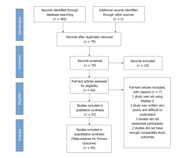
A flow diagram, in adherence with PRISMA guidelines, summarizing the screening of studies can be found in Figure 1. A total of 404 studies were identified from a literature search completed using both English and Chinese databases. After screening study titles and abstracts against basic eligibility criteria and searching for duplicates, 325 studies were discarded and 79 studies remained. Further screening of the study titles, abstracts and obtaining of the full study text eliminated 19 additional studies. After review of the full study texts of 60 articles, seven were eliminated and 53 RCTs were identified as eligible for the review[31-83]. The remaining 26 articles were excluded for various reasons. The rationale for exclusion of the 26 studies is found in Supplementary Table 1.
Figure 1.
Study selection flow diagram.
Study characteristics
The 53 Chinese RCTs included 3984 participants in our review, with 1985 and 1999 participants in control and treatment arms respectively. All studies contained a control arm, providing only conventional oral medication (aminosalicylates), and a treatment arm providing the same conventional medication with Medilac-S®. Treatment periods ranged from 4 to 96 wk. Twelve studies included a third study arm[35,47,55,60-62,66,70,72,77,82,83] which was not incorporated into the study analysis as a comparator because it incorporated the use of a different probiotic product, an herbal remedy and/or an enema. Nine studies included a post-treatment follow-up period of 8, 26 or 52 wk to track the maintenance of symptom remission[32,39,44,48,53,57,63,68,74] and one study, by Wang et al[66] 2016 evaluated the maintenance of remission alone. Most studies[31,33-37,40,41,43-48,51,53-57,59-62,65-67,69,71-77,79-83] included participants with mild-to-moderate UC symptoms (75.4%; 40/53), one study included participants with severe UC[49] and the remaining studies[32,38,39,42,50,52,58,63,64,68,70,78] did not describe the severity of all participants, however the levels of severity between control and treatment groups at baseline were stated as not significantly different. Study characteristics and outcomes are presented in Table 1.
Table 1.
Characteristics of included studies
| Trial reference | Participants evaluated | Medication | Probiotic dose | Treatment duration | Follow-up period | Outcomes analyzed |
| Bu et al[31] 2017 | 68 | Mesalazine | 3.0 × 10⁹ cfu/d | 16 wk | NR | 1, 6 |
| Chen[32] 2007 | 47 | SASP | 3.0 × 10⁹ cfu/d | 12 wk | 26 wk | 1, 4, 5, 6 |
| Chen[33] 2014 | 100 | Mesalazine | 3.0 × 10⁹ cfu/d | 6 wk | NR | 1, 4 |
| Chen et al[34] 2017 | 68 | Mesalazine | 3.0 × 10⁹ cfu/d | 8 wk | NR | 1, 6, 7 |
| Duan et al[35] 2015 | 64 | SASP | 3.0 × 10⁹ cfu/d | 4 wk | NR | 2, 3, 6, 7 |
| Gu[36] 2012 | 62 | Mesalazine | 3.0 × 10⁹ cfu/d | 12 wk | NR | 1, 6 |
| Guo and Sun[37] 2009 | 92 | Mesalazine | 3.0 × 10⁹ cfu/d | 12 wk | NR | 1, 6 |
| He et al[38] 2016 | 52 | Mesalazine | 3.0 × 10⁹ cfu/d | 12 wk | NR | 1, 6 |
| Jiang[39] 2013 | 110 | Mesalazine | 3.0 × 10⁹ cfu/d | 16 wk | 52 wk | 1, 5, 6 |
| Jin et al[40] 2014 | 226 | Mesalazine | 3.0 × 10⁹ cfu/d | 12 wk | NR | 1, 6 |
| Li[41] 2011 | 62 | Mesalazine | 60 mg/d | 12 wk | NR | 1 |
| Li[42] 2013 | 124 | SASP | 3.0 × 10⁹ cfu/d | 12 wk | NR | 1, 6 |
| Li[43] 2014 | 147 | SASP | 3.0 × 10⁹ cfu/d | 4 wk | NR | 1 |
| Li et al[44] 2006 | 50 | SASP | 3.0 × 10⁹ cfu/d | 12 wk | 26 wk | 1, 5 |
| Li et al[45] 2016 | 100 | Mesalazine | 3.0 × 10⁹ cfu/d | 8 wk | NR | 1, 4, 6 |
| Liang et al[46] 2016 | 92 | Mesalazine | 3.0 × 10⁹ cfu/d | 16 wk | NR | 1 |
| Liu[47] 2014 | 62 | SASP | 3.0 × 10⁹ cfu/d | 4 wk | NR | 1, 2, 3 |
| Liu and Yao[48] 2012 | 139 | Mesalazine | 3.0 × 10⁹ cfu/d | unknown | unknown | 1, 5 |
| Liu et al[49] 2009 | 43 | SASP | 3.0 × 10⁹ cfu/d | 8 wk | NR | 1, 6 |
| Liu and Li[50] 2014 | 101 | Mesalazine | 3.0 × 10⁹ cfu/d | 6 wk | NR | 1, 2, 4 |
| Luo[51] 2016 | 56 | Mesalazine | 3.0 × 10⁹ cfu/d | 8 wk | NR | 1, 4, 6 |
| Lu and Lei[52] 2011 | 132 | Olsalazine | 3.0 × 10⁹ cfu/d | 12 wk | NR | 1, 6 |
| Miao[53] 2014 | 72 | Mesalazine | 3.0 × 10⁹ cfu/d | 8 wk | 26 wk | 1, 5, 6 |
| Meng[54] 2012 | 90 | SASP | 3.0 × 10⁹ cfu/d | 8 wk | NR | 1, 2 |
| Qin[55] 2015 | 56 | Mesalazine | 3.0 × 10⁹ cfu/d | 8 wk | NR | 4 |
| Qin et al[56] 2010 | 20 | SASP | 3.0 × 10⁹ cfu/d | 4 wk | NR | 3, 7 |
| Qin et al[57] 2010 | 64 | Mesalazine | 3.0 × 10⁹ cfu/d | 8 wk | 26 wk | 5, 6 |
| Shen[58] 2014 | 96 | Mesalazine | 3.0 × 10⁹ cfu/d | 6 wk | NR | 1, 4, 6 |
| Su[59] 2015 | 120 | Mesalazine | 3.0 × 10⁹ cfu/d | 12 wk | NR | 1, 6 |
| Tan et al[60] 2008 | 20 | SASP | 3.0 × 10⁹ cfu/d | 4 wk | NR | 2, 3, 6, 7 |
| Tan et al[61] 2014 | 20 | SASP | 3.0 × 10⁹ cfu/d | 4 wk | NR | 2, 3, 6, 7 |
| Tang[62] 2008 | 104 | SASP | 3.0 × 10⁹ cfu/d | 4 wk | NR | 1 |
| Wang[63] 2013 | 84 | Mesalazine | 3.0 × 10⁹ cfu/d | 16 wk | 52 wk | 1, 5 |
| Wang and Liu[64] 2007 | 36 | SASP | 3.0 × 10⁹ cfu/d | 4 wk | NR | 1, 6 |
| Wang and Li[65] 2014 | 100 | Mesalazine | 3.0 × 10⁹ cfu/d | 8 wk | NR | 1, 2 |
| Wang et al[66] 2016 | 65 | Mesalazine | 3.0 × 10⁹ cfu/d | 26 wk | NR | 5, 6 |
| Xiang and Feng[67] 2006 | 46 | SASP | 3.0 × 10⁹ cfu/d | 4 wk | NR | 1, 4, 6 |
| Xiao[68] 2014 | 63 | SASP | 3.0 × 10⁹ cfu/d | 8 wk | 8 wk | 5, 6 |
| Xu[69] 2014 | 60 | Balsalazide | 3.0 × 10⁹ cfu/d | 12 wk | NR | 1, 4, 6 |
| Xu and Cui[70] 2009 | 56 | Mesalazine | 3.0 × 10⁹ cfu/d | 4 wk | NR | 1 |
| Yang[71] 2014 | 80 | Mesalazine | 3.0 × 10⁹ cfu/d | Unknown | NR | 1 |
| Yang et al[72] 2008 | 52 | SASP | 3.0 × 10⁹ cfu/d | 4 wk | NR | 1 |
| Yuan et al[73] 2008 | 40 | SASP | 3.0 × 10⁹ cfu/d | 12 wk | 26 wk | 1, 4, 6 |
| Zeng[74] 2008 | 49 | SASP | 3.0 × 10⁹ cfu/d | 12 wk | NR | 1, 5 |
| Zhang[75] 2013 | 78 | SASP | 3.0 × 10⁹ cfu/d | 4 wk | NR | 1, 4, 6 |
| Zhang[76] 2013 | 68 | Olsalazine | 3.0 × 10⁹ cfu/d | 12 wk | NR | 1, 6 |
| Zhang et al[77] 2010 | 54 | Mesalazine | 3.0 × 10⁹ cfu/d | 12 wk | NR | 1 |
| Zhang et al[78] 2016 | 70 | Mesalazine | 60 mg/d | 12 wk | NR | 1 |
| Zhang et al[79] 2016 | 60 | Mesalazine | 3.0 × 10⁹ cfu/d | 12 wk | NR | 1 |
| Zhao and Zhang[80] 2016 | 62 | Mesalazine | 3.0 × 10⁹ cfu/d | 24 wk | NR | 1 |
| Zheng et al[81] 2016 | 118 | Mesalazine | 3.0 × 10⁹ cfu/d | 4 wk | NR | 1, 6 |
| Zhu et al[82] 2013 | 44 | Olsalazine | 3.0 × 10⁹ cfu/d | 96 wk | NR | 2, 3 |
| Zhuo et al[83] 2016 | 40 | Mesalazine | 3.0 × 10⁹ cfu/d | 8 wk | NR | 1, 3, 6 |
Outcomes analyzed: (1) Clinical Efficacy; (2) Histological Assessment; (3) Endoscopy Assessment; (4) Clinical Symptoms; (5) Maintenance of Remission; (6) Adverse Events; (7) Sutherland Index. NR: Not reported.
Risk of bias in included studies
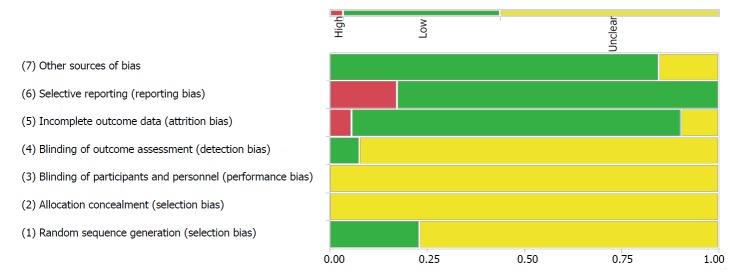
Results of the risk of bias analysis are presented in Figure 2. Although all included trials are RCTs, most fail to report the method used for randomization, resulting in unclear risk of bias in 77.4% (41/53) of studies[31-34,36,38,40-44,46-52,54,55,57-59,62-71,73,74,76,77,80-83]. The twelve remaining studies report appropriate methods to randomize participants, such as random number tables, and are ranked as low risk of bias for random sequence generation[35,37,39,45,53,56,60,61,72,75,78,79].
Figure 2.
Risk of bias mosaic plot showing proportion of studies deemed to be at high (red), unclear (yellow) or low (green) risk in each bias category.
Only 7.55% (4/53) of studies report the implemented levels of blinding[40,56,60,61]. These four studies reported a blind assessment of participant samples, thereby receiving a low risk of detection bias rating. No other forms of blinding for participants, personnel or assessors were reported, nor was allocation concealment reported in any studies, resulting in unclear levels of bias for allocation concealment and detection bias amongst all studies.
After evaluating for incomplete outcome data and selective reporting, 83.0% (44/53) of studies[31-41,44-49,51-55,62-65,67-81,83] were ranked as low risk of attrition bias and 81% (43/53) of studies[32,34-47,49-51,55,56,58-62,64,66-82] were ranked as low risk of reporting bias. Three trials[42,43,50] were rated as high risk of attrition bias because of inconsistencies in subject reporting and the remaining six studies[56,57,60,61,66,82] were rated as unclear risk of attrition bias due to missing information regarding possible participant withdrawal. The ten studies[31,33,48,52-54,57,63,65,83] which were not ranked as low risk of reporting bias were rated as high risk of reporting bias because results were either reported without pre-specification or expected outcomes failed to be included.
Other potential sources of bias included an evaluation of the characteristic similarity and prognostic indicators of disease at baseline in study groups. In trials of UC, it is important for studies to limit significant differences between disease severity of participants at baseline because the medication and dosage differ for those suffering from varying stages of disease, ranging from mild and moderate to severe. Most studies[31-43,45-49,52,54-56,58,59,61,63-71,75-83] (83.0%; 44/53) highlighted no significant differences between the control and treatment groups at baseline, thereby being labelled as low risk bias. The nine remaining studies not ranked as low risk of bias[44,51,55,57,60,62,72-74] had insufficient information to determine the presence of other types of bias and therefore are ranked as unclear risk. Overall, studies were better ranked in their provision of study results and participant withdrawals, and were ranked more poorly in their explanation of randomization techniques blinding.
Effects of intervention - primary outcome
Clinical remission: Forty-six studies reported clinical remission as a primary outcome, however, only 45 studies[31-34,36-55,58,59,62-65,67,69-81,83] were included in the meta-analysis because one study[57] failed to report enough data. Studies included 3624 participants, with 1808 in the control group and 1816 in the treatment group. The proportion of subjects for which the treatment was effective was analyzed, with “effective” defined as any response not considered “ineffective”- including participants with “completely effective”, “very effective” or “somewhat effective” responses. Further analysis was then conducted by comparing induction of clinical remission using specific drug-probiotic combinations to assess the most effective combination. One study used balsalazide as the conventional drug[69], two studies used olsalazine[52,76], 14 studies used SASP[32,42,43,45,47,49,54,62,64,67,72-75] and 28 studies used mesalazine[31,33-41,44,46,48,50,51,53,55-61,63,65,66,68,70,71,77-84].
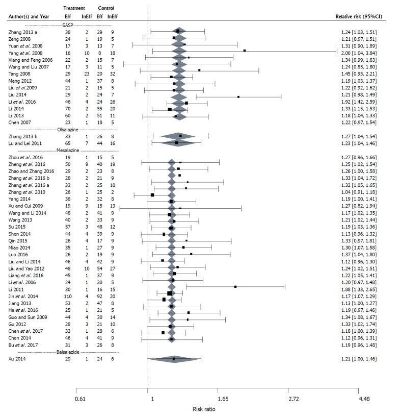
The first analysis with the 45 studies was conducted using a fixed effects model since the test for heterogeneity was not significant (P = 0.8102). Results showed a positive effect of Medilac-S® treatment (RR = 1.21, CI: 1.18-1.24, P < 0.0001) with the “risk” of treatment being effective estimated to be, on average, 21% higher for those on combination Medilac-S® therapy, than conventional drug therapy alone.
Upon comparing effect means for different drug types, evidence of significant variability (P < 0.0001) was seen. In the balsalazide study the predicted mean RR was the same as in the study, RR = 1.21 (CI: 1.00-1.46). In studies using olsalazine, the estimated mean was RR = 1.25 (CI: 1.10-1.42). Finally, the estimated mean RR for SASP drug therapy was RR = 1.26 (CI: 1.19-1.33) and the mean RR for mesalazine was RR = 1.19 (CI: 1.15-1.23) (Figure 3). Since there were sufficient numbers of studies using mesalazine and SASP, further comparison of the mean effect sizes was performed, and a significant difference between mean effect sizes of studies using mesalazine and SASP was observed (P < 0.0001), with SASP outperforming mesalazine.
Figure 3.
Forest plot of the results of a fixed effects meta-analysis with a moderator for concomitant drug therapy with 45 studies evaluating the effect of Medilac-S® in combination with conventional drug therapy on clinical efficacy. “Eff” is the number of subjects in the study for which the treatment was effective and “InEff” is the number of subjects in the study for which the treatment was ineffective. The relative risk (RR) and its 95%CI for each study are listed on the right hand side of the graph. The 95%CI for the estimated mean RR for each concomitant drug therapy category is shown as a shaded diamond with the endpoints of the diamond being the CI endpoints and the location of the maximum width of the diamond being at the estimated mean RR for that drug type. The vertical dashed line at 1 indicates a RR of 1 which occurs when there is no observed difference between the treatment and the control.
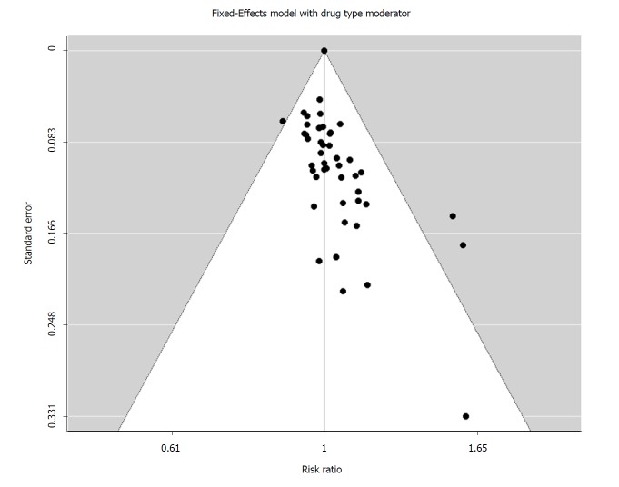
Publication bias: A funnel plot was constructed based on the results of the fixed effects model with drug type moderator (Figure 4). A rank correlation test, Kendall’s tau, tested for asymmetry in the plot by evaluating whether the observed effect sizes and the corresponding sampling errors are correlated. Kendall’s tau was 0.5366 (P < 0.0001), providing strong evidence of publication bias. The funnel plot suggests a few studies with smaller sample size present larger SEs for Medilac-S® consumption with greater RR values than studies with larger sample sizes.
Figure 4.
Funnel plot showing the relationship between the relative risk and its standard error for each of 45 studies used in a fixed effects meta-analysis with a moderator for concomitant drug therapy evaluating the effect of Medilac-S® in combination with conventional drug therapy on clinical efficacy.
In addition, the number of missing studies from the meta-analysis due to the suppression of the more extreme results to one side of the funnel plot, were estimated using the trim and fill method of Duval and Tweedie[27,28]. Results indicate that the funnel plot would be made symmetric with the addition of 18 (SE = 4.0663) studies on the left side of the plot. With the addition of the studies, the average log(RR) would decrease to approximately 0.16 (SE = 0.0128; estimated RR = 1.17), which still indicates a positive impact of adding Medilac-S® to conventional drug therapy.
Effects of intervention - secondary outcomes
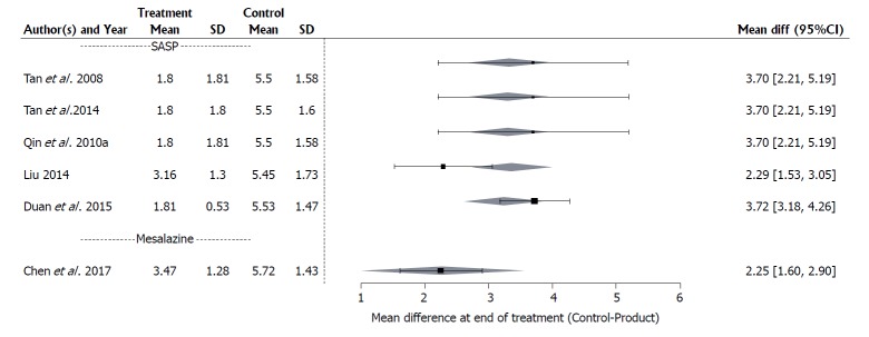
Sutherland index: A mixed effects meta-analysis was completed on six studies[34,35,47,57,60,61] to evaluate changes in the Sutherland index score, comprised of three clinical symptom scores (stool frequency, rectal bleeding and mucosal appearance) and a physician’s global rating of disease. Analysis was also conducted with a moderator variable for drug type to determine which drug-probiotic combination is most effective in improving the Sutherland index score. Studies included 254 participants, with 127 participants in both control and treatment groups. One study used mesalazine as concomitant medication[34] and five studies used SASP[35,43,56,60,61].
The effect size analyzed is the difference in means between treatment and control groups at the end of the intervention period because no differences were found between groups at baseline using the random effects model without the moderator variable for conventional drug type (P = 0.9999). Differences were seen using a random effects model run on the post-intervention treatment group means, both with the moderator variable (P = 0.0428) and without (P = 0.0032).
Results indicate a positive effect of Medilac-S® treatment on the Sutherland Index. Without the moderator variable for conventional drug therapy, the mean difference between the control and treatment at the end of the intervention period was 3.10 (95%CI: 2.41, 3.78), indicating that the control mean is, on average, 3.10 units greater than the treatment arm. With the moderator variable, results suggest that the effect size may be drug-type dependent (P < 0.0001), with SASP outperforming mesalazine. The mean difference in the index between treatment and control arms was 3.33 (CI: 2.63-4.03) for the drug SASP and 2.25 (CI: 0.95-3.55) for mesalazine (Figure 5).
Figure 5.
Forest plot of the results of a mixed effects meta-analysis with a moderator for concomitant drug therapy, with 6 studies evaluating the effect of Medilac-S® in combination with conventional drug therapy on the change in mean Sutherland index score evaluating three clinical symptoms and a global physician’s assessment. “Mean” is the mean index and “SD” is the standard deviation for each study; the difference between the treatment and control of the mean indices at the end of the study and its 95%CI is listed on the right hand side of the graph; the 95%CI for the estimated mean difference for each concomitant drug therapy category is shown as a shaded diamond with the endpoints of the diamond being the CI endpoints and the location of the maximum width of the diamond being at the estimated mean difference for that drug type.
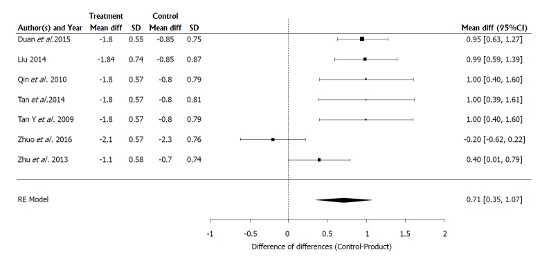
Endoscopy scores: Seven clinical studies[35,43,56,60,61,82,83] with 270 participants, 135 in both control and treatment arms, were included in a meta-analysis to evaluate changes in the endoscopic scores assessed using a Chinese version of the Modified Baron Score evaluating mucosal friability, hyperemia, granulation, spontaneous bleeding and ulceration[21,22,84]. Reporting was done as the average of the change in score. The effect size analyzed is the difference between treatment and control arms of the change in mean scores between final and baseline measurements. As the test for heterogeneity was significant (P = 0.0001), a random effects model was used for the analysis. Further analysis to determine if the results are dependent on concomitant drug therapy was not conducted due to insufficient data.
Results present a positive effect of Medilac-S® treatment for the improvement of the endoscopic score (P = 0.0001). The difference from baseline in the control and treatment arms was 0.7139 (95%CI: 0.3537-1.0742), indicating that the average decrease of endoscopy scores for the control arm was 0.71 units smaller than the average decrease of endoscopy scores for the treatment arm (Figure 6). The mean drop in scores for those in the control arm was approximately 1.01, while the mean drop for those on combination therapy was approximately 1.72.
Figure 6.
Forest plot of the results of a random effects meta-analysis with 7 studies evaluating the effect of Medilac-S® in combination with conventional drug therapy on the change in mean endoscopic score evaluating mucosal friability, hyperemia, granulation, spontaneous bleeding and ulceration. “Mean Diff” is the change of the treatment mean score from the baseline mean score and “SD” is the standard deviation of the mean change; the difference between the treatment and control of the change in the mean score and its 95%CI for each study is listed on the right hand side of the graph; the 95%CI for the estimated mean difference in the change from baseline for each concomitant drug therapy category is shown as a shaded diamond with the endpoints of the diamond being the CI endpoints and the location of the maximum width of the diamond being at the estimated mean difference for that drug type; the vertical dashed line at 0 indicates a difference of 0 which occurs when there is no observed difference between the change from baseline for the treatment and the control.
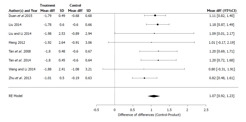
Histological scores: Eight clinical studies[35,43,50,54,60,61,65,82] included 501 participants, with 251 in the control arm and 250 in the treatment arm, in a meta-analysis to evaluate changes in the histological scores assessed using a Chinese version of the Truelove and Richards Index evaluating colonic histological specimens for inflammation and crypt distortion[21-22,85]. Reporting was done as the average of the change in score. The effect size used in the analysis is the difference between treatment and control of the change in mean scores between final and baseline measurements. The test for heterogeneity was not significant (P = 0.8427) and so a fixed effects model was used for the analysis. Further analysis to determine if the results are dependent on concomitant drug therapy was not conducted due to insufficient data.
The test for difference between the change from baseline for treatment and control arms was significant (P < 0.0001) and favors results of Medilac-S® treatment. The difference in the change from baseline of the control and treatment arms was 1.07 (95%CI: 0.9189-1.2300) indicating that the average decrease in histology scores between baseline and final measurements for the control arm was approximately 1.1 units smaller than the average decrease between baseline and final measurements for the treatment arm (Figure 7). The mean drop in histology scores for those in the control arm was approximately 0.9 while the mean drop for those on treatment was approximately 1.9.
Figure 7.
Forest plot of the results of a random effects meta-analysis with 8 studies evaluating the effect of Medilac-S® in combination with conventional drug therapy on the change in mean histological score, evaluating colonic histological specimens for inflammation and crypt distortion. “Mean Diff” is the change of the treatment mean score from the baseline mean score and “SD” is the standard deviation of the mean change; the difference between the treatment and control of the change in the mean score and its 95%CI for each study is listed on the right hand side of the graph; the 95%CI for the estimated mean difference in the change from baseline for each concomitant drug therapy category is shown as a shaded diamond with the endpoints of the diamond being the CI endpoints and the location of the maximum width of the diamond being at the estimated mean difference for that drug type; the vertical dashed line at 0 indicates a difference of 0 which occurs when there is no observed difference between the change from baseline for the treatment and the control.
Clinical symptoms: A meta-analysis of 11 studies[32,33,45,50,51,55,58,67,69,73,75] was conducted to evaluate changes in any of the following patient-reported symptoms: Abdominal pain, tenesmus, blood and mucous in stool, and/or diarrhea. Analysis was also conducted with a moderator variable for drug type to determine which drug-probiotic combination was most effective in reducing clinical symptoms. Studies included 780 participants, with 386 participants in the control arm and 394 participants in the treatment arm.
The effect size is the ratio of the proportion of individuals reporting symptoms in the treatment group as compared to the proportion of individuals reporting symptoms in the control group. Values less than one for the RR or less than zero for log (RR) indicate that subjects receiving combination therapy with Medilac-S® have a lower probability of reporting clinical symptoms relative to subjects receiving drug therapy alone.
Using the random effects model, no significant differences were reported at baseline between control and treatment groups for any of the clinical symptoms and no significant evidence for heterogeneity was seen in the proportion of individuals reporting symptoms between treatment arms. Consequently, fixed effects models were used to identify variability in mean RRs after intervention.
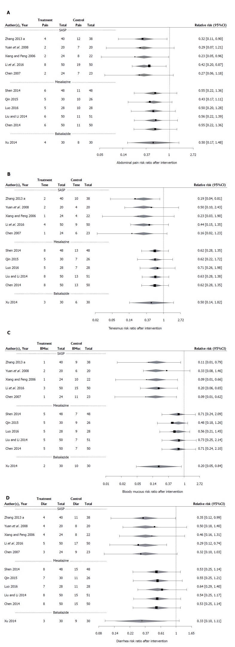
Results of the fixed effects meta-analyses demonstrate a significant decrease (P < 0.0001) in the proportion of individuals reporting abdominal pain (RR = 0.44, CI: 0.32-0.59), tenesmus (RR = 0.53, CI: 0.38-74), blood and mucous in stool (RR = 0.40, CI: 0.28-0.58) and diarrhea (RR = 0.47, CI: 0.36-0.42) post-intervention after using combination therapy. Hence, the proportions of individuals who received combination therapy are 44%, 53%, 40% and 47% respectively of the proportion of individuals in the control group reporting the same symptoms.
When the moderator variable is applied, significant differences (P < 0.0001) are indicated for at least one pair of mean RRs among drug types for every clinical symptom. One study[69] used balsalazide, five studies[33,50,51,55,58] used mesalazine and five studies[32,45,67,73,75] used SASP as the concomitant medications with Medilac-S®.
The calculated predicted mean RRs for combination therapy with balsalazide drug include: RR = 0.5 (95%CI: 0.17-1.48) for abdominal pain, RR = 0.53 (CI: 0.14-0.82) for tenesmus, RR = 0.2 (CI: 0.05-0.84) for blood and mucous in stool, and RR = 0.33 (CI: 0.10-1.11) for diarrhea. The estimated mean RRs calculated for SASP drug include: RR = 0.34 (CI: 0.21-0.55) for abdominal pain, RR = 0.31 (CI: 0.16-0.62) for tenesmus, RR = 0.17 (CI: 0.08-0.34) for blood and mucous in stool, and RR = 0.37 (CI: 0.23-0.59) for diarrhea. The estimated mean RR for mesalazine drug therapy include: RR = 0.51 (CI: 0.34-0.78) for abdominal pain, RR = 0.63 (CI: 0.43-0.93) for tenesmus, RR = 0.62 (CI: 0.39-0.98) for blood and mucous in stool, and RR = 0.56 (CI: 0.39-0.79) for diarrhea. Results of RRs and CIs are presented in Figure 8A-D. Because of the greater number of studies using SASP and mesalazine, tests for differences in mean RRs for SASP and mesalazine were also performed for each clinical symptom. A significant difference was seen between the effects of the two medications (P < 0.0001) for all clinical symptoms with SASP outperforming mesalazine.
Figure 8.
Forest plots of the results of fixed effects meta-analysis with a moderator for concomitant drug therapy for 11 studies evaluating the effect of Medilac-S® in combination with conventional drug therapy on number of participants reporting clinical symptoms including: (A) abdominal pain; (B) tenesmus; (C) blood and mucous in stool; and (D) diarrhea. “Pain” is the number of participants reporting abdominal pain in each study, “Tene” is the number of participants reporting tenesmus in each study, “BMuc” is the number of participants reporting blood and mucous in stool in each study, “Diar” is the number of participants reporting diarrhea in each study, and “Total” is the total number of participants in each study. The relative risk (RR) and its 95%CI for each study is listed on the right hand side of the graph. The 95%CI for the estimated mean RR for each concomitant drug therapy category is shown as a shaded diamond with the endpoints of the diamond being the CI endpoints and the location of the maximum width of the diamond being at the estimated mean RR for that drug type. The vertical dashed line at 1 indicates a RR of 1 which occurs when there is no observed difference between the treatment and the control.
Maintenance of clinical remission: Ten studies[32,39,44,48,53,57,63,65,68,74] enrolled 379 patients in the control group and 364 in the treatment group for a total of 743 participants, excluding any participants in a third arm. These studies evaluated the ability of Medilac-S® treatment, in conjunction with conventional medication, mesalazine or SASP, to maintain clinical remission of UC symptoms. A meta-analysis was not conducted for these ten studies as there was insufficient data to evaluate relapse rate as a function of follow-up time.
Nine studies first aimed to induce UC remission in patients and included a follow-up period to observe symptom recurrence. Of the nine studies, one had a follow-up period of 8 wk[68], five studies[32,44,53,57,74] had a 26 wk follow-up period, two studies[39,63] had a 52-wk follow-up period and one study had a follow-up period of unknown length[48]. One study by Wang et al[66] did not aim to induce UC remission as only patients in remission were recruited. The study evaluated a maintenance of remission for a period of 26 wk.
In 80% (8/10) of studies the symptom recurrence rate for participants receiving conventional therapy with Medilac-S® was significantly lower (P < 0.05) than participants taking conventional medication alone. In contrast, one study, by Qin et al[56] 2010, showed a greater number of participants in the treatment group experiencing symptom recurrence, with 17.2% (5/29) experiencing symptom recurrence during post-treatment evaluation at 26 wk and only 15.8% (3/19) in the control group. However, the study reports no significant difference between the two recurrence rates. The study by Wang et al[66] also reported no significant difference (P = 0.753) between the recurrence rates of the control and treatment group at 26 wk, however, the treatment group experienced less symptom recurrence than the control group, at 9.09% and 12.5% respectively.
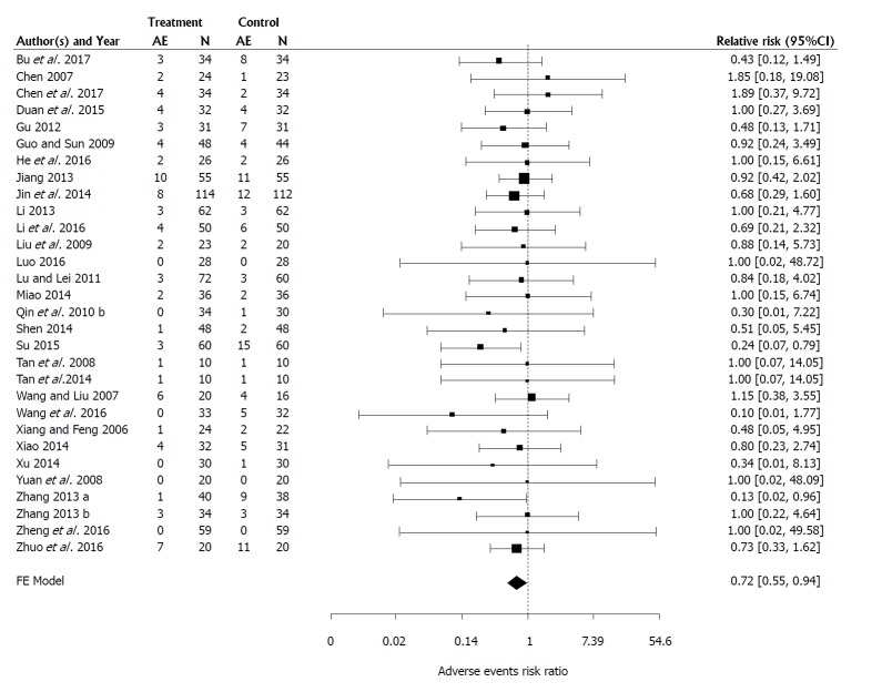
AEs: A meta-analysis of 30 RCTs reporting AEs which included 2430 participants, with 1195 in the control group and 1235 in the treatment group, was completed. As the test for heterogeneity was not significant (P = 0.9914) analysis was conducted with a fixed effects model[31,32,34-40,42,45,49,51-53,57-61,64,66-69,73,75,76,81,83]. As shown on Figure 9, on average, the proportion of individuals in the treatment arm reporting an AE is estimated to be 72% of the proportion of individuals reporting an AE in the control arm (RR = 0.72, 95%CI: 0.55-0.94, P = 0.0175).
Figure 9.
Forest plots of the results of fixed effects meta-analysis with 30 studies evaluating the effect of Medilac-S® in combination with conventional drug therapy on number of participants reporting adverse events. “AE” is the number of participants reporting adverse events within a study and “N” is the total number of participants within the study. The relative risk (RR) and its 95%CI for each study are listed on the right hand side of the graph. The 95%CI for the estimated mean RR for each concomitant drug therapy category is shown as a shaded diamond with the endpoints of the diamond being the CI endpoints. The vertical dashed line at 1 indicates a RR of 1 which occurs when there is no observed difference between the treatment and the control.
Publication bias for secondary outcomes
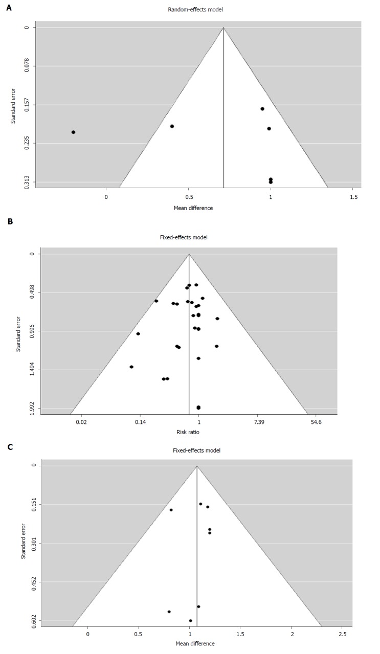
Publication bias for the secondary outcomes was included in the meta-analyses. In studies reporting the endoscopy scores, histology scores, and AEs, no evidence of publication bias was seen [Kendall’s tau -0.30 (P = 0.3567)], which may be related to the very small sample sizes (Figure 10A-C).
Figure 10.
Funnel plots showing the relationship between the relative risk and its standard error for (A) 7 studies used in a random effects meta-analysis evaluating the efficacy of Medilac-S® in combination with conventional drug therapy on change in mean endoscopic score (B) 8 studies used in a fixed effects meta-analysis evaluating the efficacy of Medilac-S® in combination with conventional drug therapy on change in mean endoscopic score and (C) 30 studies used in a fixed effects meta-analysis evaluating the efficacy of Medilac-S® in combination with conventional drug therapy on change in the number of reported adverse events.
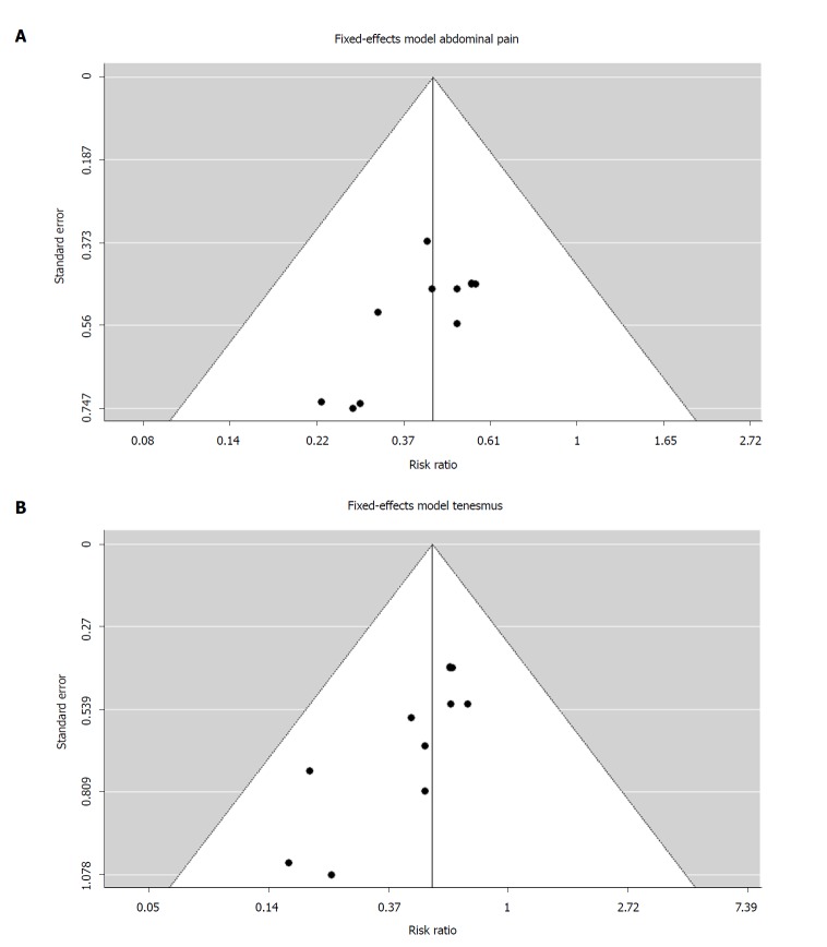
For the clinical symptoms, outcome results indicate some evidence of publication bias. Kendall’s tau was significant for two clinical symptom outcomes -abdominal pain [-0.4909 (P = 0.0405)] and tenesmus [-0.45273 (P = 0.0264)] (Figure 11A and B). The trim and fill method of Duval and Tweedie[27,28] suggests the funnel plot for studies reporting abdominal pain would be made symmetric with the addition of two studies on the left side of the plot (SE = 2.308). The same method suggests that one study be added on the right side of the plot for studies reporting tenesmus (SE = 2.2944). The average log(RR) would be reduced to -0.78 (SE = 0.1478; RR = 0.46) for abdominal pain and -0.60 (SE = 0.1634; RR = 0.55) for tenesmus, which still indicates a positive impact of adding Medilac-S® to conventional drug therapy.
Figure 11.
Funnel plot showing the relationship between the relative risk and its standard error for each of 11 studies used in a fixed effects meta-analysis with a moderator for concomitant drug therapy evaluating the effect of Medilac-S® in combination with conventional drug therapy on number of reported clinical symptoms for (A) abdominal pain and (B) tenesmus.
DISCUSSION
Currently available therapies for UC, such as pharmaceutical anti-inflammatories, elicit high response rates, however, due to the potential for high-risk side effects, costs and non-adherence, alternative treatments such as probiotics are being considered[10]. Growing evidence illustrates the pivotal role of gut microflora in UC pathogenesis[2,9], and studies have also shown the influence of the gut microflora on drug pharmacokinetics[86,87], particularly drugs consumed per os. Thus, identifying specific probiotics which mediate symptoms of UC may improve responses to, and decrease potential side effects of, currently available therapies.
The present systematic review and meta-analysis evaluates 53 RCTs to assess the efficacy of the probiotic Medilac-S® in combination with aminosalicylates to induce UC clinical remission within a Chinese population. Results show that combination therapy improves clinical remission rates, reduces symptom severity within the GI tract, and decreases incidence of UC symptoms and AEs. A review of studies evaluating maintenance of remission rates also shows reduced symptom recurrence of participants in the probiotic combination therapy groups as compared to conventional therapy alone. Prior studies have demonstrated similar positive effects of probiotics in UC patients through probiotic combination therapy[15,88] or probiotic therapy alone[89]. However, some studies show limited evidence of probiotics as clinically beneficial for the induction and maintenance of UC remission[90,91]. Consequently, it would appear that not all probiotic products will be effective and each product must be uniquely evaluated in the target population.
Some concerns with several prior systematic reviews and meta-analyses are the relatively small numbers of pooled participants analyzed and significant heterogeneity amongst studies due to pooling of data from mixed populations (i.e., adults and children), various probiotic combinations, and the use of different concomitant therapies, which makes it difficult to interpret results accurately. Additionally, many of these reviews fail to incorporate studies outside of North America and Europe, where different probiotics are routinely used and accepted in combination with standard care. For example, many Asian countries have readily accepted probiotics as dietary supplements and pharmaceuticals for a number of years[19]. This results in very few alternative probiotic therapies highlighted amongst systematic reviews assessing probiotics and IBD, which may be misleading.
This systematic review and meta-analysis is distinctive because it focuses on one disease state, one product and one population type, thereby allowing for a more focused analysis. In the past, only one other review has been completed to evaluate the efficacy of Medilac-S® on symptoms of UC[24]. However, due to the unconventional methodology and the abundance of new evidence, it was important to reconstruct the process using stricter review guidelines and improved sub-analyses.
By conducting a more focused review, we were also able to analyze specific probiotic-drug combinations to elucidate that which is most effective. Medilac-S® was shown to be the most effective in inducing remission when partnered with SASP. We hypothesize this may be due to the stability of SASP in the upper GI tract, which allows for greater quantities of the drug to reach the damaged epithelium[92]. The exact dosage and duration of use of the probiotic remains to be elucidated due to insufficient variation amongst studies to test the effects of these variables, however the majority of studies in our review (94.3%; 50/53) used the recommended dose of 3.0 x 10⁹ colony forming units (cfu)/d.
SASP remains the most common anti-inflammatory drug used in China for mild-to-moderate UC[93], however, long term use presents many side-effects[94]. Our results found that the incorporation of probiotic Medilac-S® with SASP and other aminosalicylates (mesalazine, olsalazine, balsalazide) reduced the risk of AEs, suggesting the role of probiotics in the prevention of AEs associated with anti-inflammatory drug use. Prior studies evaluating the effects of probiotics on IBD commonly report no significant differences between probiotic-treated and conventionally-treated participants[15,95]. Due to limited assessments and systematic reporting of AEs from studies evaluating probiotic-treated IBD, further evidence is required to effectively assess the benefit of probiotic intervention on drug-induced side effects. Further study is also required to assess the effects of probiotic adjunctive therapy on varying disease severity of UC, however, probiotics are most commonly used, and most effective, in individuals with mild-to-moderate UC[14].
Researchers have suggested various mechanisms of action for probiotics in the prevention and treatment of IBD and UC, including maintenance of microbial gut microflora[15], reduced GI inflammation[96], protection against pathogens[97], and improving immune system function[98]. Although few studies have elucidated the potential mechanisms for the beneficial effects of the two Medilac-S® strains specifically (E. faecium R0026 and B. subtilis R0179), a study by Zhong et al[99], found the probiotic decreased and prevented the growth of various enteric bacterial pathogens and Tompkins et al[19] found both strains to adhere to human intestinal cells (HT-29) in culture.
Research also suggests the gut microflora and, subsequently, probiotics which influence the gut microflora, may play an important role in drug pharmacokinetics[86,87,100]. Gut microflora are a determinant for azo-containing compounds such as SASP and other aminosalicylate pro-drugs[86]. Orally ingested pro-drugs are broken down in the large intestine through a two-step azo-bond reduction mediated by the azoreductase enzyme found in the natural gut microflora[101]. Many probiotic strains, including those found in Medilac-S®, also contain the azoreductase enzyme, and a recent unpublished evaluation by one of the authors (TT) demonstrated that the bacterial strains in Medilac-S®, particularly the E. faecium R0026, in vitro facilitate the breakdown of SASP into its active moiety 5-ASA and sulphapyridine.
These findings are consistent with studies, such as Lee et al[102], conducted in animal models which found probiotic administration significantly improved the breakdown of SASP to 5-ASA and sulphapyridine. Significant metabolic breakdown of SASP was not observed in a 2010 study by Lee et al[103] evaluating the influence of a 9 × 10⁸ CFU multi-strain probiotic (Lactobacillus acidophilus, Bifidobacterium lactis and Streptococcus salivarius) in patients suffering from rheumatoid arthritis. However, the probiotic was given only twice daily for a short one-week treatment period. Therefore, further exploration on the mechanism of action of probiotics and the natural gut microflora in the breakdown of pro-drugs such as SASP is required, with different treatment dosages and durations reviewed.
Limitations
We cannot rule out the potential for some risk of bias amongst included studies and possible publication bias, however, results concerning publication bias should be considered exploratory because neither unpublished literature nor the potentially missing articles were located. Most studies presented an unclear risk of bias in assessed categories, such as techniques for randomization, allocation concealment, and the blinding of participants or study personnel. Some included studies also demonstrated a high risk of bias in reporting because results were either reported without pre-specification or expected outcomes failed to be included. We hypothesize this style of reporting is a result of the guidelines and trends in China[104-107].
Furthermore, the use of Chinese diagnostic guidelines from different years of publication, which are appropriate and independently validated in their own right, results in diagnostic techniques and testing scales which are not completely uniform or analogous to the more commonly seen Western guidelines. Finally, in reporting the efficacy of different drug-probiotic combinations, results indicated SASP outperformed mesalazine, however, due to the limited number of studies using balsalazide and olsalazine, further evidence is required to draw firm conclusions on other Medilac-S® and aminosalicylate combinations.
In conclusion, moderate-quality evidence, as seen by improvements in the Sutherland index, endoscopy and histology scores, a decrease in patient-reported clinical symptoms of UC (abdominal pain, diarrhea, tenesmus and blood/mucous in the stool), and a decrease in AEs suggests Medilac-S®, in conjunction with conventional aminosalicylates, is effective in inducing clinical remission of UC and improving symptoms of the GI tract in Chinese populations. Therefore, for this application, this probiotic should be considered as best practice for standard care in clinical practice. Additional work is required in non-Chinese populations to substantiate its use. Further analytical evidence is also required to determine the benefit of Medilac-S® combination therapy for the maintenance of UC remission.
Due to the unknown risk of bias amongst many study categories, more robust and well conducted RCTs are needed to draw further conclusions about which combination of Medilac-S® and conventional UC treatment is most effective. Future studies should also attempt to evaluate the impact on SASP, SP and 5-ASA levels with varying Medilac-S® dosages. It is suggested that authors of clinical studies in China include more detailed information on the study design and technique implementation for future clinical trials, particularly when tracking the maintenance of symptom remission, to further improve upon study quality.
ARTICLE HIGHLIGHTS
Research background
Ulcerative colitis (UC), an inflammatory bowel disease (IBD) of the colonic gastrointestinal tract, is steadily increasing across the globe, particularly in Asian countries such as China, where rapid industrialization and urbanization are contributing factors to the growing onset. Mild-to-moderate UC is the most prevalent amongst diagnosed cases, and although targeted pharmacological therapies, such as aminosalicylates, have been at the forefront of UC therapy, growing evidence suggests the integral role of intestinal microflora in UC pathogenesis. Subsequently, researchers are interested in the use and development of alternative therapies, such as probiotics, for the management and treatment of this colonic disease. This systematic review and meta-analysis addresses the use of a probiotic product, Medilac-S®, as adjunctive therapy for the induction of clinical UC remission and improvement of UC symptoms in a defined Chinese population, through the evaluation of 53 randomized, controlled trials (RCTs). Past reviews of probiotics for the induction of UC remission have described the positive effects of probiotic combination therapy in patients, however, many of these reviews demonstrated significant variability in study populations and probiotic treatments, making it difficult to interpret results accurately. This study therefore aims to provide a more focused analysis through the evaluation of one disease state, one probiotic and one population.
Research motivation
The incidence of UC has increased rapidly around the globe and although current pharmacological therapies elicit high response rates, they also present high-risk side effects or adverse events (AEs), growing rates of non-adherence and high costs to patients. Growing evidence also illustrates the important role of gut microflora in UC pathogenesis and the influence of the intestinal microbiome on drug pharmacokinetics. Therefore, it is important to identify treatments, such as probiotics, which can mediate the gut microflora to improve symptoms of UC and also improve responses to currently available therapies, whilst mitigating potential side effects.
Research objectives
Our primary objective was to conduct an up-to-date systematic review and meta-analysis to assess the efficacy of Medilac-S® as an adjunctive to conventional oral drugs for the induction of UC clinical remission within a Chinese population. One prior systematic review and meta-analysis, published by Hu et al, had, to date, discussed the efficacy of the probiotic Medilac-S® on the induction and maintenance of remission in UC patients. However, since its publication, a large number of new studies had been published and remained to be evaluated in a meta-analytic setting. In this review, we assessed 53 RCTs which highlighted the efficacy of the probiotic Medilac-S®, in combination with conventional aminosalicylates, to induce UC clinical remission within a Chinese population, improve UC symptoms and decrease AEs. This supports suggestions of the important role of the gut microbiome in the modulation of IBDs and presents a new potential treatment to mitigate the effects of the microflora on worsening UC symptoms.
Research methods
The review protocol was registered in PROSPERO and the Preferred Reporting Items for Systematic Reviews and Meta-Analyses (PRISMA) guidelines were followed for the review to ensure up-to-date methodology and study reasoning. Nine databases, both English and Chinese, were searched from 2000 to 2017, unrestricted by language or trial size to identify RCTs evaluating the therapeutic effects of Medilac-S® combination therapy with conventional aminosalicylate drugs in the treatment of UC within a Chinese population. If the study met the inclusion criteria, then outcome data extraction and assessment of both study quality and risk of bias was independently performed by two authors. Any disagreement was resolved by discussion and if consensus could not be reached, a third author was addressed. The included studies were evaluated on clinical remission, changes in patient-reported clinical symptoms, maintenance of remission and relapse rate, Sutherland index, endoscopic score, histological score and AEs. Meta-analysis was conducted on each outcome of interest using a fixed effect or a random effect model, depending on the significance of the heterogeneity. For dichotomous outcomes (e.g., clinical efficacy), risk ratios (RR) were calculated while the mean difference was used for continuous variables (e.g., histology scores). CIs were calculated at 95% and, when possible, the type of the conventional drug was applied as a moderator in the model of analysis. The overall quality of evidence supporting the outcome was assessed through the use of the Cochrane Collaboration tool and a risk of bias analysis was conducted on each study. The risk of bias parameters included the type of randomization method, allocation concealment, blinding of participants, personnel and outcome assessments, incomplete outcome data, selective reporting and other sources of bias. Additionally, selective reporting (reporting bias) and other types of bias were also considered. The potential for publication bias across studies was analyzed using the rank correlation test Kendall’s Tau, which tested for asymmetry in a funnel plot showing the relationship of the effect size [log (RR)] and its standard error (SE) among studies.
Research results
Fifty-three studies involving 3984 patients were identified and included in the systematic review. Forty-five studies were analyzed for the primary outcome of clinical remission and results demonstrated that combination Medilac-S® therapy is significantly more effective than conventional drug therapy alone (RR = 1.21, CI: 1.18-1.24, P < 0.0001). Moreover, sulfasalazine (SASP) outperformed mesalazine as a concomitant drug. Further meta-analysis also provided evidence that the combination of Medilac-S® significantly improved the Sutherland index score (P < 0.05), endoscopic score (P = 0.0001), histological score (P < 0.0001) and the number of patient reported symptoms (P < 0.0001), which include, abdominal pain, tenesmus, blood and mucous in stool, and diarrhea. The proportions of individuals who received combination therapy and complained of the aforementioned symptoms were 44%, 53%, 40% and 47% respectively of the proportion of individuals in the control group reporting the symptoms. The test for difference in the RR with different drugs also revealed that SASP in combination with Medilac-S® is more effective than mesalazine as a concomitant drug. The meta-analysis comparing the number of AE’s found that addition of Medilac-S® to conventional drug plays a role in significantly reducing (P = 0.0175) AE incidence, with the proportion of individuals in the treatment arm reporting an AE estimated to be 72% of the proportion of individuals reporting an AE in the control arm. Due to the insufficient data to evaluate relapse rate as a function of follow-up time, a descriptive analysis was conducted on the maintenance of remission. It was found that 80% (8/10) of the studies presenting the outcome showed that the recurrence rate of UC was significantly lower in the Medilac-S® combination group. The quality and risk of bias amongst included studies found the majority of the studies presented a low risk of attrition bias, reporting bias and other potential sources of bias. However, failure to report the methods for randomization or implementation of blinding resulted in unclear risk of bias evaluations. Thus, evidence from studies is considered of moderate-quality. Since no restrictions on the severity of UC were made, this confounding factor was not considered in the analysis because of the very limited number of studies with severe UC patients. Additionally, due to the limited number of studies which included the aminosalicylate drugs olsalazine and balsalazide, sub-analysis could not be conducted to compare their efficacy as concomitant medications.
Research conclusions
This systematic review and meta-analysis is the most up-to date, comprehensive review evaluating the effectiveness of probiotic Medilac-S® adjunctive therapy in treatment of UC. It critically examined currently available data and found evidence that Medilac-S® as adjunctive therapy to conventional oral aminosalicylate medications significantly increases UC clinical remission and leads to improvements in the Sutherland index, endoscopy and histology scores, patient-reported clinical symptoms, and AEs. Although review has provided further insight to the global community on the application of this probiotic therapy for inducing symptom remission, further analytical evidence is also required to determine the benefit of Medilac-S® combination therapy for the maintenance of UC remission. The uniformity in study design of the included studies, such as per os administration of Medilac-S® and concomitant therapies, minimizes the heterogeneity of medication delivery method across studies and facilitates a more applicable future global clinical application due to ease of practical use. The most effective combination: Medilac-S® with SASP, was also identified through sub-analysis presented in this review. In addition, it was concluded that Medilac-S® has significant effect in reducing the incidence of AEs in UC treatment and thus, indicates another option for patients with low tolerance of conventional drug treatments. This new finding is of importance because the side effects and tolerance of the anti-inflammatory drugs in UC treatment is a big concern of both physicians and patients and the previous studies have shown conflicting results in the function of probiotics in reducing the occurrence of AEs. This meta-analysis has great value for clinicians as evidence from this study implies that Medilac-S® in conjunction with conventional oral therapy, predominantly, SASP, should be considered as standard care for UC in the Chinese population.
Research perspectives
Through the use of a focused meta-analysis and sub-analyses, evaluating Medilac-S® for UC remission within the Chinese population, heterogeneity across studies was limited, which allowed for greater accuracy when defining results. Although evidence from the study suggests the incorporation of Medilac-S® into standard UC therapy for Chinese populations, future studies should aim to conduct additional work with the probiotic in non-Chinese populations to substantiate its use. Additional research can also be conducted to evaluate variables of Medilac-S® treatment such as optimal treatment dosage and treatment duration in participants with varying levels of UC severity. In addition, future Chinese clinical trials evaluating probiotics are recommended to use large sample sizes and incorporate rigorous study design methodology, which includes reporting of blinding, the techniques for randomization and allocation concealment, as it limits risk of bias, and will aid researchers in drawing firmer conclusions on the benefits of probiotics, like Medilac-S® for IBD.
ACKNOWLEDGMENTS
We would like to thank analysts Brenna Preve, Phil Thorne and Erin Wiswall from Mascoma (Lebanon, NH) for their work on probiotic mediated drug hydrolysis. We also extend our thanks to Simin Wang, Shuyu Ma, Yanting Wang and Yan Liu for their help in locating the RCTs literature.
Footnotes
Conflict-of-interest statement: Sohail G, Xu X and Tompkins TA declare that they are paid employees of Lallemand Health Solutions Inc. (Montreal, QC), a company that studies, manufactures and sells probiotics globally, business-to-business, but not to consumers.
PRISMA 2009 Checklist statement: This systematic review with meta-analysis was prepared and revised according to the PRISMA 2009 guidelines and checklist.
Manuscript source: Unsolicited manuscript
Peer-review started: August 28, 2018
First decision: October 5, 2018
Article in press: November 15, 2018
Specialty type: Medicine, research and experimental
Country of origin: Canada
Peer-review report classification
Grade A (Excellent): A
Grade B (Very good): 0
Grade C (Good): C, C
Grade D (Fair): 0
Grade E (Poor): 0
P- Reviewer: Eleftheriadis NP, Suzuki H, Watanabe T S- Editor: Ji FF L- Editor: A E- Editor: Tan WW
Contributor Information
Ghania Sohail, Lallemand Health Solutions Inc., Montreal, QC H4P 2R2, Canada.
Xiaoyu Xu, Lallemand Health Solutions Inc., Montreal, QC H4P 2R2, Canada.
Mary C Christman, MCC Statistical Consulting, Gainesville, FL 32605, United States.
Thomas A Tompkins, Lallemand Health Solutions Inc., Montreal, QC H4P 2R2, Canada. ttompkins@lallemand.com.
References
- 1.Adams SM, Bornemann PH. Ulcerative colitis. Am Fam Physician. 2013;87:699–705. [PubMed] [Google Scholar]
- 2.Ford AC, Moayyedi P, Hanauer SB. Ulcerative colitis. BMJ. 2013;346:f432. doi: 10.1136/bmj.f432. [DOI] [PubMed] [Google Scholar]
- 3.Guinane CM, Cotter PD. Role of the gut microbiota in health and chronic gastrointestinal disease: understanding a hidden metabolic organ. Therap Adv Gastroenterol. 2013;6:295–308. doi: 10.1177/1756283X13482996. [DOI] [PMC free article] [PubMed] [Google Scholar]
- 4.Sekirov I, Russell SL, Antunes LC, Finlay BB. Gut microbiota in health and disease. Physiol Rev. 2010;90:859–904. doi: 10.1152/physrev.00045.2009. [DOI] [PubMed] [Google Scholar]
- 5.Hansen J, Gulati A, Sartor RB. The role of mucosal immunity and host genetics in defining intestinal commensal bacteria. Curr Opin Gastroenterol. 2010;26:564–571. doi: 10.1097/MOG.0b013e32833f1195. [DOI] [PMC free article] [PubMed] [Google Scholar]
- 6.Ng SC, Shi HY, Hamidi N, Underwood FE, Tang W, Benchimol EI, Panaccione R, Ghosh S, Wu JCY, Chan FKL, et al. Worldwide incidence and prevalence of inflammatory bowel disease in the 21st century: a systematic review of population-based studies. Lancet. 2018;390:2769–2778. doi: 10.1016/S0140-6736(17)32448-0. [DOI] [PubMed] [Google Scholar]
- 7.Ng SC. Emerging Trends of Inflammatory Bowel Disease in Asia. Gastroenterol Hepatol (N Y) 2016;12:193–196. [PMC free article] [PubMed] [Google Scholar]
- 8.Williams C, Panaccione R, Ghosh S, Rioux K. Optimizing clinical use of mesalazine (5-aminosalicylic acid) in inflammatory bowel disease. Therap Adv Gastroenterol. 2011;4:237–248. doi: 10.1177/1756283X11405250. [DOI] [PMC free article] [PubMed] [Google Scholar]
- 9.Sasaki M, Klapproth JM. The role of bacteria in the pathogenesis of ulcerative colitis. J Signal Transduct. 2012;2012:704953. doi: 10.1155/2012/704953. [DOI] [PMC free article] [PubMed] [Google Scholar]
- 10.Orel R, Kamhi Trop T. Intestinal microbiota, probiotics and prebiotics in inflammatory bowel disease. World J Gastroenterol. 2014;20:11505–11524. doi: 10.3748/wjg.v20.i33.11505. [DOI] [PMC free article] [PubMed] [Google Scholar]
- 11.Food and Agriculture Organization and World Health Organization Expert Consultation. Evaluation of health and nutritional properties of powder milk and live lactic acid bacteria. Proceedings of the Joint FAO/WHO Expert Consultation on Evaluation of Health and Nutritional Properties of Probiotics in Food including Powder Milk with Live Lactic Acid Bacteria; 2001;Oct 1-4; Cordoba, Argentina. Rome:Food and Agriculture Organization of the United Nations, 2001: 1–30. [Google Scholar]
- 12.Macho Fernandez E, Valenti V, Rockel C, Hermann C, Pot B, Boneca IG, Grangette C. Anti-inflammatory capacity of selected lactobacilli in experimental colitis is driven by NOD2-mediated recognition of a specific peptidoglycan-derived muropeptide. Gut. 2011;60:1050–1059. doi: 10.1136/gut.2010.232918. [DOI] [PubMed] [Google Scholar]
- 13.Gareau MG, Sherman PM, Walker WA. Probiotics and the gut microbiota in intestinal health and disease. Nat Rev Gastroenterol Hepatol. 2010;7:503–514. doi: 10.1038/nrgastro.2010.117. [DOI] [PMC free article] [PubMed] [Google Scholar]
- 14.Mardini HE, Grigorian AY. Probiotic mix VSL#3 is effective adjunctive therapy for mild to moderately active ulcerative colitis: a meta-analysis. Inflamm Bowel Dis. 2014;20:1562–1567. doi: 10.1097/MIB.0000000000000084. [DOI] [PubMed] [Google Scholar]
- 15.Shen J, Zuo ZX, Mao AP. Effect of probiotics on inducing remission and maintaining therapy in ulcerative colitis, Crohn’s disease, and pouchitis: meta-analysis of randomized controlled trials. Inflamm Bowel Dis. 2014;20:21–35. doi: 10.1097/01.MIB.0000437495.30052.be. [DOI] [PubMed] [Google Scholar]
- 16.Fujiya M, Ueno N, Kohgo Y. Probiotic treatments for induction and maintenance of remission in inflammatory bowel diseases: a meta-analysis of randomized controlled trials. Clin J Gastroenterol. 2014;7:1–13. doi: 10.1007/s12328-013-0440-8. [DOI] [PubMed] [Google Scholar]
- 17.Ganji-Arjenaki M, Rafieian-Kopaei M. Probiotics are a good choice in remission of inflammatory bowel diseases: A meta analysis and systematic review. J Cell Physiol. 2018;233:2091–2103. doi: 10.1002/jcp.25911. [DOI] [PubMed] [Google Scholar]
- 18.Kruis W, Fric P, Pokrotnieks J, Lukás M, Fixa B, Kascák M, Kamm MA, Weismueller J, Beglinger C, Stolte M, et al. Maintaining remission of ulcerative colitis with the probiotic Escherichia coli Nissle 1917 is as effective as with standard mesalazine. Gut. 2004;53:1617–1623. doi: 10.1136/gut.2003.037747. [DOI] [PMC free article] [PubMed] [Google Scholar]
- 19.Tompkins TA, Xu X, Ahmarani J. A comprehensive review of post-market clinical studies performed in adults with an Asian probiotic formulation. Benef Microbes. 2010;1:93–106. doi: 10.3920/BM2008.1005. [DOI] [PubMed] [Google Scholar]
- 20.Li DG, Liu YL, Liu HL. A memoir for the National Conference of Chronic Non-infectious Intestinal Disease. Zhonghua Xiaohua Zazhi. 1993;13:351–353. [Google Scholar]
- 21.Chinese Society of Gastroenterology, Chinese Medical Association. Suggested guidelines for the diagnosis and treatment of inflammatory bowel disease. Zhonghua Xiaohua Zazhi. 2001;21:236–239. [Google Scholar]
- 22.Gastroenterology Branch of Inflammatory Bowel Disease Collaboration Group, Chinese Medical Association. Consensus on diagnosis and treatment of inflammatory bowel disease in China. Gastroenterol. 2007;12:488–495. [Google Scholar]
- 23.Inflammatory Bowel Disease Group, Chinese Society of Digestive Diseases. Consensus opinion on diagnosis and treatment of inflammatory bowel disease in China. Zhonghua Neike Zazhi. 2012;51:818–819. [Google Scholar]
- 24.Hu Z, Chen LJ, Teng F, Liu LL, Lu HB. Systematic evaluation on Medilac-S combined conventional treatment for ulcerative colitis. Zhongguo Yaoye. 2013;22:3–7. [Google Scholar]
- 25.Moher D, Liberati A, Tetzlaff J, Altman DG; PRISMA Group. Preferred reporting items for systematic reviews and meta-analyses: the PRISMA statement. PLoS Med. 2009;6:e1000097. [PMC free article] [PubMed] [Google Scholar]
- 26.Higgins JP, Green S. Cochrane handbook for systematic reviews of interventions. Version 5.1.0. 2011;March 1. [cited 2018 Feb 12]. [Internet]. London:The Cochrane Collaboration, 2008. Available from: http://handbook.cochrane.org. [Google Scholar]
- 27.Duval SJ, Tweedie RL. Nonparametric `trim and fill’ method of accounting for publication bias in meta-analysis. J Am Stat Assoc. 2000;95:89–98. [Google Scholar]
- 28.Duval S, Tweedie R. Trim and fill: A simple funnel-plot-based method of testing and adjusting for publication bias in meta-analysis. Biometrics. 2000;56:455–463. doi: 10.1111/j.0006-341x.2000.00455.x. [DOI] [PubMed] [Google Scholar]
- 29.Viechtbauer W. Conducting Meta-Analysis in R with the metaphor Package. J Stat Softw. 2010;36:1–48. [Google Scholar]
- 30.R Core Team. R: A language and environment for statistical computing. 2013 [cited 2017 May 30] Vienna, Austria [Computer software] Available from: https://www.r-project.org. [Google Scholar]
- 31.Bu N, Wang R, Nie YY. Clinical efficacy and effects on the inflammatory responses of combined treatment of mesalazine enteric capsule and Medilac-S capsule for the ulcerative colitis. Yiyao Qianyan. 2017;7:104–105. [Google Scholar]
- 32.Chen YM. Curative effect of combined treatment with sulfasalazine and Medilac-S for ulcerative colitis. Xinan Junyi. 2007;9:32–33. [Google Scholar]
- 33.Chen C. Observations of the effects of microecological agent in the treatment of ulcerative colitis. Dangdai Yiyao Luncong. 2014;12:21–21. [Google Scholar]
- 34.Chen XX, Zhao Y, Xu XN. Efficacy and safety of probiotics combined with mesalazine in the treatment of ulcerative colitis. Hunan Shifan Daxue Xuebao (Yixue) 2017;14:43–45. [Google Scholar]
- 35.Duan ZY, Li FC, He L, Zhu XF, Huo XH. Clinical observation of sulfasalazine combined with Medilac-S or Bifidobiogen in the treatment of ulcerative colitis. Zhongguo Yiyao Daobao. 2015;12:137–140. [Google Scholar]
- 36.Gu ZX. Clinical efficacy and safety analysis on the combination of mesalazine and Medilac-S in the treatment of ulcerative colitis. Xiandai Zhongxiyi Jiehe Zazhi. 2012;21:2346–2347. [Google Scholar]
- 37.Guo SC, Sun J. Efficacy observations of Medilac-S combined with mesalazine for ulcerative colitis. Zhongguo Shiyong Yikan. 2009;36:83–84. [Google Scholar]
- 38.He BD, Ma LX, Ma XX. Analysis on the treatment of 52 cases of early-onset ulcerative colitis by combined therapy of Medilac-S and mesalazine. Yiyao Luntan Zazhi. 2016;37:132–133. [Google Scholar]
- 39.Jiang XL. Investigation on the efficacy of the combined treatment of mesalazine with bacillus subtilis and enterococcus faecium enteric capsules for ulcerative colitis. Zhongguo Yaowu Yu Linchuang. 2013;3:386–387. [Google Scholar]
- 40.Jin SL, Li ZX, Li LP. The effect of Medilac-S capsule combined with mesalazine on inflammatory factors in patients with ulcerative colitis. Henan Yixue Yanjiu. 2014;23:16–18. [Google Scholar]
- 41.Li J. Observations of the efficacy of combined mesalazine and Medilac-S in the treatment of 62 patients with ulcerative colitis. Zhongguo Shequ Yishi. 2011;13:54. [Google Scholar]
- 42.Li LY. Efficacy analysis of the combined treatment of Medilac-S and sulfasalazine for ulcerative colitis. Henan Yixue Yanjiu. 2013;22:881–882. [Google Scholar]
- 43.Li H. Analysis of combined therapy of Medilac-S and sulfasalazine for the treatment of 72 patients with mild and moderate ulcerative colitis. Shoudu Yiyao. 2014;12:148. [Google Scholar]
- 44.Li SC, Cao CY, Tang WH. Clinical observation of the combination treatment of Medilac-S and mesalazine for ulcerative colitis. Xiandai Zhongxiyi Jiehe Zazhi. 2006;15:900–901. [Google Scholar]
- 45.Li YL, Yu XD, Su L, Tang YD, Qin LL, Wang LF. Ulcerative colitis: efficacy of Mei changan plus sulfasalazine and its influence on serum TNF-α, IL-6 and IL-8. Xiandai Shengwu Yixue Jinzhan. 2016;16:1926–1929. [Google Scholar]
- 46.Liang Y, Yan LP, Su L, Li H, Yan YD. Effect of mesalazine combined with Medilac-S on clinical symptoms and inflammatory response in patients with ulcerative colitis. Xiandai Shengwu Yixue Jinzhan. 2016;16:4520–4522. [Google Scholar]
- 47.Liu WQ. Evaluation of the efficacy of supplementary treatment with probiotics for ulcerative colitis. Dangdai Yixue. 2014;20:131–132. [Google Scholar]
- 48.Liu H, Yao P. Treatment of ulcerative colitis with a microecological preparation comprising of Bacillus subtilis and Enterococcus faecium: An analysis of 58 cases. Shijie Huren Xiaohua Zazhi. 2012;20:1796–1799. [Google Scholar]
- 49.Liu WZ, Song FQ, Zhang HP, Li W. The applications of Medilac-S in the treatment of ulcerative colitis. Zhongguo Changkuang Yixue. 2009;22:590. [Google Scholar]
- 50.Liu C, Li Q. Clinical efficacy analysis of the use of microecological preparations for the treatment of ulcerative colitis. Dangdai Yiyao Luncong. 2014;12:145–146. [Google Scholar]
- 51.Luo Q. Efficacy analysis on the combination treatment of Medilac-S with mesalazine in ulcerative colitis patients. Shijie Linchuang Yixue. 2016;10:112. [Google Scholar]
- 52.Lu Y, Lei XW. Clinical effect of olsalazine combined with Meichangan in treating ulcerative colitis. Jujie Shoushuxue Zazhi. 2011;20:292–293. [Google Scholar]
- 53.Miao T. Clinical analysis of the combined mesalazine granules with bacillus subtilis and enterococcus faecium enteric capsules for the treatment of ulcerative colitis. Zhongguo Xiandai Yaowu Yingyong. 2014;6:153–154. [Google Scholar]
- 54.Meng HH. Clinical efficacy of probiotics Medilac-S for treating the patients with ulcerative colitis. Zhongguo Xiandai Yisheng. 2012;50:82–83. [Google Scholar]
- 55.Qin JY. Efficacy analysis of 56 cases of ulcerative colitis treated with Meichangan capsule combined with mesalazine. Yixue Xinxi. 2015;28:224–225. [Google Scholar]
- 56.Qin GD, Ma SY, Zhang F. Observations of the efficacy of microecological preparations for the treatment of ulcerative colitis. Changzhi Yixueyuan Xuebao. 2010;24:271–273. [Google Scholar]
- 57.Qin R, Zhu SJ, Wang GK. Bacillus subtilis and enterococcus faecium enteric capsules combined with 5-aminosalicylic acid for the treatment of 64 ulcerative colitis patients. Guangdong Yixue. 2010;31:1739–1740. [Google Scholar]
- 58.Shen QF. Clinical treatment efficacy observation in patients with ulcerative colitis. Zhonguo Weisheng Chanye. 2014:124–125. [Google Scholar]
- 59.Su W. Clinical observation of the efficacy of combined Medilac-S and mesalazine in the treatment of 120 ulcerative colitis patients. Yixue Meixue Meirong. 2015:725. [Google Scholar]
- 60.Tan Y, Zou KF, Yang T, Hou XH, Pan QH. Clinical effect of microecological pharmaceutical in ulcerative colitis. Zhonghua Xiaohua Neijing Zazhi. 2008;25:77–81. [Google Scholar]
- 61.Tan Y, Zou KF, Qian W, Chen S, Hou XH. Study of TLR9 expression changes in the patients with ulcerative colitis. Huazhong Keji Daxue Xuebao (Medical) 2014;43:520–527. doi: 10.1007/s11596-014-1353-6. [DOI] [PubMed] [Google Scholar]
- 62.Tang CY, Jiangxi J. The integrated therapy of traditional Chinese medicine and western medicine in the treatment of 52 mild and moderate ulcerative colitis. Jiangxi Zhongyi Zazhi. 2008:30. [Google Scholar]
- 63.Wang HZ. Clinical analysis of the combined Mesalazine and bacillus subtilis and enterococcus faecium enteric capsules for the treatment of ulcerative colitis. Zhongguo Xiandai Yaowu Yingyong. 2013;7:125–126. [Google Scholar]
- 64.Wang CR, Liu ZJ. Treatment of patients with ulcerative colitis by Medilac-S combined with sulfasalazine. Yiyao Luntan Zazhi. 2007;28:77–78. [Google Scholar]
- 65.Wang LH, Li HH. Clinical observation of the microecological preparation Medilac-S for the treatment of ulcerative colitis. Zhongguo Yaowu Jingjixue. 2014:236–237. [Google Scholar]
- 66.Wang W, Zhang L, Zhan YJ, Mo HH. Therapeutic effect of different dose Medilac-S on the maintenance of remission in ulcerative colitis. Weichangbingxue he Ganbingxue Zazhi. 2016;25:59–61. [Google Scholar]
- 67.Xiang JY, Feng YC. Therapeutic effect of Medilac-S combined with sulfasalazine in treatment of ulcerative colitis: an analysis of 22 cases. Shijie Huaren Xiaohua Zazhi. 2006;14:1742–1744. [Google Scholar]
- 68.Xiao JF. Sulfasalazine combined with bacillus subtilis and enterococcus faecium enteric capsules for the treatment of ulcerative colitis in 32 cases. Shanxi Yixue Zazhi. 2014;43:614–615. [Google Scholar]
- 69.Xu MM. Clinical observations of the use of balsalazide sodium with bacillus subtilis and enterococcus faecium enteric capsules in the treatment of 30 cases with ulcerative colitis. Zhongguo Yiyao Zhinan. 2014;12:167–168. [Google Scholar]
- 70.Xu G, Cui J. Efficacy analysis of mesalazine and Medilac-S combination therapy for the treatment of ulcerative colitis. Yiyao Luntan Zazhi. 2009;30:76–77. [Google Scholar]
- 71.Yang LX. Clinical observation of the combined treatment of mesalazine and Medilac-S in ulcerative colitis patients. Yiyao Qianyan. 2014:170–171. [Google Scholar]
- 72.Yang GL, Wang Q, Gao YJ, Zhang WG, Wang XH, Tong Q. Observation of the efficacy of SASP combined with Medilac-S enema in the treatment of ulcerative colitis. Zhongguo Wuzhenxue Zazhi. 2008;8:4056–4057. [Google Scholar]
- 73.Yuan SF, Mahe MT, Li K. Efficacy analysis of sulfasalazine combined with Medilac-S in the treatment of ulcerative colitis. Zhongguo Xiandai Yingyong Yaoxue Zazhi. 2008;25:749–750. [Google Scholar]
- 74.Zeng Q. Clinical observations of the combined Medilac-S and sulfasalazine for the treatment of ulcerative colitis. Shiyong Yiji Zazhi. 2008;15:1271–1272. [Google Scholar]
- 75.Zhang QC. Observations of the clinical efficacy of Medilac-S combined with SASP in the treatment of ulcerative colitis. Huaihai Yiyao. 2013;31:40–41. [Google Scholar]
- 76.Zhang H. Efficacy of combined Medilac-S for the ulcerative colitis treatment. Zhongguo Weisheng Chanye. 2013:73. [Google Scholar]
- 77.Zhang XB, Yang XW, Dang HJ, Wu GB, Li HY, Xu ZB, Wu ML. The change of expressions of IL-8 and TNF-α in the serum of the ulcerative colitis patients treated with mesalazine and Medilac-S. Hebei Yiyao. 2010;32:589–590. [Google Scholar]
- 78.Zhang R, Yuan WY. Clinical efficacy observation of the combination treatment of Medilac-S and mesalazine in ulcerative colitis. Xinli Yisheng. 2016;22:38–39. [Google Scholar]
- 79.Zhang CL, Wang LM. Observations of the clinical efficacy of combined mesalazine and Medilac-S in the treatment of ulcerative colitis. Xinli Yisheng. 2016;22:107–108. [Google Scholar]
- 80.Zhao JL, Zhang M. Efficacy observation of Bacillus subtilis and Enterococcus faecium enteric capsules in the treatment of ulcerative colitis. Xinxiang Yixueyuan Xuebao. 2016;33:983–986. [Google Scholar]
- 81.Zheng XJ, Zheng HY, Luo LH, Chen Y. Clinical efficacy of Bacillus subtilis and Enterococcus faecium enteric coated capsules combined with mesalazine enteric coated tablets in the treatment of patients with ulcerative colitis. Zhongguo Linchuang Yaolixue Zazhi. 2016;32:212–214. [Google Scholar]
- 82.Zhu BG, Chen ZH, Zhang M. The clinical effects of probiotics in the treatment of ulcerative colitis. Zhongguo Xiandai Yisheng. 2013;51:38–40. [Google Scholar]
- 83.Zhuo YH, Chen PH, Ye WZ, Zhou XG, Chen QH. Clinical study on the different microecological agents and mesalazine in the treatment of mild and moderate ulcerative colitis. Shenzhen Zhongxiyi Jiehe Zazhi. 2016;26:23–24. [Google Scholar]
- 84.Feagan BG, Greenberg GR, Wild G, Fedorak RN, Paré P, McDonald JW, Dubé R, Cohen A, Steinhart AH, Landau S, et al. Treatment of ulcerative colitis with a humanized antibody to the alpha4beta7 integrin. N Engl J Med. 2005;352:2499–2507. doi: 10.1056/NEJMoa042982. [DOI] [PubMed] [Google Scholar]
- 85.Truelove SC, Richards WC. Biopsy studies in ulcerative colitis. Br Med J. 1956;1:1315–1318. doi: 10.1136/bmj.1.4979.1315. [DOI] [PMC free article] [PubMed] [Google Scholar]
- 86.Stojančević M, Bojić G, Salami HA, Mikov M. The Influence of Intestinal Tract and Probiotics on the Fate of Orally Administered Drugs. Curr Issues Mol Biol. 2014;16:55–68. [PubMed] [Google Scholar]
- 87.Gavhane YN, Yadav AV. Loss of orally administered drugs in GI tract. Saudi Pharm J. 2012;20:331–344. doi: 10.1016/j.jsps.2012.03.005. [DOI] [PMC free article] [PubMed] [Google Scholar]
- 88.Kato K, Mizuno S, Umesaki Y, Ishii Y, Sugitani M, Imaoka A, Otsuka M, Hasunuma O, Kurihara R, Iwasaki A, et al. Randomized placebo-controlled trial assessing the effect of bifidobacteria-fermented milk on active ulcerative colitis. Aliment Pharmacol Ther. 2004;20:1133–1141. doi: 10.1111/j.1365-2036.2004.02268.x. [DOI] [PubMed] [Google Scholar]
- 89.Tursi A, Brandimarte G, Papa A, Giglio A, Elisei W, Giorgetti GM, Forti G, Morini S, Hassan C, Pistoia MA, et al. Treatment of relapsing mild-to-moderate ulcerative colitis with the probiotic VSL#3 as adjunctive to a standard pharmaceutical treatment: a double-blind, randomized, placebo-controlled study. Am J Gastroenterol. 2010;105:2218–2227. doi: 10.1038/ajg.2010.218. [DOI] [PMC free article] [PubMed] [Google Scholar]
- 90.Mallon P, McKay D, Kirk S, Gardiner K. Probiotics for induction of remission in ulcerative colitis. Cochrane Database Syst Rev. 2007;4:CD005573. doi: 10.1002/14651858.CD005573.pub2. [DOI] [PubMed] [Google Scholar]
- 91.Naidoo K, Gordon M, Fagbemi AO, Thomas AG, Akobeng AK. Probiotics for maintenance of remission in ulcerative colitis. Cochrane Database Syst Rev. 2011;7:CD007443. doi: 10.1002/14651858.CD007443.pub2. [DOI] [PubMed] [Google Scholar]
- 92.Caprilli R, Cesarini M, Angelucci E, Frieri G. The long journey of salicylates in ulcerative colitis: The past and the future. J Crohns Colitis. 2009;3:149–156. doi: 10.1016/j.crohns.2009.05.001. [DOI] [PubMed] [Google Scholar]
- 93.Jiang XL, Cui HF. An analysis of 10218 ulcerative colitis cases in China. World J Gastroenterol. 2002;8:158–161. doi: 10.3748/wjg.v8.i1.158. [DOI] [PMC free article] [PubMed] [Google Scholar]
- 94.Jiang XL, Cui HF. Different therapy for different types of ulcerative colitis in China. World J Gastroenterol. 2004;10:1513–1520. doi: 10.3748/wjg.v10.i10.1513. [DOI] [PMC free article] [PubMed] [Google Scholar]
- 95.Derwa Y, Gracie DJ, Hamlin PJ, Ford AC. Systematic review with meta-analysis: the efficacy of probiotics in inflammatory bowel disease. Aliment Pharmacol Ther. 2017;46:389–400. doi: 10.1111/apt.14203. [DOI] [PubMed] [Google Scholar]
- 96.Plaza-Díaz J, Ruiz-Ojeda FJ, Vilchez-Padial LM, Gil A. Evidence of the Anti-Inflammatory Effects of Probiotics and Synbiotics in Intestinal Chronic Diseases. Nutrients. 2017;9:555. doi: 10.3390/nu9060555. [DOI] [PMC free article] [PubMed] [Google Scholar]
- 97.Dongarrà ML, Rizzello V, Muccio L, Fries W, Cascio A, Bonaccorsi I, Ferlazzo G. Mucosal immunology and probiotics. Curr Allergy Asthma Rep. 2013;13:19–26. doi: 10.1007/s11882-012-0313-0. [DOI] [PubMed] [Google Scholar]
- 98.Bron PA, van Baarlen P, Kleerebezem M. Emerging molecular insights into the interaction between probiotics and the host intestinal mucosa. Nat Rev Microbiol. 2011;10:66–78. doi: 10.1038/nrmicro2690. [DOI] [PubMed] [Google Scholar]
- 99.Zhong YT, Zhang WP, Wang XL, Huang X, MA Li. The bacteriostatic effect of Medilac-Vita on enteric pathogen and conditioned pathogen and their bacterial L form. Ganan Yixueyuan Xuebao. 2006;26:487–488. [Google Scholar]
- 100.Enright EF, Gahan CG, Joyce SA, Griffin BT. The Impact of the Gut Microbiota on Drug Metabolism and Clinical Outcome. Yale J Biol Med. 2016;89:375–382. [PMC free article] [PubMed] [Google Scholar]
- 101.Ryan A. Azoreductases in drug metabolism. Br J Pharmacol. 2017;174:2161–2173. doi: 10.1111/bph.13571. [DOI] [PMC free article] [PubMed] [Google Scholar]
- 102.Lee HJ, Zhang H, Orlovich DA, Fawcett JP. The influence of probiotic treatment on sulfasalazine metabolism in rat. Xenobiotica. 2012;42:791–797. doi: 10.3109/00498254.2012.660508. [DOI] [PubMed] [Google Scholar]
- 103.Lee HJ, Waller RD, Stebbings S, Highton J, Orlovich DA, Schmierer D, Fawcett JP. The effects of an orally administered probiotic on sulfasalazine metabolism in individuals with rheumatoid arthritis: a preliminary study. Int J Rheum Dis. 2010;13:48–54. doi: 10.1111/j.1756-185X.2009.01449.x. [DOI] [PubMed] [Google Scholar]
- 104.Chinese Preventive Medicine Association. In: Yiyao Luntan Zazhi [Internet]. [Cited 2018 Apr 15] Available from: http://hyyx.cbpt.cnki.net/WKE/WebPublication/index.aspx?mid=hyyx. [Google Scholar]
- 105.Medical Science Academy of Henan. In: Henan Yixue Yanjiu [Internet]. [Cited 2018 Apr 15] Available from: http://www.hnyxyj.com/archives/19. [Google Scholar]
- 106.Xin Xiang Medical University. In: Xinxiang Yixueyuan Xuebao [Internet]. [Cited 2018 Apr 15] Available from: http://www.xxyxyxb.com/Corp/30.aspx. [Google Scholar]
- 107.Chinese Preventive Medicine Association. In: Zhongguo Chang¬kuang Yixue. [Internet]. [Cited 2018 Apr 15] Available from: http://uplw.com/gjjqkyyws/2503.html. [Google Scholar]


