Abstract
JC polyomavirus (JCV) persistently infects the urinary tract of a majority of adults. Under conditions of immune impairment, JCV causes an opportunistic brain disease, progressive multifocal leukoencephalopathy (PML). JCV strains found in the cerebrospinal fluid (CSF) of PML patients contain distinctive mutations in surface loops of the major capsid protein, VP1. We hypothesized that VP1 mutations might allow the virus to evade antibody-mediated neutralization. Consistent with this hypothesis, neutralization serology revealed that plasma samples from PML patients neutralized wild-type JCV strains but failed to neutralize patient-cognate PML-mutant JCV strains. This contrasted with serological results for healthy individuals, most of whom robustly cross-neutralized all tested JCV variants. Mice administered a JCV virus-like particle (VLP) vaccine initially showed neutralizing “blind spots” (akin to those observed in PML patients) that closed after booster immunization. A PML patient administered an experimental JCV VLP vaccine likewise showed dramatically increased neutralizing titer against her cognate PML-mutant JCV. The results indicate that deficient humoral immunity is a common aspect of PML pathogenesis and that vaccination may overcome this humoral deficiency. Thus, vaccination with JCV VLPs might prevent the development of PML.
Introduction
JC polyomavirus (JCV) is a non-enveloped DNA virus that persistently infects the urinary tract of a majority of adults. Although JCV infection is not known to be associated with overt clinical symptoms in healthy individuals, under conditions of immune dysfunction, such as HIV/AIDS, the virus can cause an opportunistic brain disease, progressive multifocal leukoencephalopathy (PML)(reviewed in (1, 2)). In recent years, PML has also increasingly been observed in patients treated with newer immunomodulatory drugs, such as the monoclonal antibody therapeutics natalizumab and rituximab (3). The mechanisms through which a common, seemingly benign viral infection leads to lethal brain disease in a minority of immunodeficient individuals remain unclear.
A recently approved ELISA-based test that detects serum antibodies specific for the JCV major capsid protein VP1 is used in clinical practice for PML risk stratification (4, 5). About 1% of JCV ELISA-seropositive individuals develop PML during long-term natalizumab therapy (6, 7). It is unclear why the JCV virion-specific antibodies detected in the ELISA fail to prevent or limit the development of PML. A possible explanation is that some or all of the antibodies detected in the ELISA fail to functionally neutralize the infectivity of the virus (8).
A series of reports have shown that JCV variants found in the cerebrospinal fluid (CSF) of PML patients carry a defined spectrum of mutations in portions of VP1 that form exposed loops on the surface of the assembled virion (9–13). Most PML-associated VP1 mutations disrupt the ability of the virion to bind sialylated glycans, which are thought to serve as infectious entry receptors for wild type (wt) JCV genotypes typically found in the urine. Maginnis and colleagues have shown that PML-associated mutations disrupt the ability of JCV to infect five transformed cell lines (14). The findings led the authors to claim that PML-mutant JCV strains are globally non-infectious on all cell types. In conflict with this claim, Kondo and colleagues have recently shown that PML-mutant JCV strains readily infect primary human oligodendrocytes, astrocytes, and glial progenitor cells, both in culture and in intact brain tissue in vivo (15). In a commentary on the findings of Kondo and colleagues, Haley and Atwood speculate that primary glial cells support an alternative sialic acid-independent infection pathway that is presumably absent in some cell lines (16). Indeed, a variety of alternative entry factors have previously been proposed for various polyomaviruses (reviewed in (17)). Using JCV reporter vectors (pseudoviruses), we identified several previously untested cell lines that are, like the primary glial cells studied by Kondo and colleagues, permissive for the infectious entry of both urine-derived wt and PML-mutant JCV genotypes (see Supplementary Materials).
The availability of cell lines permissive for transduction with pseudoviruses representing PML mutants provided us with a tractable method for performing high-throughput serological analysis of JCV-neutralizing antibodies. In this report, we use this system to perform functional neutralization serology to compare humoral immunity against JCV in healthy subjects and in patients suffering from PML.
Virus-like particle (VLP) vaccines can be remarkably effective for eliciting diverse, high-titer serum antibody responses capable of cross-neutralizing closely related viral serotypes (18–20). Neutralization serology was used to test the ability of an experimental VLP vaccine to elicit antibody responses capable of cross-neutralizing PML-mutant JCVs in a mouse model system and in a single case study of a PML patient administered an experimental JCV VLP vaccine.
Results
Neutralization testing of sera from healthy human subjects.
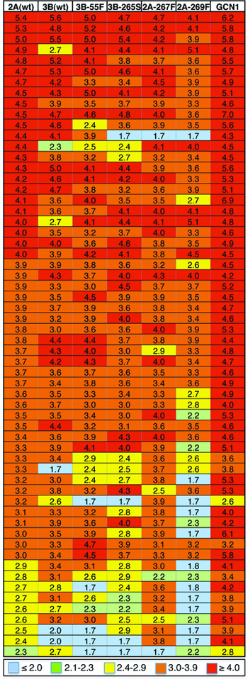
JCV-neutralization assays were used to screen a panel of sera from 96 healthy adult subjects. Sixty (63%) of the subjects neutralized a pseudovirus based on a urine-derived wt genotype 2A JCV with a reciprocal 50% neutralizing titer (EC50) of greater than 100 (Fig. 1, Table S1). This dilution was chosen as a seropositivity cutoff based on past evidence that serum dilutions of less than 1:100 can have non-specific neutralizing effects (perhaps due to the effects of serum factors on the cultured cells)(21). With respect to the current study, it is important to note that, in healthy subjects, IgG antibody concentrations in the CSF are typically 200–500 fold lower than in the serum (22, 23). Experiments with intravenously administered monoclonal antibody therapeutic agents indicate that serum IgG antibodies chronically leak across the human blood-brain barrier and accumulate in the central nervous system (CNS) at low levels (24). In this model, IgG antibodies in the CSF essentially represent a lower-concentration snapshot of antibodies found in the periphery. Thus, an individual with a serum EC50 neutralizing titer of 100 would be expected to have poorly neutralizing or non-neutralizing concentrations of antibodies in their CNS. Our seroprevalence results using a neutralizing titer cutoff of 100 are concordant with a large body prior seroprevalence studies using JCV-1A VP1 ELISA methods (reviewed in (2)).
Figure 1. JCV-neutralization serology (healthy adults).
Serum samples from 96 individual adult subjects (rows) were serially diluted and initially tested for neutralization of a wild-type JCV-2A pseudovirus. Sixty serum samples that detectably neutralized JCV-2A were tested against additional JCV pseudoviruses. Sera that failed to detectably neutralize JCV-2A were excluded from further analysis and are not shown in the figure. The inverse log10 of the calculated 50% neutralizing dilution (EC50) is indicated with a color code. EC50 values ≤2 (blue cells) are considered neutralization negative For the precise VP1 sequence identity of JCV pseudovirus, see Table S1.
Although a majority of samples that neutralized the 2A pseudovirus also neutralized all other tested JCV genotypes with similar titers, a minority of sera failed to detectably neutralize one or more PML-mutant pseudoviruses (Fig. 1). Eleven of the 60 serum samples that neutralized the 2A pseudovirus failed to neutralize the 2A-269F pseudovirus. Interestingly, the S269F mutation represents the most common variant observed in the CSF of PML patients (11, 12). The results are consistent with the idea that VP1 mutations found in the CSF of PML patients could confer a selective advantage to the virus by allowing escape from the apparently restricted spectrum of JCV-neutralizing antibodies observed in a minority of JCV-seropositive individuals.
Longitudinal neutralization analysis of PML patient plasma.
To investigate the idea that the unusual phenotype of having JCV-neutralization “blind spots” might be associated with an increased risk of developing PML under conditions of T cell immunodeficiency, we performed neutralization serology on a panel of plasma samples from PML patients. This work was confronted by two theoretical issues. First, PML patients might exhibit a narrow neutralization blind spot encompassing only the specific JCV VP1 sequence found in their CSF during PML. Second, it seems conceivable that neutralization blind spots might close during or after the development of PML. These considerations restricted our focus to patients for whom plasma samples had been collected and archived prior to the diagnosis of PML, and for whom the JCV sequences found in their CSF during PML were known. Six PML patients met these criteria. The underlying immunodeficiency in each of the six patients was HIV/AIDS, which was treated with combination antiretroviral therapy (Table S2).
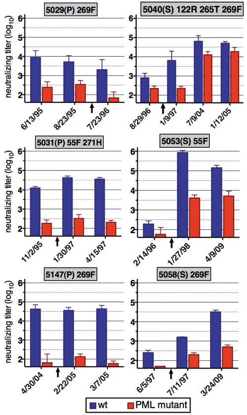
Pseudoviruses were constructed to represent the cognate mutant VP1 sequence found in individual patients’ CSF during PML (Table S1). Pseudoviruses representing inferred wt VP1 sequences were also produced. In some instances, the patient plasma samples were tested against near-cognate pseudoviruses. As shown in Fig. 2 and Fig. S4, all six PML patients exhibited little or no neutralization of their cognate PML-mutant pseudovirus at timepoints prior to PML diagnosis, even when there was robust neutralization of the wt pseudovirus. The results confirm the hypothesis that PML-specific VP1 mutations can allow the virus to escape from antibody-mediated neutralization.
Figure 2. PML patient neutralization serology.
Pre- and post-PML plasma samples from six patients were tested for neutralization of cognate (or near-cognate) wt and PML-mutant pseudoviruses. Specifically, patient 5029 was tested against viruses 5029w (cognate wt, blue) and 5029m (cognate mutant, red); 5031: 5031w/5031mb; 5040: 2A/5040m; 5053: 5053w/5053m; 5058: 2A/5147m; 5147: 5147w/5147m (see Table S1). Testing of PML patient sera against additional pseudoviruses is shown in Fig. S4. Patients whose disease progressed (left column) are indicated with (P), patients who survived (right column) are indicated with (S). Dominant PML-associated mutations observed in each patient’s CSF are indicated. Y-axes indicate neutralizing EC50. Error bars represent standard error of the mean for data from three independent experimental replicates, two of which were performed with blinding. Arrows below x-axes indicate the date of onset of PML symptoms. Date format is month/day/year.
Patients who survived PML eventually developed broader antibody responses capable of neutralizing their cognate PML-mutant pseudovirus. This suggests that at least some individuals with neutralization blind spots are ultimately capable of mounting broadly cross-neutralizing antibody responses. In contrast to patients who survived PML, the three patients with progressive (fatal) disease did not develop the ability to robustly neutralize their cognate mutant virus. Although this could simply reflect the recovery of effective cell-mediated immunity (which would, in turn, provide CD4+ T cell help for B cell responses), an important conclusion that can be drawn from the result is that individuals with JCV-neutralizing blind spots are not intrinsically incapable of mounting more broadly neutralizing antibody responses.
Remarkably, plasma from patients 5029 and 5058 robustly neutralized the 2A-269F pseudovirus at timepoints where there was poor neutralization of the patient-cognate mutant virus, which carries the S269F mutation in a slightly different genotypic background (Fig. S4 and Table S2). The results suggest that naturally occurring wt genotypic variations outside the PML mutation “hotspots” can influence neutralization-escape phenotypes. This is consistent with the observation that a few healthy subject sera that robustly neutralized the wt 2A pseudovirus showed very low titers against the wt 3B pseudovirus (Fig. 1). Taken together, the results illustrate the caveat that it is essential to analyze the neutralization of the exact VP1 sequence(s) observed in any given subject.
Evaluation of JCV VLPs as vaccine immunogens in a mouse model.
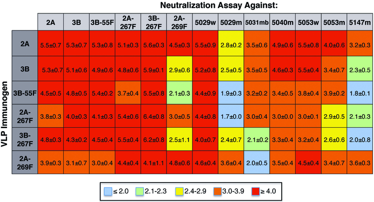
To test the idea that a VP1 VLP-based vaccine against JCV might elicit broadly neutralizing serum antibody responses, groups of mice were given a single intramuscular dose of 720 ng of a monovalent VLP preparation in alum. A single priming dose of VLP immunogen elicited high titer serum antibody responses capable of robustly neutralizing the cognate pseudovirus (Fig. 3). Interestingly, each set of primed mice failed to robustly cross-neutralize at least one non-cognate pseudovirus type. This result recapitulates the neutralization blind spot effects observed in human subjects.
Figure 3. Preclinical evaluation of a candidate JCV VLP vaccine.
Mice were given an intramuscular injection of VLPs based on the JCV genotype indicated in each row label. Four weeks later, sera from the mice were tested for neutralization of various pseudoviruses indicated in column labels. Numerical values represent EC50 neutralizing titers. Error represents standard deviation for independent neutralization assays of sera from five replicate mice. Pre-immune sera were non-neutralizing at a 1:100 dilution.
Mice were administered a booster dose of the same monovalent VLP preparation. Sera from all boosted animals cross-neutralized all tested JCV variants (Fig. S5). This shows that blind spots can be closed through prime-boost vaccination with a monovalent JCV VLP vaccine. Overall, the wt 2A VLPs elicited the most uniformly robust cross-neutralizing responses, suggesting that it is unnecessary (and perhaps undesirable) to use PML-mutant VLP immunogens to elicit antibodies capable of neutralizing PML-mutant pseudoviruses (25).
Case study of a PML patient administered an experimental JCV vaccine.
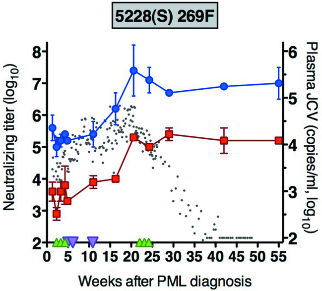
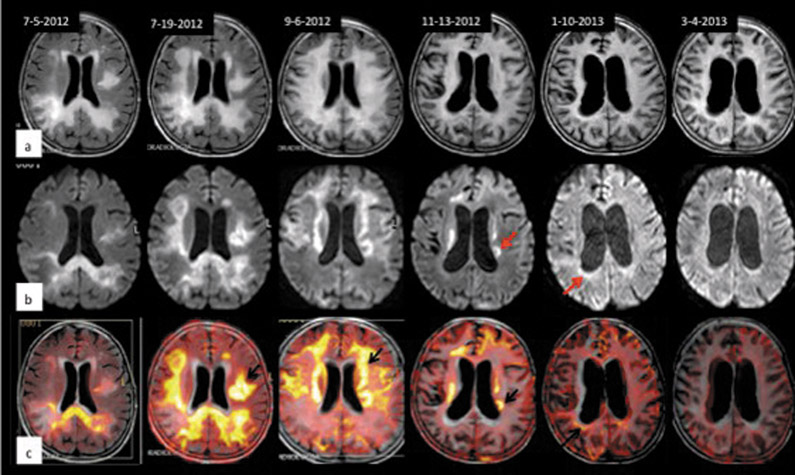
PML patient 5228 is a 75 year-old female with idiopathic CD4 lymphopenia who was admitted at the Department of Infectious Diseases of San Raffaele Hospital, Milan, Italy upon diagnosis with PML on 5/24/2012. Her case has not previously been reported. The patient’s clinical condition deteriorated rapidly after admission and she became comatose. In addition to mefloquine and mirtazapine, the patient was treated with a previously reported vaccination protocol consisting of separate administrations of interleukin-7 (IL-7) and JCV-1A VLPs combined with imiquimod (26). As shown in Fig. 4, vaccination was followed by a roughly 100-fold increase in the patient’s neutralizing titer against her cognate mutant virus. This confirms that PML patients are not intrinsically incapable of mounting new antibody responses capable of neutralizing their cognate JCV strain. After vaccination, patient 5228 showed an extraordinarily high peak titer (25 million) against her inferred wt JCV. This is particularly remarkable in the sense that the patient was suffering from intermittent lymphopenia at the time of vaccination (Fig. S6). Although it is tempting to speculate that the JCV VLP component of the treatment was a primary factor in induction of a potent humoral immune response, we note that a prior case study of a PML patient administered recombinant IL-7 alone (without JCV VLPs) also showed increasing anti-JCV antibody titers, including a pronounced IgM response (27). This suggests that improved CD4 T cell function and “auto-inoculation” with PML lesion-derived virions could have played a significant role in the response. Increases in patient 5228’s JCV-neutralizing titer preceded a gradual fall in JCV viremia (Fig. 4) and an arrest of PML lesion progression (Fig. 5). It is uncertain whether there was a causal relationship between the improved JCV-neutralizing antibody response and the resolution of disease progression.
Figure 4. JCV-neutralizing titers following vaccination of PML patient 5228.
Patient 5228 was administered JCV VLPs subcutaneously at three time points (downward purple triangles). Recombinant IL-7 was also administered for two cycles each consisting of a subcutaneous weekly dose of 10 µg/kg (upward green triangles). The patient’s neutralizing titer against her cognate PML-mutant JCV (red squares) or inferred wt JCV (blue circles) was monitored over time. JCV load in the patient’s plasma (gray dots) was also monitored over time. See Supplementary Materials for additional patient details.
Figure 5. Evolution of PML lesions by magnetic resonance imaging (MRI) in patient 5228.
The patient was initially diagnosed with PML on 5/24/2012. The date of each scan is indicated along the top of the figure. Axial fluid-attenuated inversion recovery (FLAIR, Row a), diffusion-weighted imaging (DWI, Row b) and FLAIR-DWI merged images (Row c) show the evolution of PML lesions. Row a: the first image on the left (July 2012) shows a high signal intensity lesion of the right parieto-occipital white matter extending contralaterally through the corpus callosum; another focus is present in the left subcortical temporal region. Subsequent examinations showed evolution of the signal alteration with progressive rapid extension of the lesions to the entire white matter bilaterally. In September 2012, the white matter was completely occupied by lesional and atrophic processes and enlargements of the ventricles and cortical sulci began to appear. The last examinations (January and March 2013) show progression of atrophy. Row b: axial DWI images show the evolution of the hyperintense signal alterations corresponding to the front of progression of PML lesions (red arrows). The signal alteration is substantially reduced starting from November 2012 and is no longer visible at the March 2013 examination, implying lesion stabilization. Row c: merged FLAIR and DWI sequences show, in yellow (black arrows), the initial fronts of advance of PML lesions and their regression over time.
Discussion
Our results indicate that a minority of healthy JCV-seropositive subjects are deficient in serum antibodies capable of neutralizing JCVs carrying VP1 mutations associated with PML. A simple explanation for this finding is that some individuals may have an unusually low diversity of plasma cells secreting effectively neutralizing antibodies, such that single point mutations in VP1 can allow the virus to evade neutralization. This finding is consistent with past observations in murine viral challenge systems (28) and we have recently reported a similar scenario for BKV (a close relative of JCV)(29). Neutralization-escape mutations would presumably increase the fitness of the virus under circumstances where T cell-mediated immunity in the CNS is impaired and antibody-mediated neutralization therefore serves as a last line of defense against neuropathic JCV replication. The idea that humoral vulnerability is a key element of PML pathogenesis is supported by the observation that all six PML patients we studied displayed neutralization blind spots prior to disease onset.
At present, there are no antiviral agents known to be effective for treatment of JCV disease, and reversal of immune dysfunction remains the only approach of proven utility for treatment of PML. Immune reconstitution treatments are not always timely or successful and, even if they are successful, sometimes lead to immune reconstitution inflammatory syndrome, which can be lethal (30). VLP-based vaccines against other viral families have been highly successful in humans (31, 32). In particular, VLP-based vaccines against human papillomaviruses (which share key structural features with polyomaviruses) elicit remarkably potent, diverse, and long-lasting neutralizing antibody responses (reviewed in (20)). Our testing of experimental JCV VLP immunogens demonstrates that high-titer serum antibody responses capable of broadly cross-neutralizing PML-mutant JCVs can be elicited by prime-boost vaccination with VLPs representing a single wt JCV genotype. Since antibodies are present at much lower levels in the CNS (23, 24), the apparently high immunogenic potency of JCV VLP immunogens could be important for induction of protective levels of JCV-neutralizing antibodies in the brain parenchyma. Taken together, the results suggest that a prophylactic JCV VLP vaccine could serve to boost and broaden humoral immunity against JCV and thus protect at-risk individuals against the development of PML.
Although the widespread availability of combination antiretroviral therapy has led to a dramatic decrease in the incidence of AIDS, treatment failure and/or lack of compliance with antiretroviral dosing schedules remain long-term risks for HIV-infected individuals. The availability of a preventive vaccine against PML could thus be of potential benefit for people living with HIV.
At present, treatment of multiple sclerosis patients with the highly effective monoclonal antibody (mAb) therapeutic natalizumab is generally time-limited due to the progressively increasing risk of PML side effects (2). The utility of the drug rituximab for treating rheumatoid arthritis is likewise limited by PML side effects. Efalizumab, an immunomodulatory mAb previously used for the treatment of psoriasis, was withdrawn from market due to PML side effects. Perhaps a dozen other immunomodulatory agents that are either in development or on the market are potentially associated with rare PML side effects. Thus, the availability of safe and effective measures for preventing PML could significantly increase the overall safety profiles of therapies for a wide variety of autoimmune diseases and lymphoid cancers. This would be particularly true for immunomodulatory therapies, such as natalizumab, that are compatible with vaccination (44).
A limitation of our study is the availability of only a small population of PML patients with appropriate longitudinal samples. Thus, it remains unclear whether neutralizing blind spots are a universal feature of PML development or whether, in some patients, the disease develops despite the presence of an effective humoral response. Likewise, although the single case study of patient 5228 is consistent with the possibility that IL-7/JCV VLP administration was a factor in arrest of PML progression, the fact that other patients, including another patient administered IL-7 without VLPs (27, 45), sometimes spontaneously recover leaves the question of whether JCV VLPs would be a useful therapeutic intervention for treatment of PML unclear.
An additional limitation of the study is that we did not have access to paired CSF samples for healthy subjects and only sporadic CSF samples from some of the PML patients. In the future, it will be important to test the prediction that serum JCV-neutralizing antibody responses essentially represent a 200–500 fold higher titer than would typically be observed in CSF (22–24). It will be important to address this question using passive transfer of antibodies (or bone marrow plasma cells) in the new murine PML model reported by Kondo and colleagues (15). Such experiments could test the prediction that broadly cross-neutralizing antibodies could protect mice against PML when administered at high doses, while lower doses of “blind spot” polyclonal antibodies (for example, from mice given a single priming dose of JCV-3B VLPs) might drive the evolution of the common L55F and S269F escape mutations.
Materials and Methods
Study design.
A set of 96 previously characterized (21) healthy human sera were screened for neutralization of wild-type JCV 2A pseudovirus. The JCV 2A- neutralizing sera were then tested against diverse range of JCV variants. Testing of healthy subject sera did not employ blinding. Serum samples from six PML patients for whom CSF JCV sequences were known and archived serum samples were available were initially tested against the indicated cognate pseudoviruses without blinding. Samples from the six PML patients were then randomized and re-tested in two independent additional experiments in a blinded fashion. Neutralization serology for patient 5228 (who was vaccinated with JCV VLPs) was likewise initially performed without blinding followed by two blinded repeats. Experiments with mice arbitrarily used five animals per group. Serological analysis of mice was performed without blinding.
Pseudovirus production.
Pseudovirions were produced using previously described methods, with minor modifications (21, 46). Briefly, Gaussia luciferase reporter genes were packaged into pseudovirions in 293TT cells transiently transfected with JCV VP1/2/3 expression plasmids. The resulting pseudovirions were purified over Optiprep gradients. Additional methodological details are presented in the Supplementary Materials.
Sera.
Healthy subject sera were purchased from Equitech Bio, Inc. and Innovative Research, Inc (21, 29, 47). Ethical assurances are provided on the suppliers’ websites. Samples from PML patients were either collected for diagnostic purposes or as part of internal research protocols approved by the Ethical Committee of San Raffaele Hospital, Milan. Written informed consent to use of samples and clinical information was given by patients who were still alive at the time of the start of the study. Permission from Ethical Committee was given for the use of samples and clinical information from patients who were deceased.
Serum and plasma samples were heat-inactivated at 56°C for 30 minutes, followed by brief centrifugation to sediment any aggregated material. For samples from healthy adults, serum IgG antibodies were then purified out of the serum samples using Melon Gel (Pierce) resin according to manufacturer’s instructions. Plasma samples from PML patients and mice were not subjected to Melon Gel purification.
Neutralization assays.
Generation and use of ART cells (an ovarian cancer line (48, 49)) and SFT cells (a gliosarcoma line (50)) for neutralization assays has been described previously (29). In initial validation experiments, comparable JCV-neutralization results were observed using either cell line (Fig. S3) (50). Neutralization serology studies used ART cells with the dose of VP1 specified in Table S1.
Preclinical VLP vaccine.
Animal experiments were performed at NCI facilities under the approval of the Animal Care and Use Committee and according to the guidelines of the Association for Assessment and Accreditation of Laboratory Animal Care International. Procedures were carried out in accordance with the eighth edition of the National Research Council of the National Academies’ Guide for the Care and Use of Laboratory Animals. Female BALB/cAnNCr mice were subjected to intramuscular immunization with 720 ng of JCV VLPs (VP1 only) mixed with 0.2% of aluminium hydroxide (alum, InvivoGen) with a total volume of 50 µl. Five mice were immunized per JCV VLP type. One month after the single priming dose of VLPs, plasma samples were collected by sub-mandibular bleed into Microtainer lithium-heparin tubes (BD). The mice were then boosted with the same JCV VLP type intramuscularly in alum. Serum samples were collected one month after the booster dose.
Vaccination case study.
Ethical approval for compassionate administration of the IL-7/VLP combination treatment was provided by the Ethical Committee of San Raffaele Hospital. The family of patient 5228 gave written informed consent for the collection and use of research samples and for the experimental administration of the IL-7 / JCV VLP vaccine. Recombinant human IL-7 was given subcutaneously at 10 µg/kg on 6/9/, 6/16, 6/23 (first cycle), and on 10/25, 11/2, 11/9/2012 (second cycle). Subcutaneous injections of 1 mg of VLPs composed of JCV-1A VP1 were performed on 6/28, 7/6, and 8/8/2012. The VLP preparation was the same as the one previously administered to two other patients whose cases were described in a recent report by Sospedra and colleagues (26). Imiquimod cream (5%, Aldara, MEDA Pharm, Germany) was applied as a vaccine adjuvant topically at the injection site. The treatments appeared to be well tolerated. Additional clinical details for patient 5228 are provided in Supplementary Materials.
Statistical analyses:
The neutralization titers were calculated in Prism (Graphpad) using non-linear regression analyses. A sigmoidal dose-response equation (variable slope) (four parameter logistic equation) was used with top and bottom values constrained based on “no antibody” and “no virus” controls, respectively.
Supplementary Material
List of Supplementary Materials
Pseudovirus Production
Establishment of a pseudovirus-based JCV neutralization assay
Sera
Patient 5228 clinical details
Fig. S1. Transducibility of various cell lines
Fig. S2. An example of luminometry results for a pilot JCV neutralization assay using ART cells
Fig. S3. Neutralization assay validation
Fig. S4. PML patient neutralization serology (an expansion of Fig. 2)
Fig. S5. Serological analysis of mice after a booster dose of JCV VLPs
Fig. S6. JCV neutralization titers following vaccination of PML patient 5228
Table S1. Characteristics of JCV pseudovirus stocks
Table S2. Patient characteristics
Table S3. PML patient neutralization serology (source data)
Table S4. Neutralization serology of patient 5228 (source data)
Table S5. Viremia of patient 5228 (source data)
Acknowledgements
The authors thank Leonid Gorelik for critical evaluation of the manuscript.
Funding: The work was funded in part by the Intramural Research Program of the NIH with support from the Center for Cancer Research and by ISS grant 40H.83 (AIDS Research Program, Italian Ministry of Health).
Footnotes
Competing interests: Authors C.B.B.; UR and D.V.P. are coinventors on U.S. patent application (61/919,043). RM and SS are coinventors on European application – Appl. No./Patent No. 11006031.6–2406, which has been licensed by the University Zurich to Neuway GmbH, Bonn, Germany. All other authors declare no conflict of interest.
Consulting statistitian: none
References
- 1.Cinque P, Koralnik IJ, Gerevini S, Miro JM, Price RW, Progressive multifocal leukoencephalopathy in HIV-1 infection. The Lancet infectious diseases 9, 625–636 (2009). [DOI] [PMC free article] [PubMed] [Google Scholar]
- 2.Hirsch HH, Kardas P, Kranz D, Leboeuf C, The human JC polyomavirus (JCPyV): virological background and clinical implications. APMIS : acta pathologica, microbiologica, et immunologica Scandinavica 121, 685–727 (2013). [DOI] [PubMed] [Google Scholar]
- 3.Carrithers MD, Update on disease-modifying treatments for multiple sclerosis. Clinical therapeutics 36, 1938–1945 (2014). [DOI] [PubMed] [Google Scholar]
- 4.Bozic C, Richman S, Plavina T, Natarajan A, Scanlon JV, Subramanyam M, Sandrock A, Bloomgren G, Anti-John Cunnigham virus antibody prevalence in multiple sclerosis patients: baseline results of STRATIFY-1. Ann Neurol 70, 742–750 (2011). [DOI] [PubMed] [Google Scholar]
- 5.Lee P, Plavina T, Castro A, Berman M, Jaiswal D, Rivas S, Schlain B, Subramanyam M, A second-generation ELISA (STRATIFY JCV DxSelect) for detection of JC virus antibodies in human serum and plasma to support progressive multifocal leukoencephalopathy risk stratification. J Clin Virol 57, 141–146 (2013). [DOI] [PubMed] [Google Scholar]
- 6.Bloomgren G, Richman S, Hotermans C, Subramanyam M, Goelz S, Natarajan A, Lee S, Plavina T, Scanlon JV, Sandrock A, Bozic C, Risk of natalizumab-associated progressive multifocal leukoencephalopathy. The New England journal of medicine 366, 1870–1880 (2012). [DOI] [PubMed] [Google Scholar]
- 7.Keller DM, JCV Test Stratifies PML Risk in Antibody-Positive MS. http://www.medscape.com/viewarticle/806294.
- 8.Diotti RA, Mancini N, Clementi N, Sautto G, Moreno GJ, Criscuolo E, Cappelletti F, Man P, Forest E, Remy L, Giannecchini S, Clementi M, Burioni R, Cloning of the first human anti-JCPyV/VP1 neutralizing monoclonal antibody: epitope definition and implications in risk stratification of patients under natalizumab therapy. Antiviral Res 108, 94–103 (2014). [DOI] [PubMed] [Google Scholar]
- 9.Dang X, Koralnik IJ, A granule cell neuron-associated JC virus variant has a unique deletion in the VP1 gene. J Gen Virol 87, 2533–2537 (2006). [DOI] [PubMed] [Google Scholar]
- 10.Sunyaev SR, Lugovskoy A, Simon K, Gorelik L, Adaptive mutations in the JC virus protein capsid are associated with progressive multifocal leukoencephalopathy (PML). PLoS Genet 5, e1000368 (2009). [DOI] [PMC free article] [PubMed] [Google Scholar]
- 11.Gorelik L, Reid C, Testa M, Brickelmaier M, Bossolasco S, Pazzi A, Bestetti A, Carmillo P, Wilson E, McAuliffe M, Tonkin C, Carulli JP, Lugovskoy A, Lazzarin A, Sunyaev S, Simon K, Cinque P, Progressive multifocal leukoencephalopathy (PML) development is associated with mutations in JC virus capsid protein VP1 that change its receptor specificity. The Journal of infectious diseases 204, 103–114 (2011). [DOI] [PMC free article] [PubMed] [Google Scholar]
- 12.Reid CE, Li H, Sur G, Carmillo P, Bushnell S, Tizard R, McAuliffe M, Tonkin C, Simon K, Goelz S, Cinque P, Gorelik L, Carulli JP, Sequencing and analysis of JC virus DNA from natalizumab-treated PML patients. The Journal of infectious diseases 204, 237–244 (2011). [DOI] [PMC free article] [PubMed] [Google Scholar]
- 13.Dang X, Vidal JE, Oliveira AC, Simpson DM, Morgello S, Hecht JH, Ngo LH, Koralnik IJ, JC virus granule cell neuronopathy is associated with VP1 C terminus mutants. J Gen Virol 93, 175–183 (2012). [DOI] [PMC free article] [PubMed] [Google Scholar]
- 14.Maginnis MS, Stroh LJ, Gee GV, O’Hara BA, Derdowski A, Stehle T, Atwood WJ, Progressive multifocal leukoencephalopathy-associated mutations in the JC polyomavirus capsid disrupt lactoseries tetrasaccharide c binding. mBio 4, e00247–00213 (2013). [DOI] [PMC free article] [PubMed] [Google Scholar]
- 15.Kondo Y, Windrem MS, Zou L, Chandler-Militello D, Schanz SJ, Auvergne RM, Betstadt SJ, Harrington AR, Johnson M, Kazarov A, Gorelik L, Goldman SA, Human glial chimeric mice reveal astrocytic dependence of JC virus infection. The Journal of clinical investigation 124, 5323–5336 (2014). [DOI] [PMC free article] [PubMed] [Google Scholar]
- 16.Haley SA, Atwood WJ, An animal model for progressive multifocal leukoencephalopathy. The Journal of clinical investigation 124, 5103–5106 (2014). [DOI] [PMC free article] [PubMed] [Google Scholar]
- 17.O’Hara SD, Stehle T, Garcea R, Glycan receptors of the Polyomaviridae: structure, function, and pathogenesis. Current opinion in virology 7, 73–78 (2014). [DOI] [PubMed] [Google Scholar]
- 18.Kemp TJ, Safaeian M, Hildesheim A, Pan Y, Penrose KJ, Porras C, Schiller JT, Lowy DR, Herrero R, Pinto LA, Kinetic and HPV infection effects on cross-type neutralizing antibody and avidity responses induced by Cervarix. Vaccine 31, 165–170 (2012). [DOI] [PMC free article] [PubMed] [Google Scholar]
- 19.Draper E, Bissett SL, Howell-Jones R, Waight P, Soldan K, Jit M, Andrews N, Miller E, Beddows S, A randomized, observer-blinded immunogenicity trial of Cervarix and Gardasil Human Papillomavirus vaccines in 12–15 year old girls. PloS one 8, e61825 (2013). [DOI] [PMC free article] [PubMed] [Google Scholar]
- 20.Schiller JT, Lowy DR, Raising Expectations For Subunit Vaccine. The Journal of infectious diseases 211, 1373–1375 (2015). [DOI] [PMC free article] [PubMed] [Google Scholar]
- 21.Pastrana DV, Tolstov YL, Becker JC, Moore PS, Chang Y, Buck CB, Quantitation of human seroresponsiveness to Merkel cell polyomavirus. PLoS pathogens 5, e1000578 (2009). [DOI] [PMC free article] [PubMed] [Google Scholar]
- 22.Tibbling G, Link H, Ohman S, Principles of albumin and IgG analyses in neurological disorders. I. Establishment of reference values. Scandinavian journal of clinical and laboratory investigation 37, 385–390 (1977). [DOI] [PubMed] [Google Scholar]
- 23.Ahlgren C, Oden A, Haghighi S, Andersen O, Bergstrom T, Lycke J, The effect of live, attenuated measles vaccine and measles infection on measles antibody levels in serum and CSF of patients with multiple sclerosis or clinically isolated syndrome. J Neuroimmunol 235, 98–103 (2011). [DOI] [PubMed] [Google Scholar]
- 24.Yu YJ, Watts RJ, Developing therapeutic antibodies for neurodegenerative disease. Neurotherapeutics : the journal of the American Society for Experimental NeuroTherapeutics 10, 459–472 (2013). [DOI] [PMC free article] [PubMed] [Google Scholar]
- 25.Gorelik L, Lugovskoy A, Methods for the detection of JC polyoma virus. Patent application WO/2010/090757. (2010).
- 26.Sospedra M, Schippling S, Yousef S, Jelcic I, Bofill-Mas S, Planas R, Stellmann JP, Demina V, Cinque P, Garcea R, Croughs T, Girones R, Martin R, Treating Progressive Multifocal Leukoencephalopathy With Interleukin 7 and Vaccination With JC Virus Capsid Protein VP1. Clin Infect Dis 59, 1588–1592 (2014). [DOI] [PMC free article] [PubMed] [Google Scholar]
- 27.Alstadhaug KB, Croughs T, Henriksen S, Leboeuf C, Sereti I, Hirsch HH, Rinaldo CH, Treatment of Progressive Multifocal Leukoencephalopathy With Interleukin 7. JAMA neurology 71, 1030–1035 (2014). [DOI] [PubMed] [Google Scholar]
- 28.Purtha WE, Tedder TF, Johnson S, Bhattacharya D, Diamond MS, Memory B cells, but not long-lived plasma cells, possess antigen specificities for viral escape mutants. J Exp Med 208, 2599–2606 (2011). [DOI] [PMC free article] [PubMed] [Google Scholar]
- 29.Pastrana DV, Ray U, Magaldi TG, Schowalter RM, Çuburu N, Buck CB, BK Polyomavirus Genotypes Represent Distinct Serotypes with Distinct Entry Tropisms. J Virol 87, 10105–10113 (2013). [DOI] [PMC free article] [PubMed] [Google Scholar]
- 30.Fox R, Advances in the management of PML: focus on natalizumab. Cleveland Clinic journal of medicine 78 Suppl 2, S33–37 (2011). [DOI] [PubMed] [Google Scholar]
- 31.Zhao Q, Li S, Yu H, Xia N, Modis Y, Virus-like particle-based human vaccines: quality assessment based on structural and functional properties. Trends in biotechnology 31, 654–663 (2013). [DOI] [PubMed] [Google Scholar]
- 32.Kreimer AR, Sherman ME, Sahasrabuddhe VV, Safaeian M, The case for conducting a randomized clinical trial to assess the efficacy of a single dose of prophylactic HPV vaccines among adolescents. Journal of the National Cancer Institute 107, (2015). [DOI] [PMC free article] [PubMed] [Google Scholar]
- 33.Kelleher MB, Galutira D, Duggan TD, Nuovo GJ, Progressive multifocal leukoencephalopathy in a patient with Alzheimer’s disease. Diagnostic molecular pathology : the American journal of surgical pathology, part B 3, 105–113 (1994). [DOI] [PubMed] [Google Scholar]
- 34.Gheuens S, Wuthrich C, Koralnik IJ, Progressive multifocal leukoencephalopathy: why gray and white matter. Annual review of pathology 8, 189–215 (2013). [DOI] [PubMed] [Google Scholar]
- 35.Agnihotri SP, Wuthrich C, Dang X, Nauen D, Karimi R, Viscidi R, Bord E, Batson S, Troncoso J, Koralnik IJ, A fatal case of JC virus meningitis presenting with hydrocephalus in a human immunodeficiency virus-seronegative patient. Ann Neurol, (2014). [DOI] [PMC free article] [PubMed] [Google Scholar]
- 36.Mori M, Kurata H, Tajima M, Shimada H, JC virus detection by in situ hybridization in brain tissue from elderly patients. Ann Neurol 29, 428–432 (1991). [DOI] [PubMed] [Google Scholar]
- 37.White FA 3rd, Ishaq M, Stoner GL, Frisque RJ, JC virus DNA is present in many human brain samples from patients without progressive multifocal leukoencephalopathy. J Virol 66, 5726–5734 (1992). [DOI] [PMC free article] [PubMed] [Google Scholar]
- 38.Elsner C, Dorries K, Evidence of human polyomavirus BK and JC infection in normal brain tissue. Virology 191, 72–80 (1992). [DOI] [PubMed] [Google Scholar]
- 39.Bogdanovic G, Hammarin AL, Grandien M, Winblad B, Bergenheim AT, Nennesmo I, Dalianis T, No association of JC virus with Alzheimer’s disease or astrocytomas. Clin Diagn Virol 4, 223–230 (1995). [DOI] [PubMed] [Google Scholar]
- 40.Perez-Liz G, Del Valle L, Gentilella A, Croul S, Khalili K, Detection of JC virus DNA fragments but not proteins in normal brain tissue. Ann Neurol 64, 379–387 (2008). [DOI] [PMC free article] [PubMed] [Google Scholar]
- 41.Bayliss J, Karasoulos T, McLean CA, Frequency and large T (LT) sequence of JC polyomavirus DNA in oligodendrocytes, astrocytes and granular cells in non-PML brain. Brain pathology 22, 329–336 (2012). [DOI] [PMC free article] [PubMed] [Google Scholar]
- 42.Kitamura T, Aso Y, Kuniyoshi N, Hara K, Yogo Y, High incidence of urinary JC virus excretion in nonimmunosuppressed older patients. The Journal of infectious diseases 161, 1128–1133 (1990). [DOI] [PubMed] [Google Scholar]
- 43.Katona S, JCV vaccination could reduce the risk of developing cognitive decline, dementia, strokes and brain tumours, by preventing chronic JCV cerebral infection, and recurrent reactivation. Medical hypotheses 73, 268–269 (2009). [DOI] [PubMed] [Google Scholar]
- 44.Kaufman M, Pardo G, Rossman H, Sweetser MT, Forrestal F, Duda P, Natalizumab treatment shows no clinically meaningful effects on immunization responses in patients with relapsing-remitting multiple sclerosis. Journal of the neurological sciences 341, 22–27 (2014). [DOI] [PubMed] [Google Scholar]
- 45.Patel A, Patel J, Ikwuagwu J, A case of progressive multifocal leukoencephalopathy and idiopathic CD4+ lymphocytopenia. The Journal of antimicrobial chemotherapy 65, 2697–2698 (2010). [DOI] [PubMed] [Google Scholar]
- 46.Buck CB, Pastrana DV, Lowy DR, Schiller JT, Efficient intracellular assembly of papillomaviral vectors. J Virol 78, 751–757 (2004). [DOI] [PMC free article] [PubMed] [Google Scholar]
- 47.Pastrana DV, Brennan DC, Çuburu N, Storch GA, Viscidi RP, Randhawa PS, Buck CB, Neutralization Serotyping of BK Polyomavirus Infection in Kidney Transplant Recipients. PLoS pathogens 8, e1002650 (2012). [DOI] [PMC free article] [PubMed] [Google Scholar]
- 48.Roschke AV, Tonon G, Gehlhaus KS, McTyre N, Bussey KJ, Lababidi S, Scudiero DA, Weinstein JN, Kirsch IR, Karyotypic complexity of the NCI-60 drug-screening panel. Cancer Res 63, 8634–8647 (2003). [PubMed] [Google Scholar]
- 49.Garraway LA, Widlund HR, Rubin MA, Getz G, Berger AJ, Ramaswamy S, Beroukhim R, Milner DA, Granter SR, Du J, Lee C, Wagner SN, Li C, Golub TR, Rimm DL, Meyerson ML, Fisher DE, Sellers WR, Integrative genomic analyses identify MITF as a lineage survival oncogene amplified in malignant melanoma. Nature 436, 117–122 (2005). [DOI] [PubMed] [Google Scholar]
- 50.Rutka JT, Giblin JR, Hoifodt HK, Dougherty DV, Bell CW, McCulloch JR, Davis RL, Wilson CB, Rosenblum ML, Establishment and characterization of a cell line from a human gliosarcoma. Cancer Res 46, 5893–5902 (1986). [PubMed] [Google Scholar]
- 51.Agostini HT, Deckhut A, Jobes DV, Girones R, Schlunck G, Prost MG, Frias C, Perez-Trallero E, Ryschkewitsch CF, Stoner GL, Genotypes of JC virus in East, Central and Southwest Europe. J Gen Virol 82, 1221–1331 (2001). [DOI] [PubMed] [Google Scholar]
- 52.Pastrana DV, Buck CB, Pang YY, Thompson CD, Castle PE, FitzGerald PC, Kruger Kjaer S, Lowy DR, Schiller JT, Reactivity of human sera in a sensitive, high-throughput pseudovirus-based papillomavirus neutralization assay for HPV16 and HPV18. Virology 321, 205–216 (2004). [DOI] [PubMed] [Google Scholar]
- 53.Schowalter RM, Reinhold WC, Buck CB, Entry tropism of BK and Merkel Cell Polyomaviruses in cell culture. PloS one 7, e42181 (2012). [DOI] [PMC free article] [PubMed] [Google Scholar]
- 54.Shaw G, Morse S, Ararat M, Graham FL, Preferential transformation of human neuronal cells by human adenoviruses and the origin of HEK 293 cells. FASEB J 16, 869–871 (2002). [DOI] [PubMed] [Google Scholar]
- 55.Henriksen S, Tylden GD, Dumoulin A, Sharma BN, Hirsch HH, Rinaldo CH, The human fetal glial cell line SVG p12 contains infectious BK polyomavirus (BKPyV). J Virol, (2014). [DOI] [PMC free article] [PubMed] [Google Scholar]
- 56.Lehmann HC, Chen W, Mi R, Wang S, Liu Y, Rao M, Hoke A, Human Schwann cells retain essential phenotype characteristics after immortalization. Stem cells and development 21, 423–431 (2012). [DOI] [PMC free article] [PubMed] [Google Scholar]
- 57.Gross JL, Behrens DL, Mullins DE, Kornblith PL, Dexter DL, Plasminogen activator and inhibitor activity in human glioma cells and modulation by sodium butyrate. Cancer Res 48, 291–296 (1988). [PubMed] [Google Scholar]
- 58.Pierson TC, Sanchez MD, Puffer BA, Ahmed AA, Geiss BJ, Valentine LE, Altamura LA, Diamond MS, Doms RW, A rapid and quantitative assay for measuring antibody-mediated neutralization of West Nile virus infection. Virology 346, 53–65 (2006). [DOI] [PubMed] [Google Scholar]
- 59.Bossolasco S, Calori G, Moretti F, Boschini A, Bertelli D, Mena M, Gerevini S, Bestetti A, Pedale R, Sala S, Sala S, Lazzarin A, Cinque P, Prognostic significance of JC virus DNA levels in cerebrospinal fluid of patients with HIV-associated progressive multifocal leukoencephalopathy. Clin Infect Dis 40, 738–744 (2005). [DOI] [PubMed] [Google Scholar]
Associated Data
This section collects any data citations, data availability statements, or supplementary materials included in this article.
Supplementary Materials
List of Supplementary Materials
Pseudovirus Production
Establishment of a pseudovirus-based JCV neutralization assay
Sera
Patient 5228 clinical details
Fig. S1. Transducibility of various cell lines
Fig. S2. An example of luminometry results for a pilot JCV neutralization assay using ART cells
Fig. S3. Neutralization assay validation
Fig. S4. PML patient neutralization serology (an expansion of Fig. 2)
Fig. S5. Serological analysis of mice after a booster dose of JCV VLPs
Fig. S6. JCV neutralization titers following vaccination of PML patient 5228
Table S1. Characteristics of JCV pseudovirus stocks
Table S2. Patient characteristics
Table S3. PML patient neutralization serology (source data)
Table S4. Neutralization serology of patient 5228 (source data)
Table S5. Viremia of patient 5228 (source data)


