Otitis externa is an inflammatory disease of the external ear canal, including the ear pinna. Otitis externa may be acute or chronic (persistent or recurrent otitis lasting for 3 months or longer). Changes that occur in the external ear canal in response to chronic inflammation may include glandular hyperplasia, glandular dilation, epithelial hyperplasia, and hyperkeratosis (1). These changes usually result in increased cerumen production along the external ear canal, which contributes to increase in local humidity and pH of the external ear canal, thus predisposing the ear to secondary infection.
The bacteria most commonly isolated from ear canals of dogs affected by otitis are Staphylococcus spp. (2). Other bacteria commonly associated with otitis include Pseudomonas, Proteus, Enterococcus, Streptococcus, and Corynebacterium. Some bacteria such as Staphylococcus and Pseudomonas may produce biofilm, which can lead to persistence of infection despite adequate therapy, as the biofilm needs to be disrupted for any antimicrobial therapy to be effective in clearing the infection. Malassezia yeast is another common component of otitis externa in dogs. Some dogs appear to develop an allergic response to Malassezia spp., leading to significant discomfort and pruritus.
Acute and uncomplicated otitis externa can often be treated successfully, but chronic or recurrent otitis externa is more challenging. Typically, underlying primary factors as well as predisposing and perpetuating factors are at play, including secondary otic infection. Repeated bouts of inflammation and infection can cause secondary changes in the ear canal that can ultimately lead to further lack of success in treating otitis, and possible end-stage ear disease. Severe glandular changes, fibrosis, stenosis, and calcification along the external ear canal lead to patient discomfort as well as progression of otitis from acute to chronic, and from straightforward to complicated otic disease. These changes are indicative of end-stage ear disease, that can usually be avoided with appropriate therapy for secondary and primary disease early in its progression. Client education and regular follow-up evaluations are key to prevention of end-stage ear disease.
Clinical diagnosis and etiological factors
Otitis externa is common in dogs, and may be unilateral or bilateral. Evaluation for otitis and its diagnosis is based on ear canal palpation, visual inspection of ears, including otoscopic examination, and cytological analysis of otic contents.
Changes to the ear pinna may include alopecia, excoriation, crusting, erythema, and hyperpigmentation. The external ear canal may exhibit presence of hyperemia, ulceration, ceruminous or suppurative discharge, masses, stenosis, glandular changes, or foreign bodies. Usually more than one abnormal finding is noted within an affected ear. Evaluation of the tympanic membrane forms a key part of the otoscopic evaluation, though it may be difficult to assess the tympanum when otitis externa is present. It is reasonable to leave assessment of the tympanic membrane to a later date, after changes attributed to active otitis have been corrected.
Cytological evaluation of otic contents is the single most informative diagnostic test that helps with treatment of otitis. Otic cytological evaluations also help monitor response to therapy. Occasionally, bacterial culture sampling from the horizontal ear canal may be used to help determine treatment options and for selection of systemic antibiotic therapy, if indicated. Imaging studies such as radiographs, computed tomography (CT) scan or magnetic resonance imaging (MRI) are not routinely used but can be helpful in cases of chronic otitis, or when otitis media is of concern.
It is vital that the clinician evaluate involvement of various primary, predisposing, and perpetuating factors that may be contributing to ear disease while evaluating each individual patient affected by otitis externa. If all or most of these factors are identified, resolution of current otitis and prevention of future otitis episodes are likely.
Primary factors
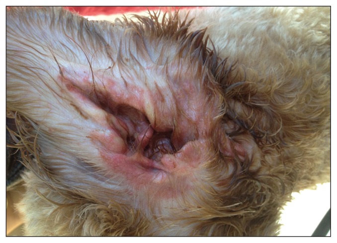
Primary factors are diseases that have a direct effect on the external ear canal and can cause otitis, including otic parasites such as Otodectes cyanotis, hypersensitivity disease [food allergy, atopic dermatitis, contact hypersensitivity (Figure 1)], endocrine disease such as hypothyroidism, otic neoplasia and foreign bodies. Underlying hypersensitivity disease is the most common primary factor leading to otitis in dogs (3).
Figure 1.
Contact hypersensitivity reaction to topical otic medication during treatment for otitis externa.
Predisposing factors
Predisposing factors are factors that alter the local ear canal environment and create an increased risk for development of otitis externa. Ears with excessive hair, stenotic ears, increased cerumen production in the canals, otic masses, frequent ear cleaning, as well as changes in external environmental temperature and humidity can all act as predisposing factors.
Perpetuating factors
Perpetuating factors are factors that do not initiate inflammation but lead to exacerbation of the inflammatory process and maintain ear disease even if the primary factor has been identified and corrected. Bacteria such has Staphylococcus and Pseudomonas, and Malassezia yeast are common perpetuating factors. If infection travels to the tympanic bulla, presence of this infection in the middle ear can also act as a perpetuating factor, leading to recurrent external ear infections. Perpetuating factors are often the main reason for treatment failure in dogs affected by recurrent otitis externa.
Treatment of otitis externa
Effective treatment of ear infection includes treatment of infection and inflammatory changes as well as determination of the underlying factors that led to development of otitis in the first place. Topical therapy is the mainstay treatment for otitis externa although systemic use of anti-inflammatory therapy and/or antimicrobial therapy may be indicated for individual patients. Most dogs with otitis, irrespective of its cause, will benefit from anti-inflammatory therapy. Glucocorticoids can be used for a short duration to help with reduction of pain and swelling, thus helping with improved compliance for ear cleaning and medication administration. Glucocorticoids can also help disrupt biofilm formation and prevent development of chronic otic changes. Long courses or dependence on glucocorticoids for management of otic disease is not encouraged, unless it is necessary. Typically, systemic antibiotic therapy for treatment of otitis externa is discouraged.
While cytological analysis is very helpful with therapeutic decision-making and for monitoring response to therapy, simply treating otic infection may not always lead to a successful outcome. Cleaning ears before topical therapy is critical in helping decrease otic cerumen, thus allowing topical therapy to be effective. Ear cleaning also helps break up the biofilm that may protect bacterial colonies from appropriate antimicrobial therapy. During treatment of otitis externa frequent patient reevaluation, including otic cytology, is encouraged to determine if any changes are needed in treatment. A follow-up evaluation (including palpation, otoscopic examination, and ear canal cytology) to confirm complete resolution is very helpful in ensuring perpetuating or primary factors are not present at the time of completion of therapy. A recurrence of the problem despite documented resolution of otitis indicates need for a thorough review of possible underlying primary disease as well as predisposing and perpetuating factors.
Otitis and hearing loss
Dogs may develop hearing loss due to presence of otitis externa. Hearing loss can be classified as conductive, sensorineural, or mixed (combination of conductive and sensorineural). Sensorineural hearing loss may occur when the neurological (cochlea nerve) pathways of the brain are damaged by otitis interna, ototoxic medications (4), presbycusis, noise, physical trauma, general anesthesia, or infection (5,6). Conductive hearing loss may result from otitis related exudates, stenosis, or glandular hyperplasia (7). When hearing is affected in dogs with otitis externa, cleaning of exudates and debris from the external ear canals results in measurable improvements in hearing (8). As otitis externa resolves, an improvement in hearing can be noticed in dogs if it was due to changes related to the otitis. Deafness has been reported in dogs due to conductive hearing loss from presence of a mineral oil-based plasticized hydrocarbon gel otic medication in the ear canal (7). Otic medication was flushed out with improvement in brain stem auditory evoked response (BAER) test values in these patients.
As BAER testing is not usually available in companion animal practice, incidence and degree of hearing loss in dogs affected by otitis externa is difficult to assess. Some pet owners may clue in to symptoms and behavior related to hearing loss affecting their pet, while others may not. A pet-owner-based questionnaire to assess hearing loss in dogs affected by otitis has been developed and may be useful in assessment of hearing loss affecting dogs with chronic otitis with moderate to severe bilateral hearing deficits (5).
Otitis media
Otitis media is a common extension of external ear disease and occurs secondary to chronic otitis externa in up to 50% of cases (9,10). Chronic suppurative otitis is more commonly associated with middle ear disease than is erythemato-ceruminous otitis (10). Neurologic abnormalities such as head tilt, nystagmus, ataxia, and cranial nerve deficits may occur with otitis media and otitis interna (11). The absence of a tympanic membrane is suggestive of otitis media but adequate otoscopic examination can often be difficult to perform when concurrent otitis externa is present. An intact tympanic membrane does not rule out otitis media (12). Chronic otitis media has been associated with higher grades of hearing loss compared to hearing loss caused by otitis externa (5). The presence of material in the middle ear can be a perpetuating factor of otitis externa and a cause of therapeutic failure.
Referral to a veterinary dermatologist should be considered for all dogs that are presented with signs of recurrent otitis, chronic otitis, or otitis media. Video-otoscopic evaluation and deep ear treatments, with or without myringotomy sampling and medicated infusions, are often helpful in obtaining an accurate diagnosis and treatments that will prevent long-term complications associated with chronic ear disease. Imaging studies should also be considered for patients affected with otitis media. Radiography is of limited benefit and advanced imaging such as CT scan studies are preferred, if available, as they help evaluate for presence of middle ear infection, masses and bony changes to the tympanic bulla. Computed tomography studies are more sensitive and specific compared to radiography in diagnosing middle ear disease (13).
Prevention of otitis externa and its complications
Few effective preventive measures exist for otitis externa. Thorough otic examination of all patients presented for a physical examination helps with early detection of mild and early cases of otitis. When dogs are presented with early ear disease, thorough client education and detailed diagnostic work-up, including frequent follow-up examinations, can help prevent development of complications that may lead to chronic otitis, hearing loss, otitis media, and end-stage ear disease.
Footnotes
This article and the upcoming features in the Veterinary Dermatology column are a collaboration of The Canadian Veterinary Journal and the Canadian Academy of Veterinary Dermatology (CAVD). The CAVD is a federally incorporated not-for-profit organization that has promoted the advancement of veterinary dermatology in Canada for over 30 years. The initiatives of the CAVD include:
- providing continuing education such as lectures and webinars to veterinarians, animal health technicians/technologists, and veterinary students;
- keeping members up-to-date with developments in the form of a member publication, electronic newsletters, and other communications;
- developing clinical tools such as the Dog and Cat Itch Scale and the Cytology Scale for Ears and Skin (see www.cavd.ca); and
- funding research.
The CAVD invites all veterinarians, veterinary technicians and technologists, and veterinary students with an interest in veterinary dermatology to join the academy to stay current with advances and challenges in this dynamic field. Memberships ($25 annually, free for students) are available at www.cavd.ca
Use of this article is limited to a single copy for personal study. Anyone interested in obtaining reprints should contact the CVMA office (hbroughton@cvma-acmv.org) for additional copies or permission to use this material elsewhere.
References
- 1.Huang HP, Little CJL, McNeil PE. Histological changes in the external ear canal of dogs with otitis externa. Vet Dermatol. 2009;20:422–428. doi: 10.1111/j.1365-3164.2009.00853.x. [DOI] [PubMed] [Google Scholar]
- 2.Malayeri HZ, Jamshidi S, Salehi TZ. Identification and antimicrobial susceptibility patterns of bacteria causing otitis externa in dogs. Vet Res Commun. 2010;34:435–444. doi: 10.1007/s11259-010-9417-y. [DOI] [PubMed] [Google Scholar]
- 3.Saridomichelakis MN, Farmaki R, Leontides LS, Koutinas AF. Aetiology of canine otitis externa: A retrospective study of 100 cases. Vet Dermatol. 2007;18:341–347. doi: 10.1111/j.1365-3164.2007.00619.x. [DOI] [PubMed] [Google Scholar]
- 4.Morgan JL, Coulter DB, Marshall AE, Goetsch DD. Effects of neomycin on the waveform of auditory-evoked brainstem potentials in dogs. Am J Vet Res. 1980;41:1077–1081. [PubMed] [Google Scholar]
- 5.Mason CL, Paterson S, Cripps PJ. Use of a hearing loss grading system and an owner-based hearing questionnaire to assess hearing loss in pet dogs with chronic otitis externa or otitis media. Vet Dermatol. 2013;24:512–e121. doi: 10.1111/vde.12057. [DOI] [PubMed] [Google Scholar]
- 6.Tsuprun V, Cureoglu S, Schachern PA, et al. Role of pneumococcal proteins in sensorineural hearing loss due to otitis media. Otol Neurotol. 2008;29:1056–1060. doi: 10.1097/MAO.0b013e31818af3ad. [DOI] [PubMed] [Google Scholar]
- 7.Cole LK, Rajala-Schultz PJ, Lorch G. Conductive hearing loss in four dogs associated with the use of ointment-based otic medications. Vet Dermatol. 2018;29:341–e120. doi: 10.1111/vde.12542. [DOI] [PubMed] [Google Scholar]
- 8.Eger CE, Lindsay P. Effects of otitis on hearing in dogs characterized by brainstem auditory evoked response testing. J Small Anim Pract. 1997;38:380–386. doi: 10.1111/j.1748-5827.1997.tb03490.x. [DOI] [PubMed] [Google Scholar]
- 9.Shell LG. Otitis media and otitis interna. Etiology, diagnosis and medical management. Vet Clin North Am Small Anim Pract. 1988;18:885–899. doi: 10.1016/s0195-5616(88)50088-8. [DOI] [PubMed] [Google Scholar]
- 10.Belmudes A, Pressanti C, Barthez PY. Computed tomographic findings in 205 dogs with clinical signs compatible with middle ear disease: A retrospective study. Vet Dermatol. 2018;29:45–e20. doi: 10.1111/vde.12503. [DOI] [PubMed] [Google Scholar]
- 11.Hnilica KA. Otitis Externa Small Animal Dermatology: A Color Atlas and Therapeutic Guide. 3rd ed. St. Louis, Missouri: Elsevier Saunders; 2011. pp. 395–398. [Google Scholar]
- 12.Cole LK, Kwochka KW, Kowalski JJ, Hillier A. Microbial flora and antimicrobial susceptibility patterns of isolated pathogens from the horizontal ear canal and middle ear in dogs with otitis media. J Am Vet Med Assoc. 1998;212:534–538. [PubMed] [Google Scholar]
- 13.Rohleder JJ, Jones JC, Duncan RB, Larson MM, Waldron DL, Tromblee T. Comparative performance of radiography and computed tomography in the diagnosis of middle ear disease in 31 dogs. Vet Radiol Ultrasound. 2006;47:45–52. doi: 10.1111/j.1740-8261.2005.00104.x. [DOI] [PubMed] [Google Scholar]


