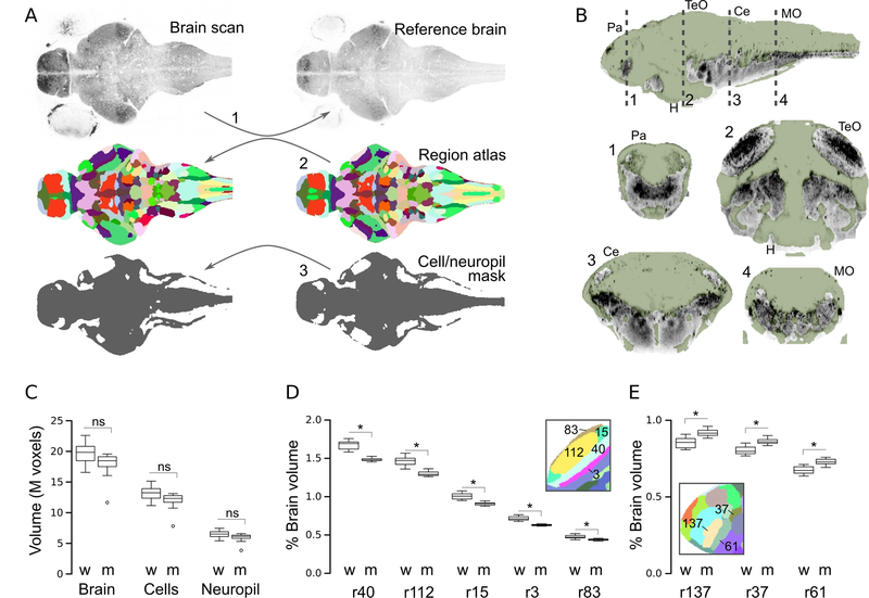
Figure 5. Atlas-volume measurements.
A.Schematic of procedure for measuring the volume of the whole brain, anatomical subdivisions and the cellular/neuropil zones. (1) Brain scans are registered to a reference. Then, the anatomical atlas (2) and map of cellular/neuropil regions (3), both generated on the reference brain, are back-transformed to the original images.
B.Cluster-derived map of cell-rich regions (green mask) and other (primarily neuropil, grey) brain regions. Dashed lines in top sagittal section indicate planes of section for corresponding coronal views below.
C. Volume of the whole brain, cellular regions and synaptic regions in wildtype larvae (w) and atoh7 morphants (m). N=11 per group.
D-E. Percent of brain occupied by regions (r) of the optic tectum neuropil (D) and forebrain (E) in wildtype (w) and atoh7 morphants (m). * p < 0.05 after Holm correction for 183 comparisons (ie.180 regions, whole brain, cell volume, neuropil volume). Insets show neuroanatomical map in the corresponding areas with the region numbers indicated.
MO:medulla oblongata; TeO:optic tectum; H:hypothalamus; Ce:cerebellum; Pa: pallium

