Abstract
Bacteria of the Roseobacter group (Rhodobacteraceae) are important members of many marine ecosystems. Similar to other Gram-negative bacteria many roseobacters produce N-acylhomoserine lactones (AHLs) for communication by quorum sensing systems. AHLs regulate different traits like cell differentiation or antibiotic production. Related N-acylalanine methyl esters (NAMEs) have been reported as well, but so far only from Roseovarius tolerans EL-164. While screening various roseobacters isolated from macroalgae we encountered four strains, Roseovarius sp. D12_1.68, Loktanella sp. F13, F14 and D3 that produced new derivatives and analogs of NAMEs, namely N-acyl-2-aminobutyric acid methyl esters (NABME), N-acylglycine methyl esters (NAGME), N-acylvaline methyl esters (NAVME), as well as for the first time a methyl-branched NAME, N-(13-methyltetradecanoyl)alanine methyl ester. These compounds were detected by GC–MS analysis, and structural proposals were derived from the mass spectra and by derivatization. Verification of compound structures was performed by synthesis. NABMEs, NAVMEs and NAGMEs are produced in low amounts only, making mass spectrometry the method of choice for their detection. The analysis of both EI and ESI mass spectra revealed fragmentation patterns helpful for the detection of similar compounds derived from other amino acids. Some of these compounds showed antimicrobial activity. The structural similarity of N-acylated amino acid methyl esters and similar lipophilicity to AHLs might indicate a yet unknown function as signalling compounds in the ecology of these bacteria, although their singular occurrence is in strong contrast to the common occurrence of AHLs. Obviously the structural motif is not restricted to Roseovarius spp. and occurs also in other genera.
Keywords: amino acid derivatives, 2-aminobutyric acid, homoserine lactones, natural products, quorum sensing
Introduction
The identification and structural elucidation of naturally occurring compounds traditionally requires isolation and NMR investigation as key method to detect novel compounds and new structural classes. Although the advent of NMR spectrometers with high frequencies and cryoprobes with small diameters enables experiments to be performed in the μg scale with pure compound, the isolation of the pure material from complex samples as well as the access to the expensive equipment still pose a considerable challenge to find new compounds. The ongoing quest for new structures also increasingly addresses minor components, requiring larger amounts of the producing organism, not always readily accessible, to isolate a targeted compound [1–3].
An alternative methodology can avoid the laborious isolation procedure. Direct analysis by mass-spectrometric methods of natural materials, e.g., extracts, may give enough information to infer the structure of an unknown compound that is finally proven by synthesis and comparison with natural material. The use of GC/EI-MS is especially advantageous because such mass spectra often reveal key structural features. Furthermore, the availability of large cross-platform databases useful for dereplication allows focussing on new compounds.
We are interested in natural compounds from Roseobacter group bacteria, an abundant class of marine bacteria occurring in diverse habitats with a broad metabolic potential [4–7]. Especially attached-living roseobacters produce diverse secondary metabolites, e.g., N-acylhomoserine lactones (AHLs) that the bacteria use for communication by quorum sensing [8–10]. AHLs are extensively investigated because of the broad knowledge on their biosynthesis, the underlying gene organization, as well as their function in many bacteria [11–13]. In the Roseobacter group, AHLs are involved, e.g., in antibiotic production [9] or cell differentiation [10]. Although many other bacterial signalling compounds must exist, only few of them have been characterized so far [14–18]. Such signalling compounds as well as many other unknown metabolites often occur in small amounts, which renders trace detecting methods like GC/MS a suitable approach for their detection and structure elucidation, provided their polarity falls into the analytical window of the method.
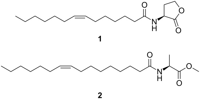
A wide variety of AHLs, e.g., widespread (Z)-N-(tetradec-7-enoyl)homoserine lactone (1, Z7-C-14:1-AHL, Figure 1), have been identified in roseobacters by these methods [19–22]. A related group of compounds occurring in Roseovarius only, are N-acylalanine methyl esters (NAMEs), e.g., (Z)-N-(hexadec-9-enoyl)alanine methyl ester (2, Z9-C16:1-NAME), the major NAME produced by Roseovarius tolerans EL 164 [23]. Although NAMEs are structurally similar to AHLs by an acyl chain linked to an amino acid derivative via an amide bond, they do not activate AHL receptors in roseobacters [21]. Instead, they show moderate antialgal activity [21]. In contrast to AHLs, the acyl chain can also be terminally oxidized [24]. During our analyses of different Roseobacter isolates, we encountered several compounds, which mass spectra show similarities to known NAMEs. These compounds proved to be either new NAMEs or constitute new classes of acylated amino acid methyl esters, derived from valine (NAVME), glycine (NAGME), or 2-aminobutyric acid (NABME). The identification of these compounds will be discussed based on the outlined approach including GC/MS analysis, interpretation of mass spectra, and verification by synthesis.
Figure 1.
N-Acylhomoserine lactones 1 (Z7-C14:1-AHL) and N-acylalanine methyl esters 2 (Z9-C16:1-NAME) occurring in Roseobacter group bacteria.
Results and Discussion
The secondary metabolites released by liquid cultures of various roseobacters were collected by extraction via Amberlite XAD-16 resin and analysed by GC/MS. Four of these strains, Roseovarius sp. D12_1.68 and Loktanella sp. F13, F14 and D3, contained low amounts of compounds with similar mass spectra to those of NAMEs [23].
Roseovarius sp. D12_1.68
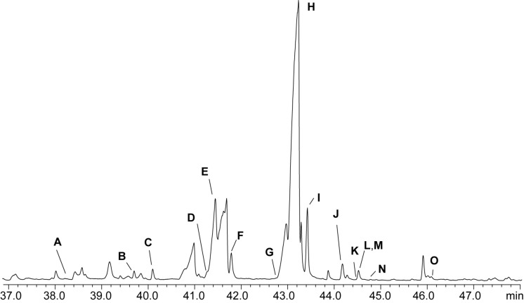
The investigation of an extract by GC/MS (Figure 2) revealed the presence of several NAMEs and AHLs due to their characteristic ions at m/z 104, 145, and 158 and m/z 102, 143, and 156, respectively [21,23]. Some of these compounds, E and L in Figure 2, were readily identified by their mass spectra and gas chromatographic retention indices Inat as known AHLs, containing saturated C12 and C14 acyl chains (Table 1).
Figure 2.
Total ion chromatogram (TIC) of an XAD extract of Roseovarius sp. D12_1.68. AHLs, NAMEs and related compounds are assigned by bold letters (Table 1).
Table 1.
Composition of the extracellular metabolites of Roseovarius sp. D12_1.68. [M]+, m/z: molecular and characteristic ions in EI mass spectrum. Inat: gas chromatographic retention index of natural compounds. HRMS: HR-mass spectral data of [M + H]+ obtained by HPLC/HR–ESI+–MS.
| peak | compound | [M]+ | m/z | Inat | HRMS [M + H]+ |
| A | C13:0-NAME | 299 | 104, 145, 158 | 2181 | |
| B | C14:1-NAME | 311 | 104, 145, 158 | 2265 | C18H34NO3 |
| C | C14:0-NAME | 313 | 104, 145, 158 | 2289 | C18H36NO3 |
| D | iso-C15:0-NAME | 327 | 104, 145, 158 | 2354 | C19H38NO3 |
| E | C12:0-AHL | 283 | 102, 143, 156 | 2367 | |
| F | C15:0-NAME | 327 | 104, 145, 158 | 2392 | C19H38NO3 |
| G | C16:1-NAME | 339 | 104, 145, 158 | 2457 | C20H38NO3 |
| H | Z9-C16:1-NAME | 339 | 104, 145, 158 | 2473 | C20H38NO3 |
| I | C16:0-NAME | 341 | 104, 145, 158 | 2497 | C20H40NO3 |
| J | 9-C16:1-NABME | 353 | 118, 159, 172 | 2548 | C21H40NO3 |
| K | C16:0-NABME | 355 | 118, 159, 172 | 2569 | C21H42NO3 |
| L | C14:0-AHL | 311 | 102, 143, 156 | 2570 | |
| M | 9-C17:1-NAME | 353 | 104, 145, 158 | 2570 | C21H40NO3 |
| N | 9-C16:1-NAVME | 367 | 132, 173, 181 | 2588 | C22H42NO3 |
| O | 11-C18:1-NAME | 367 | 104, 145, 158 | 2677 | C22H42NO3 |
Similarly, compounds C, F, H, I, and M were identified as the already known C14:0-, C15:0-, 9-C16:1-, C16:0-, and 9-C17:1-NAMEs. Compounds A, B, and O proved to be not previously reported C13:0-, C14:1- and C18:1-NAMEs, assignable by their mass spectra and gas chromatographic retention indices. These conclusions were supported by HRMS data obtained by HPLC/MS (Table 1). Localization of the double bond was established via DMDS derivatization as described previously [23]. Due to the low amounts no double bond position could be established for C14:1-NAME, while the double bond of C18:1 NAME was located at C-11. Similar as in AHLs, the double bond location in unsaturated NAMEs seems to be fixed at the ω-7 position [8,25].
Compound D (C19H37NO3) showed a mass spectrum identical to C15:0-NAME, albeit the retention index deviated by 38 units. A methyl branch at iso- or anteiso-position seemed likely. Therefore, the theoretical retention index Ic were calculated for methyl branched C15:0-NAMEs using an empirical model established in our work group [26] that had successfully been used for the detection of methyl branched AHLs [27]. The retention indices were calculated using the formula

with N indicating the number of n carbons in the chain times hundred, FG as the functional group increment depending on and Mei as an increment for the methyl branching in different positions. The increments for Mei are known [26]. The functional group increment was calculated to be 836 + 4 · n using the retention indices of C14:0, C15:0, and C16:0-NAME. Therefore, the calculated retention index for iso-C15:0 and anteiso-C15:0-NAME are Ic = 2352 and Ic = 2365, respectively, while all other methyl branch locations had a lower value. The close similarity of Inat = 2354 and Ic = 2352 suggested the methyl branch to be located in the iso-position. Consequently, iso-C15:0-NAME (6) was synthesized as shown in Scheme 1 to verify the structural proposal.
Scheme 1.
Synthesis of iso-C15:0-NAME (6). DMAP: 4-dimethylaminopyridine, EDC: 1-ethyl-3-(3-dimethylaminopropyl)carbodiimide hydrochloride, L-AME:  -alanine methyl ester.
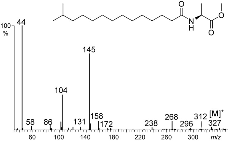
11-Bromoundecan-1-ol (3) was converted into the alcohol 13-methyltetradecan-1-ol (4) with isobutylmagnesium bromide under Li2CuCl4 catalysis according to Mori et al. [28]. After Jones oxidation, 13-methyltetradecanoic acid (5) was coupled with  -alanine methyl ester hydrochloride to deliver the desired product 6. The mass spectra and retention indices of the natural and synthetic samples were identical, proving the proposed structure (Figure 3). This compound is the first natural NAME featuring a methyl branched acyl chain.
Figure 3.
Mass spectrum of natural compound D, N-(13-methyltetradecanoyl)alanine methyl ester (iso-C15:0-NAME, 6).
Compound G showed a mass spectrum identical to C16:1-NAME and a retention index with I = 2457, eluting earlier than compound H, Z9-C16:1-NAME with I = 2473. The low amount of the material produced excluded further structural characterization of the compound that could either be methyl-branched in the acyl chain or might show a different double bond position or configuration.
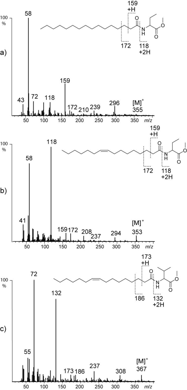
The remaining three compounds J, K, and N with the molecular composition C21H42NO3, C21H40NO3, and C22H42NO3 determined by HRMS showed related mass spectra with a characteristic mass shift compared to NAMEs. Ions m/z 44, 104, 145, and 158 where shifted, however, to m/z 58, 118, 159, and 172 in the spectra of J and K (Figure 4a,b).
Figure 4.
Mass spectra of natural compounds a) K (7, C16:0-NABME), b) J (8, C16:1-NABME), and c) N (9, C16:1-NAVME).
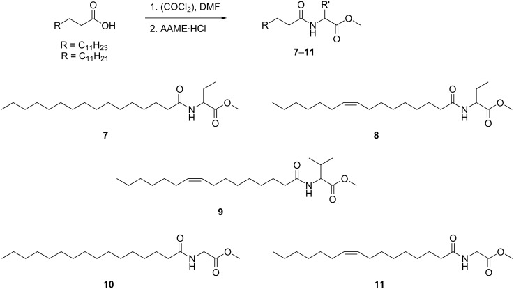
These ions can be explained by an additional CH2 group in the alanine part, leading to a 2-aminobutyric acid fragment in these compounds. The later eluting compound K with a molecular ion at m/z 355 was therefore proposed to be N-(hexadecanoyl)-2-aminobutyric acid methyl ester (7), while the earlier eluting J with m/z 353 compound was likely N-[(Z)-hexadec-9-enoyl]-2-aminobutyric acid methyl ester (8). The double bond position was determined by DMDS derivatization. The structures of both K and J were verified by synthesis according to Scheme 2. Palmitoleic acid was synthesized in g-scale by standard procedures as shown in the Supporting Information File 1, Scheme S1. This acid and palmitic acid were converted into the respective chlorides and standard acylation delivered 2-aminobutyric acid derivatives 7 and 8 (Scheme 2) that proved to be identical with the natural products. The absolute configuration of the amino acid could not be determined due to the low amount of material present. Nevertheless, because NAMEs showed the common  -configuration [23], this configuration also seems likely for the other amino acid derivatives reported here. We suggest the term NABME (N-acylated 2-aminobutyric acid methyl esters) for the new compounds that can thus be assigned as C16:0-NABME (7) and Z9-C16:1-NABME (8).
Scheme 2.
Synthesis of N-acylated amino acid methyl esters 7–11. AAME: amino acid methyl ester.
Compound N showed a mass spectrum (Figure 4c) featuring characteristic ions at m/z 72, 132, and 173, indicating a further carbon in the amino acid part. Together with the molecular ion at m/z 367 (C22H41NO3) the data indicate the presence of valine in the compound. DMDS derivatization proved the unknown compound to also carry a double bond at C-9 of the alkenoyl chain. The proposed structure N-[(Z)-hexadec-9-enoyl]valine methyl ester (9) was proven by synthesis starting from valine methyl ester as described (Scheme 2). The gas chromatographic retention indices of the synthetic material and the natural compound matched perfectly. We propose to call the N-acylated valine methyl ester 9 Z9-C16:1-NAVME.
The extract of Roseovarius sp. D12_1.68 was also investigated by HPLC/ESI+–MS to detect more polar compounds compared to GC. The NAMEs, NABMEs and NAVMEs reported here were detected by MS2 analyses based on their characteristic fragmentation (see below). The only oxygenated derivative present was 16OH-C16:1-NAME, which has been described before from Roseovarius tolerans EL-164 [24].
Loktanella sp. D3, F13 and F14
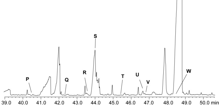
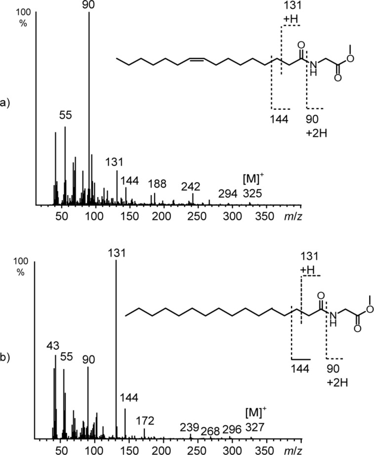
Investigation of extracts of the three isolates F14, F13, and D3 by GC–MS indicated the presence of compounds whose mass spectra were again similar to those of NAMEs (Figure 5). The spectra show characteristic ions at m/z 90, 131, and 144 (Figure 6), a loss of one methylene group compared to ions m/z 104, 145, and 158 of NAMEs. The lack of an analogous ion to m/z 44 (m/z 30 is outside the mass range of the spectrometer used) pointed this time to glycine as the core amino acid.
Figure 5.
TIC of an XAD extract of Loktanella sp. F14. Compound S is a minor component within the broad peak.
Figure 6.
Mass spectra of natural compounds a) R (11, C16:1-NAGME) and b) S (10, C16:0-NAGME).
Molecular formulas were obtained via HPLC/HRMS measurements and supported the N-acylglycine methyl ester (NAGME) structure proposed for these compounds.
Saturated and unsaturated NAGMEs can be distinguished by the intensity of the ions m/z 90 and 131. Similar to NAMEs saturated NAGMEs show a high intensity of m/z 131 whereas unsaturated NAGMEs show higher intensity of m/z 90 (Figure 6). The low amounts available did not allow to determine the position of the double bond in unsaturated NAGMEs. Nevertheless, the predominance of the (Z)-9-hexadecenoyl side chain in all NAME family compounds suggested compound R to be N-[(Z)-hexadec-9-enoyl]glycine methyl ester (11, Z9-C16:1-NAGME), while S is its saturated analogue. Therefore, both compounds were synthesized as described before from glycine methyl ester and the respective acid (Scheme 2) and their identity confirmed. The other components P, Q and T–W were also NAGMEs.
Their chain length was established using EI mass spectra and the gas chromatographic retention indices Inat of the compounds (Table 2). Overall, six saturated and unsaturated compounds with a chain length between C14 and C19 were detected. The roughly 100 retention index units between the members of this homologous series indicated that the acyl chains are unbranched. Additionally, two unsaturated glycine derivatives were present, 9-C16:1-NAGME (11) and C18:1-NAGME (Table 2).
Table 2.
NAGMEs produced by Loktanella related isolates F14, F13 and D3. Inat: gas chromatographic retention index on a HP-5 phase; [M]+: molecular mass; m/z: characteristic ions in EI mass spectra.
| peak | compound | Inat | [M]+ | m/z | Loktanella isolate | ||
| F14 | F13 | D3 | |||||
| P | C14:0-NAGME | 2312 | 299 | 131 > 90 | x | x | |
| Q | C15:0-NAGME | 2415 | 313 | 131 > 90 | x | x | |
| R | C16:1-NAGME | 2495 | 325 | 90 < 131 | x | x | |
| S | C16:0-NAGME | 2515 | 327 | 131 > 90 | x | x | x |
| T | C17:0-NAGME | 2618 | 341 | 131 > 90 | x | ||
| U | C18:1-NAGME | 2702 | 353 | 90 < 131 | x | x | |
| V | C18:0-NAGME | 2720 | 355 | 131 > 90 | x | x | |
| W | C19:0-NAGME | 2832 | 369 | 131 > 90 | x | ||
Analysis by HPLC/MS revealed no additional NAGME. Furthermore, no NAMEs, NABMEs, or NAVMEs were observed in the three strains.
Mass spectrometry
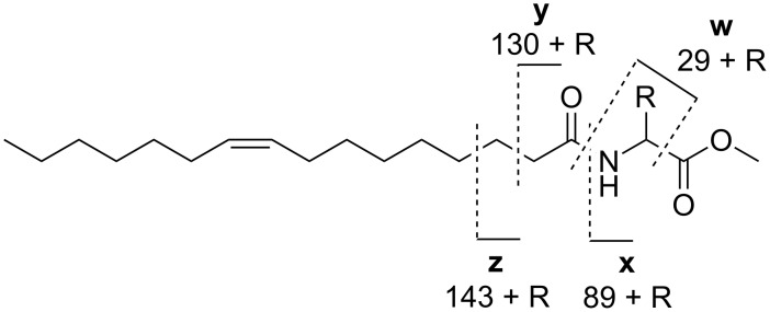
The analysis of the mass spectra of NAMEs, NABMEs, NAVMEs, and NAGMEs revealed the typical fragmentation of N-acylated amino acid methyl esters under both EI (Figure 7) and ESI ionization (Figure 8). Detailed structural information can be obtained by EI-MS. A dominant peak in the mass spectrum is the McLafferty rearrangement ion y, if the acyl chain is saturated. Together with prominent ion w [NH–CH2–R]+, often the base peak, and x it defines the amino acid, while z is usually of low abundance. Formation of x requires transfer of two H atoms to this fragment. In compounds carrying an unsaturated acyl chain the intensity of y is reduced, and x increases in intensity. In addition to these features, the molecular ion is visible, as is the loss of the carbomethoxy group [M − 59]+.
Figure 7.
EI mass spectrometric fragmentation of N-acylated amino acid derivatives. The ions w–z allow identification of the respective amino acid residue.
Figure 8.
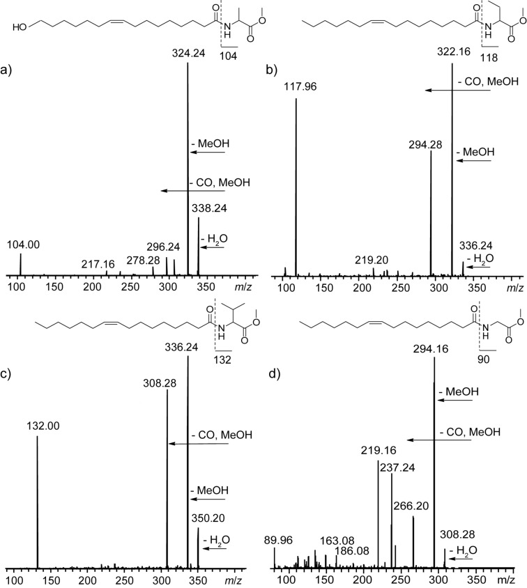
MS2 spectra in ESI positive mode of a) Z9-16OH-C16:1-NAME with [M + H − H2O]+ 356, b) Z9-C16:1-NABME (8) with [M + H]+ 354, c) Z9-C16:1-NAVME (9) with [M + H]+ 368, and d) Z9-C16:1-NAGME (11) with [M + H]+ 326.
The MS2 spectrum of the [M + H]+-ion obtained in ESI positive mode shows loss of water, an intense ion due to loss of methanol, and loss of the carbomethoxy group [24]. Often the amino acid ion can also be observed. These features indicate the presence of an amino acid methyl ester.
Biological activity
Several of the synthesized compounds were tested in a broader screening program for their antimicrobial activity (Table 3). While the valine derivative 9 was virtually inactive, the other compounds showed some activity. Glycine compound 11 showed good activity against the Gram-positive bacteria Bacillus subtilis, Staphylococcus aureus, and Micrococcus luteus, and against the filamentous fungus Mucor hiemalis. In addition, 11 displayed moderate active on Mycobacterium smegmatis and the efflux-deficient Escherichia coli TolC strain. The 2-aminobutyric acid derivative 7 was active against M. hiemalis, M. luteus and S. aureus, while the unsaturated analogue 8 was mainly active against E. coli TolC, and it was the only compound that showed moderate cytotoxicity on a human cancer cell line.
Table 3.
Antimicrobial activity (minimum inhibitory concentration, MIC, in µg/mL) and cytotoxicity (minimum inhibitory concentration, MIC, in µg/mL) of selected NAVME, NABME and NAGME derivatives. Minimal inhibitory concentration and IC50 values for cytotoxicity in μg/mL.
| strain | 11 | 9 | 8 | 7 |
| Chromobacterium violaceum DSM-30191 | >128 | >128 | nd | >128 |
| Escherichia coli DSM-1116 | >128 | >128 | nd | >128 |
| Escherichia coli (TolC-deficient) | 128 | >128 | 16 | >128 |
| Pseudomonas aeruginosa PA14 | >128 | >128 | nd | >128 |
| Bacillus subtilis DSM-10 | 4–8 | >128 | >128 | >128 |
| Micrococcus luteus DSM-1790 | 4 | >128 | 64–128 | 16 |
| Staphylococcus aureus Newman | 16 | >128 | nd | 8–16 |
| Mycobacterium smegmatis mc2155 | 64 | >128 | nd | >128 |
| Mucor hiemalis DSM-2656 | 8–16 | >128 | nd | 32 |
| Pichia anomala DSM-6766 | >128 | >128 | nd | >128 |
| Candida albicans DSM-1665 | >128 | >128 | nd | >128 |
| cytotoxicity | ||||
| HCT-116 (human colon carcinoma) | >67 | >67 | 15.8 | >67 |
The close similarity of NAMEs, NAVMEs, and NAGMEs to AHLs might indicate a function as signalling compounds, although experiments with NAMEs and AHL reporter assays did not reveal any activity on the LuxR type receptors for AHLs in bacteria [23–24]. Other potential functions are antimicrobial or cytotoxic activity of the tested derivatives. Roseovarius sp. D12_1.68 showed antimicrobial activity against a Rhodobacteraceae sp. TL and antialgal activity against Skeletonema costatum while Loktanella sp. D3 showed no antagonistic activity [29]. This observation fits well with the detection of antimicrobial or cytotoxic NAVME 9 and NABMEs 7 and 8 only in Roseovarius sp. D12_1.68.
Bacteria are known to produce acylated amino acids, although the number reported so far is small, including tyrosine, tryptophan, arginine, or phenylalanine [30–32]. They all carry long chain saturated or unsaturated acyl chains similar to those reported here. Recently, derivatives of the hydrophobic amino acids valine, leucine and isoleucine were also reported [33]. These compounds are produced by a family of acyl amino acid synthases structurally related to AHL synthases, further suggesting a function as bacterial signalling compounds [34]. In contrast to the reported compounds carrying a free acid group, all derivatives reported here are native methyl esters because no methanol was used during sample preparation
Conclusion
We have identified here new classes of acylated amino acid derivatives including previously unknown glycine and 2-aminobutyric acid derived compounds. The combination of GC/MS, HPLC/MS, retention indices and synthesis proved to be especially suited to structurally identify minor components of complex extracellular metabolite mixtures. The reported compounds are specific for Roseobacter group bacteria of the genera Roseovarius and Loktanella, in contrast to broadly distributed AHLs. Although their function as signalling compounds is not proven, the occurrence of 2-aminobutyric acid might indicate some similarity to homoserine in AHLs because both are non-proteinogenic amino acids. This similarity might also be functional, because the structures of NAMEs, NABMEs, NAGMEs and NAVMEs are similar to other bacterial signalling compounds, often carrying a lipophilic side chain and a medium polar core structure [8,16]. Nevertheless, the ecological function of NAMEs and its derivatives could also be antagonistic activity against concurrent biofilm microorganisms, suggested by the bioactivity of some of the compounds as observed in this study and the antimicrobial and antialgal activity of the producing organism [29].
Supporting Information
Experimental synthetic procedures, biological tests and NMR spectra.
Acknowledgments
We thank the Deutsche Forschungsgemeinschaft (DFG) for supporting our work through the Transregional Collaborative Research Center “Roseobacter” (SFB TRR 51/3)
References
- 1.Hüttel S, Testolin G, Herrmann J, Planke T, Gille F, Moreno M, Stadler M, Brönstrup M, Kirschning A, Müller R. Angew Chem, Int Ed. 2017;56:12760–12764. doi: 10.1002/anie.201705913. [DOI] [PubMed] [Google Scholar]
- 2.Cociancich S, Pesic A, Petras D, Uhlmann S, Kretz J, Schubert V, Vieweg L, Duplan S, Marguerettaz M, Noëll J, et al. Nat Chem Biol. 2015;11:195–197. doi: 10.1038/nchembio.1734. [DOI] [PubMed] [Google Scholar]
- 3.Ling L L, Schneider T, Peoples A J, Spoering A L, Engels I, Conlon B P, Mueller A, Schäberle T F, Hughes D E, Epstein S, et al. Nature. 2015;517:455–459. doi: 10.1038/nature14098. [DOI] [PMC free article] [PubMed] [Google Scholar]
- 4.Giebel H-A, Kalhoefer D, Lemke A, Thole S, Gahl-Janssen R, Simon M, Brinkhoff T. ISME J. 2011;5:8–19. doi: 10.1038/ismej.2010.87. [DOI] [PMC free article] [PubMed] [Google Scholar]
- 5.Freese H M, Methner A, Overmann J. Front Microbiol. 2017;8:1659. doi: 10.3389/fmicb.2017.01659. [DOI] [PMC free article] [PubMed] [Google Scholar]
- 6.Brinkhoff T, Giebel H-A, Simon M. Arch Microbiol. 2008;189:531–539. doi: 10.1007/s00203-008-0353-y. [DOI] [PubMed] [Google Scholar]
- 7.Buchan A, Gonzalez J M, Moran M A. Appl Environ Microbiol. 2005;71:5665–5677. doi: 10.1128/aem.71.10.5665-5677.2005. [DOI] [PMC free article] [PubMed] [Google Scholar]
- 8.Schulz S, Hötling S. Nat Prod Rep. 2015;32:1042–1066. doi: 10.1039/c5np00006h. [DOI] [PubMed] [Google Scholar]
- 9.Berger M, Neumann A, Schulz S, Simon M, Brinkhoff T. J Bacteriol. 2011;193:6576–6585. doi: 10.1128/jb.05818-11. [DOI] [PMC free article] [PubMed] [Google Scholar]
- 10.Patzelt D, Wang H, Buchholz I, Rohde M, Gröbe L, Pradella S, Neumann A, Schulz S, Heyber S, Münch K, et al. ISME J. 2013;7:2274–2286. doi: 10.1038/ismej.2013.107. [DOI] [PMC free article] [PubMed] [Google Scholar]
- 11.Winans S C, Bassler B L, editors. Chemical communication among bacteria. Washington, DC: ASM Press; 2008. [DOI] [Google Scholar]
- 12.Dickschat J S. Nat Prod Rep. 2010;27:343–369. doi: 10.1039/b804469b. [DOI] [PubMed] [Google Scholar]
- 13.Papenfort K, Bassler B L. Nat Rev Microbiol. 2016;14:576–588. doi: 10.1038/nrmicro.2016.89. [DOI] [PMC free article] [PubMed] [Google Scholar]
- 14.Higgins D A, Pomianek M E, Kraml C M, Taylor R K, Semmelhack M F, Bassler B L. Nature. 2007;450(7171):883–886. doi: 10.1038/nature06284. [DOI] [PubMed] [Google Scholar]
- 15.Thiel V, Vilchez R, Sztajer H, Wagner-Döbler I, Schulz S. ChemBioChem. 2009;10(3):479–485. doi: 10.1002/cbic.200800606. [DOI] [PubMed] [Google Scholar]
- 16.Brachmann A O, Brameyer S, Kresovic D, Hitkova I, Kopp Y, Manske C, Schubert K, Bode H B, Heermann R. Nat Chem Biol. 2013;9:573–578. doi: 10.1038/nchembio.1295. [DOI] [PubMed] [Google Scholar]
- 17.Chen X, Schauder S, Potier N, Van Dorsselaer A, Pelczer I, Bassler B L, Hughson F M. Nature. 2002;415(6871):545–549. doi: 10.1038/415545a. [DOI] [PubMed] [Google Scholar]
- 18.Papenfort K, Silpe J E, Schramma K R, Cong J-P, Seyedsayamdost M R, Bassler B L. Nat Chem Biol. 2017;13:551–557. doi: 10.1038/nchembio.2336. [DOI] [PMC free article] [PubMed] [Google Scholar]
- 19.Wagner-Döbler I, Thiel V, Eberl L, Allgaier M, Bodor A, Meyer S, Ebner S, Hennig A, Pukall R, Schulz S. ChemBioChem. 2005;6:2195–2206. doi: 10.1002/cbic.200500189. [DOI] [PubMed] [Google Scholar]
- 20.Neumann A, Patzelt D, Wagner-Döbler I, Schulz S. ChemBioChem. 2013;14:2355–2361. doi: 10.1002/cbic.201300424. [DOI] [PubMed] [Google Scholar]
- 21.Ziesche L, Bruns H, Dogs M, Wolter L, Mann F, Wagner-Döbler I, Brinkhoff T, Schulz S. ChemBioChem. 2015;16(14):2094–2107. doi: 10.1002/cbic.201500189. [DOI] [PubMed] [Google Scholar]
- 22.Schaefer A L, Greenberg E P, Oliver C M, Oda Y, Huang J J, Bittan-Banin G, Peres C M, Schmidt S, Juhaszova K, Sufrin J R, et al. Nature. 2008;454:595–599. doi: 10.1038/nature07088. [DOI] [PubMed] [Google Scholar]
- 23.Bruns H, Thiel V, Voget S, Patzelt D, Daniel R, Wagner-Döbler I, Schulz S. Chem Biodiversity. 2013;10:1559–1573. doi: 10.1002/cbdv.201300210. [DOI] [PubMed] [Google Scholar]
- 24.Bruns H, Herrmann J, Müller R, Wang H, Wagner Döbler I, Schulz S. J Nat Prod. 2018;81:131–139. doi: 10.1021/acs.jnatprod.7b00757. [DOI] [PubMed] [Google Scholar]
- 25.Ziesche L, Rinkel J, Dickschat J S, Schulz S. Beilstein J Org Chem. 2018;14:1309–1316. doi: 10.3762/bjoc.14.112. [DOI] [PMC free article] [PubMed] [Google Scholar]
- 26.Schulz S. Lipids. 2001;36:637–647. doi: 10.1007/s11745-001-0768-7. [DOI] [PubMed] [Google Scholar]
- 27.Thiel V, Kunze B, Verma P, Wagner-Döbler I, Schulz S. ChemBioChem. 2009;10:1861–1868. doi: 10.1002/cbic.200900126. [DOI] [PubMed] [Google Scholar]
- 28.Takikawa H, Nozawa D, Kayo A, Muto S-e, Mori K. J Chem Soc, Perkin Trans 1. 1999:2467–2477. doi: 10.1039/a904258j. [DOI] [Google Scholar]
- 29.Dogs M, Wemheuer B, Wolter L, Bergen N, Daniel R, Simon M, Brinkhoff T. Syst Appl Microbiol. 2017;40:370–382. doi: 10.1016/j.syapm.2017.05.006. [DOI] [PubMed] [Google Scholar]
- 30.Brady S F, Clardy J. J Am Chem Soc. 2000;122:12903–12904. doi: 10.1021/ja002990u. [DOI] [Google Scholar]
- 31.Brady S F, Chao C J, Clardy J. Appl Environ Microbiol. 2004;70:6865–6870. doi: 10.1128/aem.70.11.6865-6870.2004. [DOI] [PMC free article] [PubMed] [Google Scholar]
- 32.Brady S F, Clardy J. Org Lett. 2005;7:3613–3616. doi: 10.1021/ol0509585. [DOI] [PubMed] [Google Scholar]
- 33.Guo H, Rischer M, Sperfeld M, Weigel C, Menzel K D, Clardy J, Beemelmanns C. Bioorg Med Chem. 2017;25:6088–6097. doi: 10.1016/j.bmc.2017.06.053. [DOI] [PMC free article] [PubMed] [Google Scholar]
- 34.Van Wagoner R M, Clardy J. Structure. 2006;14(9):1425–1435. doi: 10.1016/j.str.2006.07.005. [DOI] [PubMed] [Google Scholar]
Associated Data
This section collects any data citations, data availability statements, or supplementary materials included in this article.
Supplementary Materials
Experimental synthetic procedures, biological tests and NMR spectra.


