Abstract
Long intergenic non-coding RNAs (lincRNAs) are >200 nucleotides long non-coding RNAs, which have been shown to be implicated in carcinogenic processes by interacting with cancer associated genes or other non-coding RNAs. However, their role in development of rare gastrointestinal stromal tumors (GISTs) is barely investigated. Therefore, the aim of this study was to define lincRNAs deregulated in GIST and find new GIST-lincRNA associations. Next-generation sequencing data of paired GIST and adjacent tissue samples from 15 patients were subjected to a web-based lincRNA analysis. Three deregulated lincRNAs (MALAT1, H19 and FENDRR; adjusted p-value < 0.05) were selected for expression validation in a larger group of patients (n = 22) by RT-qPCR method. However, only H19 and FENDRR showed significant upregulation in the validation cohort (adjusted p < 0.05). Further, we performed correlation analyses between expression levels of deregulated lincRNAs and GIST-associated oncogenes or GIST deregulated microRNAs. We found high positive correlations between expression of H19 and known GIST related oncogene ETV1, and between H19 and miR-455-3p. These findings expand the knowledge on lincRNAs deregulated in GIST and may be an important resource for the future studies investigating lincRNAs functionally relevant to GIST carcinogenesis.
Introduction
Gastrointestinal stromal tumors (GISTs) are the most common mesenchymal (non-epithelial) tumors of the gastrointestinal tract, with a majority of cases localized in stomach (55.6%) and small intestine (31.8%) [1,2]. GISTs are considered to arise from interstitial cells of Cajal and are characterized by expression of tyrosine kinase receptor protein KIT (CD117)–the main diagnostic marker for these tumors [3]. Activating mutations in KIT proto-oncogene and homologous PDGFRA receptors result in constitutive activation of these proteins and are crucial initiating events in GIST pathogenesis. Although the key aspects of GIST pathogenesis are already elucidated, the mechanisms underlying overexpression of oncogenes and the role of gene expression regulators, like non-coding RNAs, in development of GISTs are not well investigated, yet.
Long intergenic non-coding RNAs (lincRNAs) are long (>200 nucleotides) non-coding RNAs that do not overlap exons of protein coding genes and comprise more than half of human long non-coding RNAs (lncRNAs) [4]. In recent years these non-coding RNAs emerged as important regulators of multiple cellular and molecular processes such as alternative splicing, chromatin modifications, transcriptional and post-transcriptional regulation, mRNA activity, stability and degradation and were found to be implicated in pathogenesis of diseases and development of cancer [4,5]. For example, the extensively studied lincRNA metastasis-associated lung adenocarcinoma transcript 1 (MALAT1) is overexpressed in lung adenocarcinoma, breast, pancreatic, colon, prostate, and hepatocellular carcinomas while promoting cell proliferation and metastasis [5,6]. Another lincRNA, Imprinted Maternally Expressed Transcript H19, has been shown to act both as tumor suppressor and oncogene in different types of cancer, playing an important role in epithelial mesenchymal transition and tumor growth and is a precursor of oncogenic microRNA miR-675 [7–10]. LncRNAs can also interact with small non-coding RNAs, i.e. acting as competing endogenous RNAs and sponging microRNAs through their binding sites [11]. In this way, lncRNAs can suppress microRNAs from binding to their targets and modulate target gene expression or be regulated by microRNAs.
Up to now, only two lncRNAs—HOTAIR and CCDC26—were investigated in GIST in more detail and were associated with high risk grade, metastasis and sensitivity to a drug imatinib [12–14]. Therefore, in this study we aimed to investigate lincRNAs deregulated in GIST and find new GIST-lincRNA associations. We performed next-generation sequencing data analysis and validated expression of three deregulated and cancer associated lincRNAs–MALAT1, H19 and FENDRR–in GIST and adjacent tissues. Further analysis revealed correlations between overexpressed investigated lincRNAs in GIST tissue and genes involved in GIST pathogenesis, as well as with GIST deregulated microRNAs.
Materials and methods
Clinical samples, RNA material, gene expression and sequencing data
Paired formalin-fixed, paraffin-embedded (FFPE) tumor and adjacent tissue samples from 37 patients (total 74 samples) diagnosed with GIST of gastric origin were included in this study. The study group consisted of 22 women and 15 men with the average age of 66.95 (SD ± 12.39). GIST diagnosis was based on GIST morphology and positive KIT (CD117) immunohistochemical staining. Risk grade of tumors was assessed according to National Institutes of Health (NIH) GIST Consensus Criteria [15]. GIST patients included in the study were classified as: 7 high risk grade, 11 moderate, 15 low and 4 very low risk grade. Based on the GIST histological subtype, the majority of tumors in this study were of spindle cell type (n = 26), with several cases of epithelioid (n = 7) and mixed spindle and epithelioid (n = 4) cell type. KIT and PDGFRA mutational status was verified for 32 patients: 18 patients had KIT gene mutations exon 11 (n = 17), and exon 9 (n = 1), 9 patients had PDGFRA gene mutations in exon 18 (n = 6) and exon 12 (n = 3), while 5 patients were KIT/PDGFRA wild type. Distribution of all evaluated parameters did not differ in discovery and validation cohorts (p-value > 0.05). Fisher’s exact test was used for qualitative measurements and t-test was used for quantitative measurements.
RNA and DNA material was extracted from FFPE tissue samples using commercial miRNeasy FFPE and Qiamp FFPE DNA tissue Kits (Qiagen) following manufacturers recommendations.
Small RNA sequencing data (HiSeq2500, Illumina) of paired samples from 15 GIST patients was used for primary detection of deregulated lincRNAs (discovery cohort), while validation cohort for lincRNA expression analysis consisted of 22 patients’ paired samples. RNA libraries were prepared for sequencing using TruSeq Small RNA Sample Preparation Kit (Illumina).
More detailed characteristics of patients, GIST mutational status, RNA/DNA extraction, library preparation and NGS sequencing methods, as well as detailed methodology of KIT, PDGFRA, ETV1 and microRNA gene expression analysis were described in our previous publication [16]. Small RNA sequencing data can be accessed at NCBI’s Gene Expression Omnibus (series accession number GSE89051).
The study was approved by the Kaunas Regional Biomedical Research Ethics Committee (No. BE-2–8). All patients have signed an informed consent form to participate in the study.
lincRNA sequencing data analysis
Sequencing data analysis was performed using web-based tool miRMaster [17], where preprocessed sequencing reads (after adapter trimming, quality filtering and read collapsing) were mapped against the NONCODE 2016 database [18] of lncRNAs, using default parameters. P-values were generated by Kruskal-Wallis test and used for evaluation of gene expression changes after Benjamini-Hochberg adjustment for multiple testing. Changes with an adjusted probability value below 0.05 were considered as significant.
Validation of lincRNA expression by reverse transcription quantitative real-time PCR (RT-qPCR)
Paired RNA samples of 22 GIST patients (total 44 samples) were included in the lincRNA expression validation cohort. RNA was reverse transcribed using High-Capacity cDNA Reverse Transcription Kit (Thermo Fisher Scientific). Expression of lincRNAs MALAT1, H19 and FENDRR in GIST tumor and adjacent tissues was measured using commercial TaqMan Gene Expression Assays (Hs00273907_s1 for MALAT1; Hs00399294_g1 for H19; Hs05044154_s1 for FENDRR) and TaqMan Universal PCR Master Mix on the 7500 Fast Real-Time PCR system, according to the manufacturer’s protocol. Gene expression data was normalized to the expression levels of GAPDH housekeeping gene (Hs99999905_m1) and analyzed using comparative CT method.
Statistical analysis
Differences between means of lincRNA expression validation data were analyzed using paired Student’s t-test. Expression changes with a Bonferroni adjusted p-values lower than 0.05 were considered significant. Spearman’s correlation analysis was applied on log2(2-dCt) values. Correlation coefficient values were assigned to groups from “negligible correlation” to “very high positive (negative) correlation” according to Mukaka M. [19]. LincRNA-oncogene and lincRNA-microRNA expression correlations were considered significant with a p-value lower than 0.01.
Kruskal-Wallis multiple comparisons test was applied to investigate differences of lincRNA expression in different GIST phenotypes. P-values lower than 0.05 were considered significant.
All statistical analyses were performed using the statistical computing environment R (version 3.4.4) [20].
Results
LincRNA analysis using NGS data
Sequences were mapped to a total 7240 lincRNA transcripts from the NONCODE database. After application of selection criteria (Benjamini-Hochberg adjusted p-value < 0.05, 2 < log2FC < -2), 23 lincRNA transcripts (9 unique lincRNAs) were significantly deregulated in GIST tissue compared to adjacent tissue, with six lincRNAs being upregulated and three—downregulated (Table 1).
Table 1. Significantly deregulated lincRNA transcripts in GIST vs. GIST adjacent tissue (paired samples of 15 patients) obtained from small RNA sequencing data.
| Transcript | lincRNA | p-value | Benjamini-Hochberg adjusted p-valuea |
Fold Change, GIST vs. adjacent tissue | Log2(FC), GIST vs. GIST adjacent tissue |
|---|---|---|---|---|---|
| ENST00000447298.1 | H19 | 1.13×10⁻⁶ | 0.002 | 69.2 | 6.1 |
| ENST00000428066.5 | H19 | 1.13×10⁻⁶ | 0.002 | 68.9 | 6.1 |
| ENST00000442037.5 | H19 | 1.42×10⁻⁶ | 0.002 | 61.2 | 5.9 |
| ENST00000446406.5 | H19 | 2.99×10⁻⁶ | 0.002 | 34.2 | 5.1 |
| ENST00000414790.5 | H19 | 2.99×10⁻⁶ | 0.002 | 34.1 | 5.1 |
| ENST00000412788.5 | H19 | 2.99×10⁻⁶ | 0.002 | 34.0 | 5.1 |
| ENST00000431095.5 | H19 | 3.66×10⁻⁶ | 0.002 | 33.9 | 5.1 |
| ENST00000422826.1 | H19 | 2.99×10⁻⁶ | 0.002 | 33.9 | 5.1 |
| ENST00000411754.5 | H19 | 3.66×10⁻⁶ | 0.002 | 33.8 | 5.1 |
| ENST00000439725.5 | H19 | 3.66×10⁻⁶ | 0.002 | 33.7 | 5.1 |
| ENST00000417089.5 | H19 | 3.66×10⁻⁶ | 0.002 | 33.7 | 5.1 |
| ENST00000582452.1 | LINC00908 | 4.99×10⁻⁵ | 0.016 | 33.3 | 5.1 |
| ENST00000598996.2 | FENDRR | 2.20×10⁻⁵ | 0.011 | 15.8 | 4.0 |
| ENST00000519898.5 | CARMN | 1.18×10⁻⁴ | 0.028 | 11.1 | 3.5 |
| ENST00000610481.1 | MALAT1 | 2.34×10⁻⁴ | 0.046 | 8.2 | 3.0 |
| ENST00000544868.2 | MALAT1 | 3.28×10⁻⁵ | 0.012 | 6.6 | 2.7 |
| ENST00000508832.2 | MALAT1 | 1.47×10⁻⁴ | 0.034 | 6.0 | 2.6 |
| ENST00000534336.1 | MALAT1 | 8.87×10⁻⁵ | 0.022 | 5.3 | 2.4 |
| ENST00000619449.1 | MALAT1 | 8.55×10⁻⁵ | 0.022 | 5.0 | 2.3 |
| ENST00000637098.1 | LINC00862 | 8.29×10⁻⁵ | 0.022 | 4.6 | 2.2 |
| ENST00000582320.2 | AC024267.1–201 | 6.26×10⁻⁵ | 0.019 | 0.1 | -2.9 |
| ENST00000607097.1 | AC084346.2–201 | 4.58×10⁻⁶ | 0.002 | 0.1 | -3.2 |
| ENST00000413053.2 | MIR194-2HG | 3.67×10⁻⁵ | 0.013 | 0.01 | -6.6 |
a The difference is significant when Benjamini-Hochberg adjusted p-value is < 0.05.
FC–fold change
Overexpression of lincRNAs MALAT1, H19 and FENDRR verified in an independent group of GIST patients
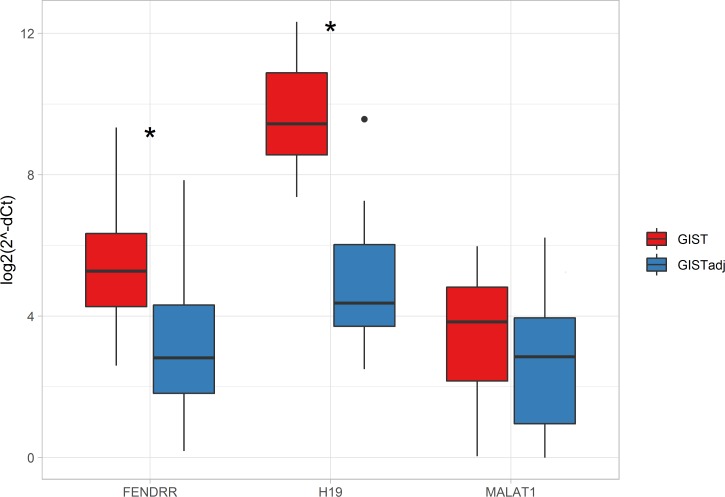
Since the NGS analysis was based on small RNA sequencing, we further validated differential expression of lincRNAs in an independent group of 22 GIST patients (paired GIST and adjacent tissue samples), using RT-qPCR analysis. Three lincRNAs–MALAT1, H19 and FENDRR shown to be significantly deregulated in GIST vs. adjacent tissue in our NGS data and previously associated with oncogenic processes, were selected for this validation step. Gene expression analysis results were in line with NGS data. All three investigated lincRNAs were upregulated in GIST tissue compared to adjacent tissue (Fig 1 and S1 Table). Significant upregulation was observed in expression levels of H19 and FENDRR, with fold changes 25.8 (Bonferroni adjusted p-value = 3.70×10⁻¹¹) and 4.7 (Bonferroni adjusted p-value = 4.66×10⁻⁴), respectively; while MALAT1 was overexpressed nominally (1.7-fold, Bonferroni adjusted p-value = 0.141).
Fig 1. LincRNAs FENDRR, H19 and MALAT1 expression in GIST vs. adjacent tissue (paired samples of 22 patients).
* p-value < 0.05 (Bonferroni adjusted p-value); whiskers–min. and max. values. LincRNA expression was normalized to expression levels of GAPDH.
Correlation analysis of MALAT1, H19, FENDRR and GIST associated oncogenes
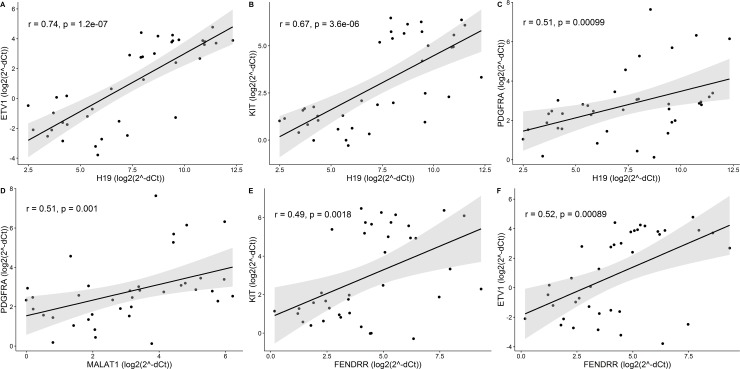
To examine potential effects of changes in lincRNAs expression on GIST pathogenesis, association analysis between lincRNAs and GIST associated oncogenes KIT, PDGFRA and ETV1 was performed. Expression data of KIT, PDGFRA and ETV1 has been previously described [16] and all three genes were shown to be significantly overexpressed in GIST tissue of our study group. High positive correlation was observed between expression levels of ETV1 and H19 (r = 0.74, p = 1.2×10⁻⁷) (Fig 2A, S2 and S3 Tables), while expression levels between H19 and KIT or PDGFRA, MALAT1 and PDGFRA, FENDRR and KIT or ETV1 correlated moderately (r-values between ±0.5 and ±0.7, p-value < 0.01) (Fig 2B–2F, S2 and S3 Tables).
Fig 2. Significant correlations between expression levels of lincRNAs and GIST associated oncogenes KIT, ETV1 and PDGFRA.
(A) High positive correlation between H19 and ETV1, (B-F) Moderate correlation scatter plots. r–Spearman’s rank correlation coefficient; correlation is significant when p < 0.01.
Correlation analysis of MALAT1, H19, FENDRR and microRNAs differentially expressed in GIST
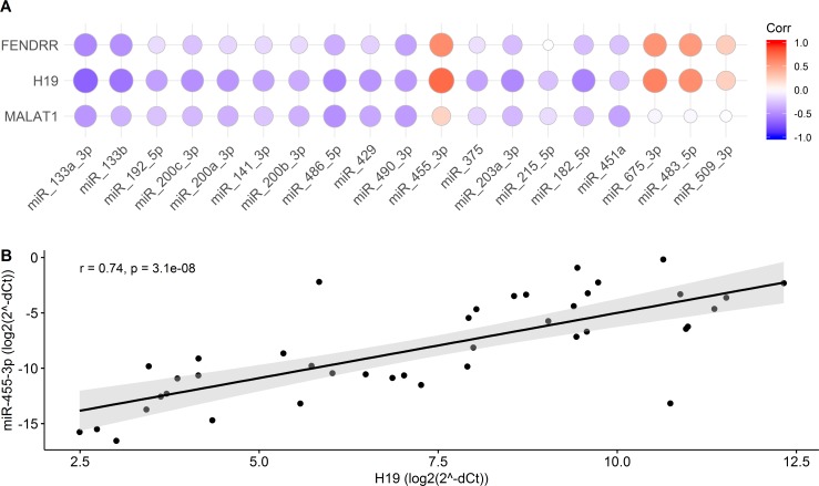
Since it has been shown that lncRNAs can interact with other non-coding RNAs and participate in regulation of tumorigenic processes, lincRNA-microRNA correlation analysis was performed using expression data of 19 microRNAs, shown to be differentially expressed in GIST tissue compared to adjacent tissue in our previous study [16]. Spearman’s correlation test revealed high positive correlation between H19 and miR-455-3p (r = 0.74, p = 3.1×10⁻⁵) (Fig 3B, S2 and S3 Tables), while expression of miR-133a-3p, miR-133b, miR-486-5p, miR-203a-3p, miR-182-5p, miR-675-3p, and miR-483-5p correlated moderately with expression levels of H19 (r-values between ±0.5 and ±0.7, p-value < 0.01) (Fig 3A, S2 and S3 Tables). Significant moderate positive correlations were also observed between FENDRR and miR-455-3p, miR-675-3p, miR-483-5p (0.5 < r < 0.7, p-value < 0.01). MALAT1 showed only low or no correlation with microRNAs differentially expressed in GIST (Fig 3A, S2 and S3 Tables).
Fig 3. Correlation of lincRNAs and microRNAs.
(A) Correlogram of GIST deregulated validated microRNAs and lincRNAs FENDRR, H19 and MALAT1. The size and color intensity of the circle reflects the strength of Spearman correlation. (B) Significant high positive correlation between lincRNA H19 and miR-455-3p. Spearman’s correlation coefficient and p value are shown in the plot.
MALAT1, H19, FENDRR and GIST phenotype association analysis
To investigate possible clinical significance of validated lincRNAs in GIST (validation group n = 22 patients), subphenotype analysis based on histological types (spindle cell type (n = 14), epithelioid cell type (n = 4), mixed spindle and epithelioid cell type (n = 4)), disease risk grade (high (n = 3), moderate (n = 7), low (n = 9) and very low grade (n = 3)) and GIST mutational status (KIT mutant (n = 9), PDGFRA mutant (n = 6), KIT/PDGFRA wild type (n = 2)) was performed (S4 Table). However, multiple group comparison analyses did not reveal any significant differences in expression levels of MALAT1, H19 or FENDRR between different histological subtypes, GIST risk grades or KIT/PDGFRA mutational status (S4 and S5 Tables).
Discussion
Although a number of studies have elucidated the importance of lncRNAs in regulation of various biological processes and development of diseases, their profile and role in pathogenesis of GISTs are scarcely investigated. In the present study, we determined the profile of lincRNAs -a subset of lncRNAs–deregulated in GISTs. Further, we examined expression of three upregulated lincRNAs in a bigger group of independent patients and investigated associations with changes in expression of GIST-associated oncogenes and GIST-deregulated microRNAs.
Analysis of NGS data revealed 9 unique and strongly deregulated lincRNAs in gastric GIST tissue compared to adjacent gastric tissue. Among the top deregulated lincRNAs, three previously cancer associated lincRNAs—MALAT1, H19 and FENDRR, were significantly upregulated in a discovery cohort. MALAT1 and H19 have been mostly described as oncogenic lincRNAs. Their overexpression, contributing to cell proliferation, metastasis, epithelial mesenchymal transition and poor prognosis, has been observed in esophageal squamous cell carcinoma, osteosarcoma, colorectal, gastric and other cancer types [6,8,21–25]. However, in our replication study only H19 showed significant 25.8-fold overexpression in GIST tissue, while MALAT1 was slightly overexpressed, but did not reach the required significance level. Both NGS and validation cohorts indicated FENDRR to be overexpressed in gastric GIST. Previous studies indicated FENDRR to be downregulated in a number of cancers, including osteosarcoma, gastric, breast, prostate cancers, and to be negatively associated with higher tumor stage, deeper invasion, shorter overall survival, suppression of apoptosis, promotion of cell proliferation and migration [26–29], but has been strongly overexpressed in an infantile hemangioma [30]. Furthermore, another study has shown that FENDRR was co-expressed with KIT–an oncogene crucial in GIST development–in a gallbladder cancer [31]. These findings are consistent with our results, where FENDRR expression significantly correlated with expression of KIT, and could explain the strong overexpression of FENDRR in GIST. However, the mechanisms of possible interactions between FENDRR and KIT have not been described yet and more thorough examination is needed in order to determine the role of these oncogenic molecules.
LincRNAs may affect expression of the coding genes through a variety of mechanisms such as regulation of chromatin, scaffolding RNA and proteins or acting as protein and RNA decoys [4]. Therefore, we tested possible correlations between differentially expressed lincRNAs and GIST-associated oncogenes. Analysis revealed a high positive correlation between expression levels of H19 and ETV1. ETV1 is an ETS family transcription factor, which is highly expressed in all GISTs and is crucial for GIST growth and survival [32]. It has been shown that ETV1 is stabilized by another GIST-associated oncogene tyrosine kinase receptor KIT through the MAP kinase signaling pathway and they together form a positive feedback circuit to regulate GIST tumorigenesis [32,33]. Interestingly, another study revealed that H19 might increase phosphorylation levels of components of MAP kinase pathway, such as pMEK and pERK, in colorectal cancer [34]. Moreover, a moderate, but significant correlation was observed between expression levels of H19 and KIT or PDGFRA oncogenes in our study. This supports the theory of the positive feedback circuit between KIT and ETV1 [33], and the possible role of PDGFRA in stabilizing ETV1 [35]. These findings altogether suggest that H19 might be an important factor in pathogenesis of GIST and is worth further investigation.
It has been reported that lncRNAs can interact with microRNAs by masking their targets’ binding sites, acting as competing endogenous RNAs and “sponge” microRNAs away from their mRNA targets, interacting with other microRNA related factors or being precursors for microRNAs [4,36]. Therefore, we used microRNA profiling data from our previous study [16] and evaluated their correlation with the expression of the three lincRNAs. A strong positive correlation was observed between expression levels of H19 and miR-455-3p. Studies of this microRNA have demonstrated inconsistent results, where miR-455-3p has been shown to be overexpressed and/or have oncogenic properties in KIT/PDGFRA mutant GISTs and esophageal squamous cell carcinoma [37,38], but acted as a tumor suppressor in melanoma, pancreatic and non-small cell lung cancers [39–41]. Both H19 and miR-455-3p were shown to be involved in the regulation of cancer related p53 signaling [42,43], which has been shown to be important in GIST oncogenesis, as well [44]. However, no direct association between H19 and miR-455-3p could be found in literature. We also observed a moderate positive association between expression levels of oncogenic miR-675 and H19, as well as a moderate negative correlation between H19 and miR-133 family members and other tumor suppressive microRNAs like miR-486-5p, miR-182-5p or miR-203a-3p [45–48]. MiR-675 is already known to be derived from H19 [9,49] and has been shown to be implicated in the development of colorectal and breast cancers [50,51], while both miR-203a and H19 have been shown to interact with E2F Transcription factor 1 (E2F1) [52–54]. This transcription factor is essential for cell proliferation [55] and can be upregulated by H19 in pancreatic ductal adenocarcinoma [54] or contrarily activates the promoter of H19 in breast cancer cells [53]. Furthermore, E2F1 binds to the promoter and activates miR-203a, while miR-203a can decrease expression of E2F1 through inhibition of CDK6, forming a feed-back loop [52]. Direct interactions between moderately correlated H19 and miR-182, miR-486 or miR-133b have also been previously predicted from Argonaute-crosslinking and immunoprecipitation (AGO-CLIP) data [56] and requires additional experimental validation. As for the other identified correlations between H19 or FENDRR and deregulated microRNAs no data has been found in the literature. Therefore, future studies are needed in order to confirm possible links between these oncogenic molecules and identify their interaction mode and possible role in oncogenesis.
We are aware that our study has certain limitations that need to be acknowledged. Firstly, due to the limited availability of fresh-frozen samples, FFPE samples were used for lincRNA and gene expression analyses in this study. Although, higher levels of RNA degradation can occur in FFPE tissues, previous studies have demonstrated that FFPE tissue can be a feasible material for gene expression analysis [57]. Secondly, we performed lincRNA profile analysis on small-RNA sequencing data. The purified RNA from FFPE is highly affected by hydrolysis and is fragmented into small (on average 100 nucleotide long) sequences [58–60]. These RNA fragments contain 5’-PO4 and 3’-OH end groups, which are used for selective ligation of sequencing adapters in Illumina Truseq small RNA sequencing protocol. Due to these features of FFPE-purified RNA (length and the end groups), small RNA-seq is sufficient to detect long non-coding RNAs in FFPE samples. However, to ensure the reliability of sequencing results, the findings were additionally validated by RT-qPCR method. We also admit that further functional experiments are needed to confirm the role of investigated lincRNAs in GISTs.
In summary, we performed lincRNA expression profile analysis in GISTs and confirmed significant overexpression of H19 and FENDRR in GIST tissue compared to adjacent non-cancerous tissue. Association analyses revealed strong correlations between expression levels of lincRNA H19 and GIST-related oncogene ETV1 and cancer associated miR-455-3p. Despite the limitations described above, lincRNAs MALAT1, H19 and FENDRR were not previously investigated in GISTs and we believe that our findings expand the knowledge in GISTs biology and elucidate possibly important components of GIST tumorigenesis for future studies.
Supporting information
(XLSX)
(XLSX)
(XLSX)
(XLSX)
(XLSX)
Data Availability
All relevant data are within the manuscript and its Supporting Information files. All sequencing data files are available from the NCBI’s Gene Expression Omnibus database (accession number GSE89051).
Funding Statement
This work of UG and JS was supported by the Research Council of Lithuania (https://www.lmt.lt/en/) under the initiative of Researcher teams' projects, Grant no. MIP-006/2014. The funder had no role in study design, data collection and analysis, decision to publish, or preparation of the manuscript.
References
- 1.Hirota S, Isozaki K, Moriyama Y, Hashimoto K, Nishida T, Ishiguro S, et al. Gain-of-function mutations of c-kit in human gastrointestinal stromal tumors. Science. American Association for the Advancement of Science; 1998;279: 577–80. 10.1126/SCIENCE.279.5350.577 [DOI] [PubMed] [Google Scholar]
- 2.Søreide K, Sandvik OM, Søreide JA, Giljaca V, Jureckova A, Bulusu VR. Global epidemiology of gastrointestinal stromal tumours (GIST): A systematic review of population-based cohort studies. Cancer Epidemiol. Elsevier Ltd; 2016;40: 39–46. 10.1016/j.canep.2015.10.031 [DOI] [PubMed] [Google Scholar]
- 3.Corless CL, Barnett CM, Heinrich MC. Gastrointestinal stromal tumours: Origin and molecular oncology. Nat Rev Cancer. Nature Publishing Group; 2011;11: 865–878. 10.1038/nrc3143 [DOI] [PubMed] [Google Scholar]
- 4.Ransohoff JD, Wei Y, Khavari PA. The functions and unique features of long intergenic non-coding RNA. Nat Rev Mol Cell Biol. Nature Publishing Group; 2018;19: 143–157. 10.1038/nrm.2017.104 [DOI] [PMC free article] [PubMed] [Google Scholar]
- 5.Zhang X, Hamblin MH, Yin KJ. The long noncoding RNA Malat1: Its physiological and pathophysiological functions. RNA Biol. Taylor & Francis; 2017;14: 1705–1714. 10.1080/15476286.2017.1358347 [DOI] [PMC free article] [PubMed] [Google Scholar]
- 6.Huarte M. The emerging role of lncRNAs in cancer. Nat Med. 2015;21: 1253–1261. 10.1038/nm.3981 [DOI] [PubMed] [Google Scholar]
- 7.Matouk IJ, DeGroot N, Mezan S, Ayesh S, Abu-lail R, Hochberg A, et al. The H19 non-coding RNA is essential for human tumor growth. PLoS One. Public Library of Science; 2007;2: e845 10.1371/journal.pone.0000845 [DOI] [PMC free article] [PubMed] [Google Scholar]
- 8.Raveh E, Matouk IJ, Gilon M, Hochberg A. The H19 Long non-coding RNA in cancer initiation, progression and metastasis—a proposed unifying theory. Mol Cancer. BioMed Central; 2015;14: 184 10.1186/s12943-015-0458-2 [DOI] [PMC free article] [PubMed] [Google Scholar]
- 9.Cai X, Cullen BR. The imprinted H19 noncoding RNA is a primary microRNA precursor. RNA. 2007;13: 313–6. 10.1261/rna.351707 [DOI] [PMC free article] [PubMed] [Google Scholar]
- 10.Zhang L, Zhou Y, Huang T, Cheng A, Yu J, Kang W, et al. The Interplay of LncRNA-H19 and Its Binding Partners in Physiological Process and Gastric Carcinogenesis. Int J Mol Sci. 2017;18: 450 10.3390/ijms18020450 [DOI] [PMC free article] [PubMed] [Google Scholar]
- 11.Yamamura S, Imai-Sumida M, Tanaka Y, Dahiya R. Interaction and cross-talk between non-coding RNAs. Cell Mol Life Sci. Springer International Publishing; 2018;75: 467–484. 10.1007/s00018-017-2626-6 [DOI] [PMC free article] [PubMed] [Google Scholar]
- 12.Niinuma T, Suzuki H, Nojima M, Nosho K, Yamamoto H, Takamaru H, et al. Upregulation of miR-196a and HOTAIR Drive Malignant Character in Gastrointestinal Stromal Tumors. Cancer Res. 2012;72: 1126–1136. 10.1158/0008-5472.CAN-11-1803 [DOI] [PubMed] [Google Scholar]
- 13.Lee NK, Lee JH, Kim WK, Yun S, Youn YH, Park CH, et al. Promoter methylation of PCDH10 by HOTAIR regulates the progression of gastrointestinal stromal tumors. Oncotarget. 2016;7: 75307–75318. doi: 10.18632/oncotarget.12171 [DOI] [PMC free article] [PubMed] [Google Scholar]
- 14.Cao K, Li M, Miao J, Lu X, Kang X, Zhu H, et al. CCDC26 knockdown enhances resistance of gastrointestinal stromal tumor cells to imatinib by interacting with c-KIT. Am J Transl Res. 2018;10: 274–282. Available: http://www.ncbi.nlm.nih.gov/pubmed/29423012 [PMC free article] [PubMed] [Google Scholar]
- 15.Fletcher CDM, Berman JJ, Corless C, Gorstein F, Lasota J, Longley BJ, et al. Diagnosis of gastrointestinal stromal tumors: A consensus approach. Hum Pathol. 2002;33: 459–65. Available: http://www.ncbi.nlm.nih.gov/pubmed/12094370 [DOI] [PubMed] [Google Scholar]
- 16.Gyvyte U, Juzenas S, Salteniene V, Kupcinskas J, Poskiene L, Kucinskas L, et al. MiRNA profiling of gastrointestinal stromal tumors by next generation sequencing. Oncotarget. 2017;8: 37225–37238. doi: 10.18632/oncotarget.16664 [DOI] [PMC free article] [PubMed] [Google Scholar]
- 17.Fehlmann T, Backes C, Kahraman M, Haas J, Ludwig N, Posch AE, et al. Web-based NGS data analysis using miRMaster: a large-scale meta-analysis of human miRNAs. Nucleic Acids Res. Oxford University Press; 2017;45: 8731–8744. 10.1093/nar/gkx595 [DOI] [PMC free article] [PubMed] [Google Scholar]
- 18.Zhao Y, Li H, Fang S, Kang Y, wu W, Hao Y, et al. NONCODE 2016: an informative and valuable data source of long non-coding RNAs. Nucleic Acids Res. 2016;44: D203–D208. 10.1093/nar/gkv1252 [DOI] [PMC free article] [PubMed] [Google Scholar]
- 19.Mukaka MM. Statistics corner: A guide to appropriate use of correlation coefficient in medical research. Malawi Med J. Medical Association of Malawi; 2012;24: 69–71. Available: http://www.ncbi.nlm.nih.gov/pubmed/23638278 [PMC free article] [PubMed] [Google Scholar]
- 20.R Development Core Team. R: a language and environment for statistical computing In: R foundation for Statistical computing; [Internet]. 2018. [cited 25 Sep 2018]. Available: http://www.r-project.org/. [Google Scholar]
- 21.Hu L, Wu Y, Tan D, Meng H, Wang K, Bai Y, et al. Up-regulation of long noncoding RNA MALAT1 contributes to proliferation and metastasis in esophageal squamous cell carcinoma. J Exp Clin Cancer Res. BioMed Central; 2015;34: 7 10.1186/s13046-015-0123-z [DOI] [PMC free article] [PubMed] [Google Scholar]
- 22.Ji Q, Zhang L, Liu X, Zhou L, Wang W, Han Z, et al. Long non-coding RNA MALAT1 promotes tumour growth and metastasis in colorectal cancer through binding to SFPQ and releasing oncogene PTBP2 from SFPQ/PTBP2 complex. Br J Cancer. 2014;111: 736–748. 10.1038/bjc.2014.383 [DOI] [PMC free article] [PubMed] [Google Scholar]
- 23.Yang F, Bi J, Xue X, Zheng L, Zhi K, Hua J, et al. Up-regulated long non-coding RNA H19 contributes to proliferation of gastric cancer cells. FEBS J. Wiley/Blackwell (10.1111); 2012;279: 3159–3165. 10.1111/j.1742-4658.2012.08694.x [DOI] [PubMed] [Google Scholar]
- 24.Xia H, Chen Q, Chen Y, Ge X, Leng W, Tang Q, et al. The lncRNA MALAT1 is a novel biomarker for gastric cancer metastasis. Oncotarget. Impact Journals, LLC; 2016;7: 56209–56218. doi: 10.18632/oncotarget.10941 [DOI] [PMC free article] [PubMed] [Google Scholar]
- 25.Min L, Garbutt C, Tu C, Hornicek F, Duan Z. Potentials of Long Noncoding RNAs (LncRNAs) in Sarcoma: From Biomarkers to Therapeutic Targets. Int J Mol Sci. Multidisciplinary Digital Publishing Institute (MDPI); 2017;18 10.3390/ijms18040731 [DOI] [PMC free article] [PubMed] [Google Scholar]
- 26.Li Y, Zhang W, Liu P, Xu Y, Tang L, Chen W, et al. Long non-coding RNA FENDRR inhibits cell proliferation and is associated with good prognosis in breast cancer. Onco Targets Ther. 2018;Volume 11: 1403–1412. 10.2147/OTT.S149511 [DOI] [PMC free article] [PubMed] [Google Scholar]
- 27.Xu T, Huang M, Xia R, Liu X, Sun M, Yin L, et al. Decreased expression of the long non-coding RNA FENDRR is associated with poor prognosis in gastric cancer and FENDRR regulates gastric cancer cell metastasis by affecting fibronectin1 expression. J Hematol Oncol. 2014;7: 63 10.1186/s13045-014-0063-7 [DOI] [PMC free article] [PubMed] [Google Scholar]
- 28.Zhang G, Han G, Zhang X, Yu Q, Li Z, Li Z, et al. Long non-coding RNA FENDRR reduces prostate cancer malignancy by competitively binding miR-18a-5p with RUNX1. Biomarkers. 2018;23: 435–445. 10.1080/1354750X.2018.1443509 [DOI] [PubMed] [Google Scholar]
- 29.Kun-Peng Z, Xiao-Long M, Chun-Lin Z. LncRNA FENDRR sensitizes doxorubicin-resistance of osteosarcoma cells through down-regulating ABCB1 and ABCC1. Oncotarget. 2017;8: 71881–71893. doi: 10.18632/oncotarget.17985 [DOI] [PMC free article] [PubMed] [Google Scholar]
- 30.Liu X, Lv R, Zhang L, Xu G, Bi J, Gao F, et al. Long noncoding RNA expression profile of infantile hemangioma identified by microarray analysis. 10.1007/s13277-016-5434-y [DOI] [PubMed] [Google Scholar]
- 31.Zhang L, Geng Z, Meng X, Meng F, Wang L. Screening for key lncRNAs in the progression of gallbladder cancer using bioinformatics analyses. Mol Med Rep. Spandidos Publications; 2018;17: 6449–6455. 10.3892/mmr.2018.8655 [DOI] [PMC free article] [PubMed] [Google Scholar]
- 32.Chi P, Chen Y, Zhang L, Guo X, Wongvipat J, Shamu T, et al. ETV1 is a lineage survival factor that cooperates with KIT in gastrointestinal stromal tumours. Nature. NIH Public Access; 2010;467: 849–53. 10.1038/nature09409 [DOI] [PMC free article] [PubMed] [Google Scholar]
- 33.Ran L, Sirota I, Cao Z, Murphy D, Chen Y, Shukla S, et al. Combined inhibition of MAP kinase and KIT signaling synergistically destabilizes ETV1 and suppresses GIST tumor growth. Cancer Discov. American Association for Cancer Research; 2015;5: 304–15. 10.1158/2159-8290.CD-14-0985 [DOI] [PMC free article] [PubMed] [Google Scholar]
- 34.Yang W, Redpath RE, Zhang C, Ning N. Long non-coding RNA H19 promotes the migration and invasion of colon cancer cells via MAPK signaling pathway. Oncol Lett. Spandidos Publications; 2018;16: 3365–3372. 10.3892/ol.2018.9052 [DOI] [PMC free article] [PubMed] [Google Scholar]
- 35.Hayashi Y, Bardsley MR, Toyomasu Y, Milosavljevic S, Gajdos GB, Choi KM, et al. Platelet-Derived Growth Factor Receptor-α Regulates Proliferation of Gastrointestinal Stromal Tumor Cells With Mutations in KIT by Stabilizing ETV1. Gastroenterology. NIH Public Access; 2015;149: 420–32.e16. 10.1053/j.gastro.2015.04.006 [DOI] [PMC free article] [PubMed] [Google Scholar]
- 36.Kung JTY, Colognori D, Lee JT. Long noncoding RNAs: past, present, and future. Genetics. Genetics Society of America; 2013;193: 651–69. 10.1534/genetics.112.146704 [DOI] [PMC free article] [PubMed] [Google Scholar]
- 37.Pantaleo MA, Ravegnini G, Astolfi A, Simeon V, Nannini M, Saponara M, et al. Integrating miRNA and gene expression profiling analysis revealed regulatory networks in gastrointestinal stromal tumors. Epigenomics. 2016;8: 1347–1366. 10.2217/epi-2016-0030 [DOI] [PubMed] [Google Scholar]
- 38.Liu A, Zhu J, Wu G, Cao L, Tan Z, Zhang S, et al. Antagonizing miR-455-3p inhibits chemoresistance and aggressiveness in esophageal squamous cell carcinoma. Mol Cancer. 2017;16: 106 10.1186/s12943-017-0669-9 [DOI] [PMC free article] [PubMed] [Google Scholar]
- 39.Zhan T, Huang X, Tian X, Chen X, Ding Y, Luo H, et al. Downregulation of MicroRNA-455-3p Links to Proliferation and Drug Resistance of Pancreatic Cancer Cells via Targeting TAZ. Mol Ther—Nucleic Acids. 2018;10: 215–226. 10.1016/j.omtn.2017.12.002 [DOI] [PMC free article] [PubMed] [Google Scholar]
- 40.Chai L, Kang X-J, Sun Z-Z, Zeng M-F, Yu S-R, Ding Y, et al. MiR-497-5p, miR-195-5p and miR-455-3p function as tumor suppressors by targeting hTERT in melanoma A375 cells. Cancer Manag Res. 2018;Volume 10: 989–1003. 10.2147/CMAR.S163335 [DOI] [PMC free article] [PubMed] [Google Scholar]
- 41.Gao X, Zhao H, Diao C, Wang X, Xie Y, Liu Y, et al. miR-455-3p serves as prognostic factor and regulates the proliferation and migration of non-small cell lung cancer through targeting HOXB5. Biochem Biophys Res Commun. 2018;495: 1074–1080. 10.1016/j.bbrc.2017.11.123 [DOI] [PubMed] [Google Scholar]
- 42.Li Z, Meng Q, Pan A, Wu X, Cui J, Wang Y, et al. MicroRNA-455-3p promotes invasion and migration in triple negative breast cancer by targeting tumor suppressor EI24. Oncotarget. 2017;8: 19455–19466. doi: 10.18632/oncotarget.14307 [DOI] [PMC free article] [PubMed] [Google Scholar]
- 43.Baldassarre A, Masotti A. Long non-coding RNAs and p53 regulation. Int J Mol Sci. Multidisciplinary Digital Publishing Institute (MDPI); 2012;13: 16708–17. 10.3390/ijms131216708 [DOI] [PMC free article] [PubMed] [Google Scholar]
- 44.Zong L, Chen P, Xu Y. Correlation between P53 expression and malignant risk of gastrointestinal stromal tumors: Evidence from 9 studies. Eur J Surg Oncol. W.B. Saunders; 2012;38: 189–195. 10.1016/J.EJSO.2011.12.012 [DOI] [PubMed] [Google Scholar]
- 45.Jiang N, Jiang X, Chen Z, Song X, Wu L, Zong D, et al. MiR-203a-3p suppresses cell proliferation and metastasis through inhibiting LASP1 in nasopharyngeal carcinoma. J Exp Clin Cancer Res. 2017;36: 138 10.1186/s13046-017-0604-3 [DOI] [PMC free article] [PubMed] [Google Scholar]
- 46.Shao Y, Shen Y-Q, Li Y-L, Liang C, Zhang B-J, Lu S-D, et al. Direct repression of the oncogene CDK4 by the tumor suppressor miR-486-5p in non-small cell lung cancer. Oncotarget. 2016;7: 34011–21. doi: 10.18632/oncotarget.8514 [DOI] [PMC free article] [PubMed] [Google Scholar]
- 47.Gong Y, Ren J, Liu K, Tang L-M. Tumor suppressor role of miR-133a in gastric cancer by repressing IGF1R. World J Gastroenterol. Baishideng Publishing Group Inc; 2015;21: 2949–58. 10.3748/wjg.v21.i10.2949 [DOI] [PMC free article] [PubMed] [Google Scholar]
- 48.Hu J, Lv G, Zhou S, Zhou Y, Nie B, Duan H, et al. The Downregulation of MiR-182 Is Associated with the Growth and Invasion of Osteosarcoma Cells through the Regulation of TIAM1 Expression. 2015; 10.1371/journal.pone.0121175 [DOI] [PMC free article] [PubMed] [Google Scholar] [Retracted]
- 49.Keniry A, Oxley D, Monnier P, Kyba M, Dandolo L, Smits G, et al. The H19 lincRNA is a developmental reservoir of miR-675 that suppresses growth and Igf1r. Nat Cell Biol. 2012;14: 659–665. 10.1038/ncb2521 [DOI] [PMC free article] [PubMed] [Google Scholar]
- 50.Tsang WP, Ng EKO, Ng SSM, Jin H, Yu J, Sung JJY, et al. Oncofetal H19-derived miR-675 regulates tumor suppressor RB in human colorectal cancer. Carcinogenesis. 2010;31: 350–358. 10.1093/carcin/bgp181 [DOI] [PubMed] [Google Scholar]
- 51.Vennin C, Spruyt N, Dahmani F, Julien S, Bertucci F, Finetti P, et al. H19 non coding RNA-derived miR-675 enhances tumorigenesis and metastasis of breast cancer cells by downregulating c-Cbl and Cbl-b. Oncotarget. Impact Journals, LLC; 2015;6: 29209–23. doi: 10.18632/oncotarget.4976 [DOI] [PMC free article] [PubMed] [Google Scholar]
- 52.Zhang K, Dai L, Zhang B, Xu X, Shi J, Fu L, et al. miR-203 Is a Direct Transcriptional Target of E2F1 and Causes G1 Arrest in Esophageal Cancer Cells. J Cell Physiol. 2015;230: 903–910. 10.1002/jcp.24821 [DOI] [PubMed] [Google Scholar]
- 53.Berteaux N, Lottin S, Monté D, Pinte S, Quatannens B, Coll J, et al. H19 mRNA-like noncoding RNA promotes breast cancer cell proliferation through positive control by E2F1. J Biol Chem. American Society for Biochemistry and Molecular Biology; 2005;280: 29625–36. 10.1074/jbc.M504033200 [DOI] [PubMed] [Google Scholar]
- 54.Ma L, Tian X, Wang F, Zhang Z, Du C, Xie X, et al. The long noncoding RNA H19 promotes cell proliferation via E2F-1 in pancreatic ductal adenocarcinoma. Cancer Biol Ther. 2016;17: 1051–1061. 10.1080/15384047.2016.1219814 [DOI] [PMC free article] [PubMed] [Google Scholar]
- 55.Wu L, Timmers C, Maiti B, Saavedra HI, Sang L, Chong GT, et al. The E2F1–3 transcription factors are essential for cellular proliferation. Nature. 2001;414: 457–462. 10.1038/35106593 [DOI] [PubMed] [Google Scholar]
- 56.Hao Y, Wu W, Li H, Yuan J, Luo J, Zhao Y, et al. Database update NPInter v3.0: an upgraded database of noncoding RNA-associated interactions. Database. 2016;2016: 57 10.1093/database/baw057 [DOI] [PMC free article] [PubMed] [Google Scholar]
- 57.Walter RFH, Mairinger FD, Wohlschlaeger J, Worm K, Ting S, Vollbrecht C, et al. FFPE tissue as a feasible source for gene expression analysis–A comparison of three reference genes and one tumor marker. Pathol—Res Pract. 2013;209: 784–789. 10.1016/j.prp.2013.09.007 [DOI] [PubMed] [Google Scholar]
- 58.Evers DL, Fowler CB, Cunningham BR, Mason JT, O’Leary TJ. The Effect of Formaldehyde Fixation on RNA: Optimization of Formaldehyde Adduct Removal. J Mol Diagnostics. Elsevier; 2011;13: 282–288. 10.1016/J.JMOLDX.2011.01.010 [DOI] [PMC free article] [PubMed] [Google Scholar]
- 59.Kashofer K, Viertler C, Pichler M, Zatloukal K. Quality Control of RNA Preservation and Extraction from Paraffin-Embedded Tissue: Implications for RT-PCR and Microarray Analysis. Yendamuri S, editor. PLoS One. Public Library of Science; 2013;8: e70714 10.1371/journal.pone.0070714 [DOI] [PMC free article] [PubMed] [Google Scholar]
- 60.von Ahlfen S, Missel A, Bendrat K, Schlumpberger M. Determinants of RNA quality from FFPE samples. PLoS One. Public Library of Science; 2007;2: e1261 10.1371/journal.pone.0001261 [DOI] [PMC free article] [PubMed] [Google Scholar]
Associated Data
This section collects any data citations, data availability statements, or supplementary materials included in this article.
Supplementary Materials
(XLSX)
(XLSX)
(XLSX)
(XLSX)
(XLSX)
Data Availability Statement
All relevant data are within the manuscript and its Supporting Information files. All sequencing data files are available from the NCBI’s Gene Expression Omnibus database (accession number GSE89051).


