A 4-year-old boy was presented who fell from a height of about a meter; 3 weeks prior to the presentation. He suffered no loss of consciousness and was active. For about a week prior to the presentation, he was feeling unwell and noted by the family to be less active and complained of occasional abdominal pain. On the day of presentation, while febrile and lying on the couch, he sled and fell on his head. He was brought by his family to the emergency room, conscious but somewhat lethargic and febrile. There was a small bruise over his left eye. The neurological examination was nonfocal.
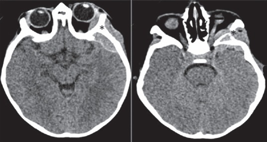
He was admitted for observation and further investigation. His initial noncontrast enhanced computed tomography (NECT) of the head is shown in Figure 1.
Figure 1.

What are the findings?
QUESTIONS
What are your imaging findings?
What is the most likely diagnosis?
What is your recommendation?
ANSWER
1. What are your findings?
Two selected axial cuts from a NECT of the brain show an epidural mass arising from the lateral wall of the left orbit. The mass abuts the left optic nerve and causes left proptosis [Figure 2].
Figure 2.

Two selected axial cuts from a noncontrast enhanced computed tomography of the brain show an epidural mass arising from the lateral wall of the left orbit (astrix) (Figure 1A). The mass abuts the left optic nerve (small black arrow) and causes left proptosis (Figure 1B).
2. What is the most likely diagnosis?
Metastatic neuroblastoma to the left orbit.
Temporal epidural hematoma could be generated as a differential diagnosis, given the history of trauma; but should be eliminated, given the orbital findings and the bony involvement.[1]
3. What is your recommendation?
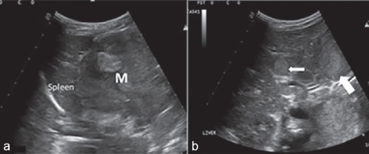
Given the consideration of metastatic neuroblastoma, full blood workup should be sought, and imaging of the abdomen should be requested early to assess the adrenal glands and abdominal metastasis.2 It was done and revealed an adrenal mass and multiple liver lesions and enlarged the retroperitoneal lymph nodes in keeping with the diagnosis [Figure 3].
Figure 3.

Two selected ultrasound images. (a) Large left adrenal mass (M). (b) Liver metastasis (small white arrow) and retroperitoneal lymph nodes (large white arrow).
CONCLUSION
Neuroblastoma is the most common extracranial solid tumor among the pediatric patients, and orbital metastatic disease is not uncommon in these children. Physical signs as a consequence of orbital metastases such as proptosis and periorbital ecchymosis, frequently are encountered and should raise concern in any pediatric patient with imaging appearances which is suggestive of epidural mass.[1,2]
REFERENCES
- 1.Ahmed S, Goel S, Khandwala M, Agrawal A, Chang B, Simmons IG. Neuroblastoma with orbital metastasis: ophthalmic presentation and role of ophthalmologists. Eye (Lond) 2006;20:466–70. doi: 10.1038/sj.eye.6701912. [DOI] [PubMed] [Google Scholar]
- 2.Brodeur GM, Seeger RC, Barrett A, Berthold F, Castleberry RP, D′Angio G, et al. International criteria for diagnosis, staging, and response to treatment in patients with neuroblastoma. J Clin Oncol. 1988;6:1874–81. doi: 10.1200/JCO.1988.6.12.1874. [DOI] [PubMed] [Google Scholar]


