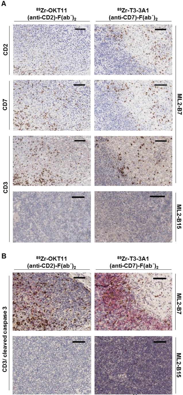
Figure 6.
Immunohistochemistry confirmed the presence of CD2+ and CD7+ T cells in the ML2-B7 tumor specifically recognized by TCR2.5D6 transduced TCM. Tumor tissue from 89Zr-OKT11 (anti-CD2)-F(ab´)2 (left panel) or 89Zr-T3-3A1 (anti-CD7)-F(ab´)2 (right panel) injected mice was harvested after imaging. The bars in the upper right corner represent 100 µm. (A) ML2-B7 tumor slides stained for CD2, CD7, and CD3, as well as ML2-B15 tumor slides stained for CD3 as control. (B) ML2-B7 and ML2-B15 tumor slides stained for CD3 (brown)/ cleaved caspase 3 (red).

