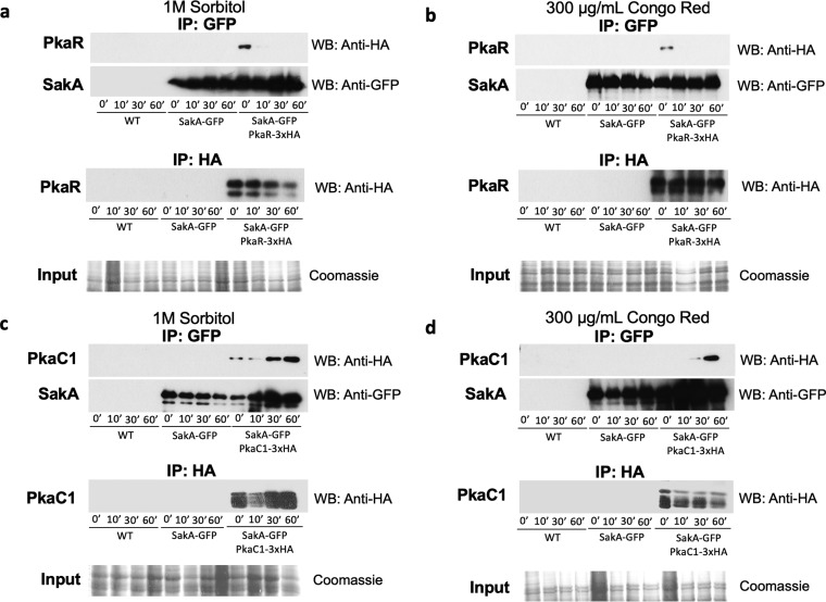
FIG 5.
PKA and the HOG MAPK SakA physically interact under osmotic and cell wall integrity stress conditions. The wild-type (WT), SakA-GFP PkaR-3×HA, and SakA-GFP PkaC1-3×HA strains were grown in either YG or YPD for 20 h before 1 M sorbitol or 300 μg/ml Congo red (CR) was added. Total protein was extracted from mycelia, and immunoprecipitation (IP) of SakA-GFP was carried out before the Western blot was run and analyzed with anti-GFP or anti-HA antibody for the (a and b) PkaR-3×HA interactions with SakA-GFP and (c and d) PkaC1-3×HA interactions with SakA-GFP. IP of PkaR-3×HA or PkaC1-3×HA was carried out, and Western blots were analyzed with anti-HA antibody (lower panel). Coomassie-stained protein gels of the total protein extracts were used as a loading control.

