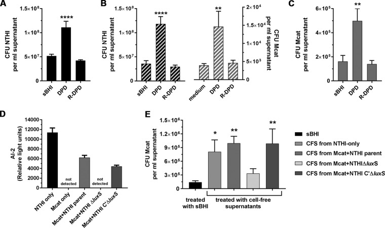
FIG 6.
AI-2 produced by NTHI induced Mcat dispersion from dual-species biofilms. (A to C) NTHI+Mcat biofilms established at 37°C were exposed to medium, DPD, or R-DPD (negative control), and the dispersal of each bacterial species was enumerated. (A) DPD induced significantly greater dispersal of NTHI from monospecies biofilms compared to R-DPD or medium alone. (B) CFU of NTHI (black striped bars) and Mcat (gray striped bars) were significantly greater in supernatants collected from 16-h NTHI+Mcat biofilms exposed to DPD, compared to R-DPD. (C) CFU of Mcat were significantly greater in supernatants taken from 24-h monospecies biofilms treated with DPD, but not R-DPD. (D) Biofilms formed at 37°C for 16 h were exposed to anti-rsPilA for 6 h at 37°C. AI-2 was present in cell-free supernatants (CFS) from biofilms formed by NTHI only, Mcat+NTHI parent strain, or Mcat+complemented ΔluxS strain, but not by Mcat-only or Mcat+NTHI ΔluxS. (E) Mcat-only biofilms established for 24 h were exposed to sBHI or CFS from biofilms formed by NTHI only, or Mcat and either NTHI parent strain, ΔluxS strain, or complemented ΔluxS strain. As assessed by CFU/ml supernatant, Mcat was dispersed from a biofilm in response to CFS from NTHI-only or NTHI parent+Mcat biofilms, but not CFS from NTHI ΔluxS+Mcat biofilms. CFS from dual-species biofilms formed with the complemented ΔluxS+Mcat mutant restored Mcat dispersal. Together these results indicated that the anti-rsPilA-induced Mcat dispersal from NTHI+Mcat biofilms was mediated by AI-2 produced by NTHI in response to exposure to anti-rsPilA. Data represent mean ± SEM from 3 to 4 experiments performed in duplicate. Statistical significance was determined by one-way analysis of variance with the Holm-Sidak correction. Asterisk denotes significant difference from medium alone. *, P ≤ 0.05; **, P ≤ 0.01; ****, P ≤ 0.0001.

