



ABSTRACT
To investigate the molecular mechanism underlying taurine-stimulated proliferation and differentiation of cochlear neural stem cells (NSCs) and potential involvement of Sonic Hedgehog (Shh) pathway. The NSCs were characterized with immunofluorescence stained with nestin antibody. Cell viability was determined by MTT assay. The relative proliferation was measured by BrdU incorporation assay. The morphologic index was measured under light microscope. The relative protein level was determined by immunoblotting. Here we presented our findings that taurine stimulated proliferation and neurite outgrowth of NSCs, which was completely abolished by Shh inhibitor cyclopamine. In addition, cyclopamine antagonized taurine’s effect on glutamatergic and GABAergic neuron population via suppressing expressions of Ptc-1, Smo and Gli-1. Our data supported the critical role of Shh pathway underlying the protective effect of taurine on auditory neural system.
KEYWORDS: Cochlear neural stem cells, cyclopamine, differentiation, Sonic Hedgehog, Taurine
INTRODUCTION
Taurine is the most abundant and widely distributed amino acid in human tissues with fundamental physiological functions.1 The endogenous taurine is mainly synthesized in pancreas via the cysteine sulfinic acid pathway,2 while exogenous source can be obtained from regular diet including egg, meat and seafood.3 It’s been disclosed that relatively high concentration of intrinsic taurine localizes in the heart and retina, which underlies its essential roles in cardiovascular function, development and function of skeletal muscle, retina and the central nervous system.4 Accumulative evidences indicated the protective effects on neural system of supplemented taurine. For example, the intracerebral taurine concentration was significantly induced in the neurons impaired by hypoxia-ischemia in rodents.5 Transient deprivation of oxygen and glucose impaired the neural progenitor cell differentiation into functional neurons, which was ameliorated by taurine treatment. Taurine simultaneously improved cell viability and proliferation of the neural progenitor cells.6 In addition, taurine in vitro administration was shown to promote the proliferative index of neural progenitor cells isolated from mouse embryonic mesencephalon, dentate gyrus and human embryonic brain.7–9 In line with these neural protective effects, our previous study demonstrated that taurine treatment significantly stimulated proliferation, differentiation and neurite outgrowth of cochlea neural stem cells (NSCs).10 However, the molecular mechanism underlying this phenotype is yet to be elucidated.
The Hedgehog signaling pathway transmits information to embryonic cells for proper cell differentiation, and is one of the key regulators of animal development in all bilaterians.11 Members of the hedgehog family play key roles in a wide range of developmental processes. Moreover, it’s increasingly recognized that Hedgehog signaling remains critical in adults and the Sonic hedgehog (Shh) has been shown to be indispensable for the proliferation of adult stem cells in various tissues, including hematopoietic cells, mammary and NSCs.12 For instance, persistent Shh signaling in adult brain determines NSC positional identity.13 Sox2-dependent regulation of Shh dictates hippocampal development and NSC maintenance.14 The neural Hedgehog signaling is required for stem cell renewal in the sensory touch dome epithelial.15 Likewise, assembling evidences suggested fundamental roles of Shh signaling in auditory neural progenitor cells. Recently, in a cDNA profiling interrogation study, the transcripts in Shh pathway were shown to be significantly up-regulated by 1.5 to 4 fold in the taurine-stimulated neural progenitor cells, which indicated that Shh pathway might be activated upon taurine treatment and in turn contributed to the protective effect.16 Therefore, here we sought to verify this hypothesis. The protection on auditory progenitor cells and hearing capacity conferred by taurine was first evaluated in vitro, and the detailed molecular events along Shh pathway were cautiously characterized. Moreover, the Shh-specific inhibitor cyclopamine was employed to efficiently block the relevant signaling with concomitant taurine treatment, and mechanistically involvement of Shh pathway in the setting was evaluated. Our study concluded that Shh activation by taurine treatment predominately mediated its protective effect on auditory progenitor cells.
MATERIALS AND METHODS
Isolation and differentiation of cochlea nscs
The experimental protocol was approved by the Institutional Committee of Ethics on Experimental Animals at The Second Xiangya Hospital, Central South University in strict accordance with the recommendations from the Guide for the Care and Use of Laboratory Animals of the National Institute of Health. All surgical operations were performed under sodium pentobarbital anesthesia and cochlear NSCs were isolated following the well-established protocol.17 Briefly, the one-day old Balb/C mice were sacrificed via decapitation and temporal bone was removed and transferred into pre-chilled Hank’s balanced salt solution (Invitrogen, Carlsbad, CA, USA). The otic capsule was subsequently separated from the otic bulla and opened to expose the membranous labyrinth of the cochlea. The spiral ganglia were obtained from the modiolus via complete removal of the cochlear duct including Corti, spiral ligament and stria vascularis, which was in turn enzymatically digested with 0.125% trypsin/EDTA in phosphate buffered saline (PBS) at 37°C for 5 min. The excess enzymatic activity was quenched by the cocktail of 10 mg/ml soybean trypsin inhibitor (Worthington, USA) and 1 mg/ml DNase I (Worthington) dissolved in DMEM/F12. The homogenous single-cell suspension was archived via passing-through a 70 μm cell strainer (BD Falcon, USA). Cells were sub-cultured every 7 days. For proliferation assay, NSCs were maintained in DMEM-F12 medium supplemented with 2% B-27, 20 ng/ml EGF and 20 ng/ml FGF-2 (R&D Systems, Minneapolis, MN, USA). For differentiation assay, NSCs were maintained in DMEM-F12 medium supplemented with 2% B-27, 10% fetal bovine serum (FBS, Gibco, Grand Island, NY, USA) and 1mM retinoic acid (Sigma, St. Louis, MO, USA). For treatment, taurine (10 mM based on our preliminary experiments) was purchased from Sigma and added into the culture medium at indicated concentrations.
Immunofluorescence
The log phase cells with indicated treatment were washed with PBS and fixed with 4% paraformaldehyde at room temperature for 30 min. The cells were then blocked and permeabilized with 10% fetal calf serum and 0.1% Triton X100 at room temperature for one hour and processed to indicated primary antibody incubation at 4°C overnight. After rigorous wash with PBS, the cells were hybridized with fluorochrome-conjugated secondary antibodies. The cover slips were counterstained with 4,6-diamidino-2-phenylindole (DAPI) in dark at room temperature for 5 min and mounted with Prolong Gold Antifade Reagent (CST#9071, USA). For immunostaining with nestin, VGLUT1 and GAT1, the cochlea was harvested, fixed and decalcified in 2% EDTA solution for 3 weeks, which was then sectioned into 6 m thickness and stained with respective antibodies following the manufacturer’s instruction. The images were acquired under fluorescence microscope (Nikon, USA).
MTT
The cell proliferation assay was performed with the commercial Vybrant MTT Cell Proliferation Assay Kit (ThermoFisher, Waltham, MA, USA) following the manufacturer’s instruction. Briefly, the exponentially growing cells were seeded into 96-well plate and subjected to indicated treatment. 10 μL of MTT stock solution was added into each well and incubated at 37°C for 4 hours. The formed formazan was then dissolved by mixture with 100 μL of the SDS-HCl solution and allowed for incubation in the humidified chamber at 37°C for 4 hours. The absorbance at 570nm was recorded using the microplate reader (Molecular Devices, Sunnyvale, CA, USA).
Brdu incorporation
The DNA synthesis-based cell proliferation assay was performed with the commercial BrdU Cell Proliferation Assay Kit (CST#6813, Cell Signaling Technology, Danvers, MA, USA) in accordance with the manufacturer’s instruction. Briefly, the log phase cells were seeded into 96-well plate and subjected to indicated treatment. The freshly prepared BrdU solution was added to each well for a final 1× concentration and incubated for 2 hours. The medium was completely aspirated and replaced with 100 μL of the Fixing/Denaturing Solution per well and incubated at room temperature for 30 min. The cells were then incubated with 1× detection antibody solution at room temperature for 1 hour. After rigorous wash with 1× wash buffer for 3 times, the cells were subjected to HRP-conjugated secondary antibody incubation at room temperature for another 30 min. The solution was completely removed and 100 μL TMB substrate was added and reaction was allowed for 30 min at room temperature until terminated by 100 μL STOP solution. The absorbance at 450 nm was recorded and relative incorporation of BrdU was calculated.
Western blot
The cell lysate was prepared in RIPA lysis buffer on ice and the cell debris was discarded by refrigerated centrifugation. The protein concentration was determined using the BCA Protein Assay Kit (ThermoFisher, USA) following the manufacturer’s instruction. The equal amount of protein was resolved by SDS-PAGE and transferred onto PVDF membrane on ice. The membrane was briefly blocked with 5% skim milk in TBST buffer and incubated with indicated primary antibodies at 4°C overnight. After rigorous wash with TBST for 30 min, the PVDF membrane was hybridized with specific secondary antibodies at room temperature for one hour. The residual antibody solution was completely removed and washed off with TBST. The protein blots were visualized using the enhanced chemiluminescence method (ECL, Millipore, Billerica, MA, USA).
Statistical analysis
All data presented in this study was acquired from at least three independent experiments. The statistical analysis was performed with the SPSS 23.0 software. The one-way ANOVA was used for multiple groups comparison. The significance was calculated as P value, and P < 0.05 was considered as significantly different.
RESULTS
Cyclopamine inhibits the effect of taurine on the viability and proliferation of mouse cochlea nscs
Our previous study demonstrated that exogenous taurine significantly stimulated proliferation and neurite outgrowth of NSCs with unappreciated mechanisms. Recently, the transcriptome interrogation revealed 1.5 ~ 4-fold increase in the transcripts of Shh pathway in response to taurine (Figure 1(A)) treatment,16 and forced over-activation of Shh pathway remarkably promoted the proliferation of neural progenitor cells in vitro,18 which immediately prompted us to investigate the potential involvement of Shh pathway in taurine-elicited beneficial effect on NSCs.
FIGURE 1.

Cyclopamine inhibited the effect of taurine on the viability and proliferation of mouse cochlea NSCs in vitro. (A) Chemical structure of taurine. (B) The identification of NSCs, revealed by immunostaining against nestin (green), and cell nuclei were counterstained with DAPI (blue). (C) Representative photomicrographs of NSCs to three groups of the control (0 mM, Ctrl), taurine treatment (10 mM, Tau), taurine (10 mM) combined with cyclopamine (2.5 mM) treatment (Tau+ Cyc). Cells were immunostained by tubulin (red) and nuclei were counterstained with DAPI (blue). (D, E) Relative cell viability and proliferation of the NSCs to the control following taurine treatment, OR taurine combined with cyclopamine treatment, measured by MTT assay (D) and BrdU incorporation assay (E). (F) Representative photomicrographs of BrdU labeled cells with their nuclei counterstained by the nuclear fluorescent stain DAPI in three experimental groups. Scale bar: 50 μm. Histogram bars represent mean ± SEM (*P < 0.05, **P < 0.01). n = 8 each group. Cyc, cyclopamine. Tau, taurine.
The cochlea NSCs were isolated from adult mice. We further confirmed this population of cells by employing immunofluorescence staining with antibody against nestin, a specific marker of neural progenitor cells. Our results demonstrated that neural progenitor cells accounted for the majority of cell population isolated (Figure 1(B)), which was appropriate for the following experiments. Taurine treatment remarkably stimulated NSCs cells proliferation and neurite outgrowth as indicated by the morphology immunostained with tubulin antibody. However, the stimulatory effect was readily abolished by co-treatment with Shh-specific inhibitor cyclopamine, which suggested the predominate role of Shh activity downstream of taurine treatment (Figure 1(C)). The cell viability and proliferation was further determined in vitro by MTT and BrdU incorporation assays. Cell viability was significantly increased by approximate 30% upon taurine treatment, which was reversed by addition of cyclopamine (Figure 1(D)). Similarly, cell proliferation interrogated by BrdU incorporation during S replicative phase demonstrated remarkable increase elicited by taurine treatment, and was abrogated by co-treatment with cyclopamine (Figure 1(E, F)).
Cyclopamine inhibits the effect of taurine on the neurite outgrowth of mouse cochlea nscs
We further characterized the impact of taurine on differentiation of the NSCs, and demonstrated taurine treatment promoted differentiation in addition to its pro-proliferative effect. Consistently, this effect apparently depended on Shh pathway since cyclopamine treatment blocked the taurine-induced cell differentiation (Figure 2(A)). The stimulatory effect of taurine on neurite outgrowth was further determined with respect to the number of primary dendrites per cell, the number of dendritic end tips and the average length of neurite in formed neurospheres in conditioned medium. As shown in Figure 2(B-D)), taurine treatment markedly increased the numbers of both primary dendrites, dendrite end tips and the average length of neurites. Again, the inducing effect was abolished by co-treatment with cyclopamine. The GAP-43 protein was significantly induced upon taurine treatment as well, which was antagonized by cyclopamine co-treatment (Figure 2(E)). Consistent with our previous data, here we consolidated that taurine treatment stimulated proliferation and differentiation of NSCs. Furthermore, the above-mentioned effects were readily abrogated by cyclopamine co-treatment, which suggested the predominate role of Shh pathway underlying taurine’s effects.
FIGURE 2.

Cyclopamine inhibited the effect of taurine on the neurite outgrowth of mouse cochlea NSCs. (A) Representative images of the NSCs culture at DIV14 after taurine and cyclopamine treatment compared to control. Cells were stained by tuj-1. Scale bar, 20 μm. (B-D) Statistical analysis of the number of primary dendrites per cell (B), number of dendritic end tips number (C) and average length of neurite (D) in experimental groups on DIV14. (E-F) Western blot analysis of GAP-43 expression and the relative optical densities in the experimental groups. Histogram bars represent mean ± SEM (*P < 0.05, **P < 0.01). Cyc, cyclopamine. Tau, taurine.
Cyclopamine antagonizes taurine’s effect on glutamatergic and gabaergic neuron population
The excitatory and inhibitory neurotransmitters were a pair of counterparts and orchestrated in regulation of cochlea neural impulses. Next, we sought to investigate the differentiation of glutamatergic and GABAergic neurons, which secreted excitatory and inhibitory neurotransmitter, respectively, in NSC culture in response to taurinetreatment. Immunostaining against the glutamatergic neuron-specific marker, vesicular glutamate transporter 1 (VGLUT1), displayed a significant increase of this population by taurine treatment, whereas co-treatment with cyclopamine remarkably compromised this effect (Figure 3(A,B)). Conversely, taurine treatment resulted in obvious reduction of the GABAergic neuron percentage from 17% to 12%, which was restored by co-treatment with cyclopamine (Figure 3(C,D)). Consistent with previous reports that taurine induced glutamatergic and inhibited GABAergic neuron differentiation, our results further demonstrated that these effects greatly depended on downstream Shh pathway, which was effectively blocked by cyclopamine. The stimulated increase in VGLUT1 was further confirmed by immunoblotting with specific antibody. As shown in Figure 4(A), the protein level in NSCs subjected to taurine treatment was markedly up-regulated in comparison with control group, which was subsequently abrogated by cyclopamine (Figure 4(A,B)). Similarly, the influence of taurine on GAT1 protein was further characterized using immunoblotting, which consolidated our immunofluorescence results (Figure 4(C,D)).
FIGURE 3.

Cyclopamine inhibited the effect of taurine-increased glutamatergic neuron population and taurine-reduced GABAergic neuron population in vitro. (A, C) Control, taurine alone or taurine combined with cyclopamine treated cultures in vitro, glutamatergic neurons (A) and GABAergic neurons (C) were indicated by positive staining by VGLUT1 (green) (A) and GAT1 (green) (C). Part of the cell population was nestin-positive (red). Nuclei were stained DAPI (blue). (B, D) Percentage of glutamatergic neurons (B) and GABAergic neurons (D) in culture relative to the control after taurine alone or taurine combined with cyclopamine treatment. Scale bar, 100 μm. Histogram bars represent mean ± SEM (*P < 0.05, **P < 0.01). n = 6 each group. Cyc, cyclopamine. Tau, taurine.
FIGURE 4.
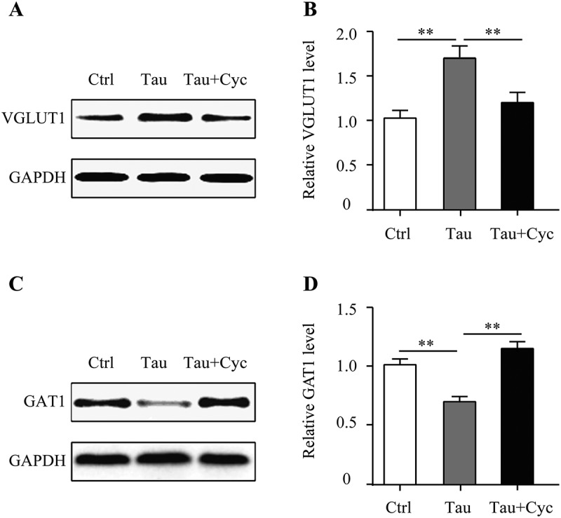
Cyclopamine inhibited the effect of taurine on VGLUT1 and GAT1 proteins expression in NSCs in vitro. (A, C) VGLUT1 and GAT1 protein levels, as detected by western blotting. (B, D) Quantitative analysis of VGLUT1 and GAT1 protein levels. GAPDH was used as a loading control. Histogram bars represent mean ± SEM (*P < 0.05, **P < 0.01). n = 6 each group. Cyc, cyclopamine. Tau, taurine.
Cyclopamine suppresses shh, ptc-1, smo, gli-1 expression induced by taurine in nscs
Our pervious investigations suggested that Shh pathway was mechanistically involved in taurine-induced proliferation and differentiation, which was readily suppressed by specific inhibitor. Next, we sought to characterize the detailed molecular profile along Shh pathway in response to taurine and cyclopamine. Four critical factors including Shh, Patched-1 (Ptc-1), Smoothened (Smo) and Gli-1 were quantitatively interrogated in NSCs treated with either taurine alone or combined with cyclopamine. As shown in Figure 5, taurine treatment greatly stimulated up-regulation of Shh, Ptc-1, Smo and Gli-1, which was abolished by addition of cyclopamine. Our data disclosed that taurine activated Shh pathway via up-regulation of Shh, Ptc-1, Smo and Gli-1 proteins, and cyclopamine significantly reversed this effect.
FIGURE 5.
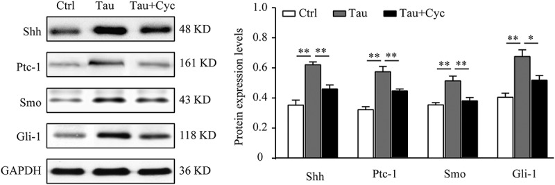
Cyclopamine inhibited the effect of taurine on the expression of Shh, Ptc-1, Smo, Gli-1 proteins in NSCs in vitro. (A) Shh, Ptc-1, Smo, and Gli-1 protein levels, as detected by western blotting. (B) Quantitative analysis of Shh, Ptc-1, Smo, and Gli-1 levels. Histogram bars represent mean ± SEM (*P < 0.05, **P < 0.01). n = 5 each group. Cyc, cyclopamine. Tau, taurine.
DISCUSSION
Our previous study demonstrated that taurine treatment promoted proliferation, differentiation and neurite outgrowth of NSCs in vitro with undefined mechanism.10 In this study, we have investigated the potential mechanistic involvement of Shh pathway in taurine-stimulated proliferation and differentiation of NSCs. Our results first consolidated the previous finding that taurine treatment significantly promoted the proliferative index of cultured NSCs.10 Morphologic inspection displayed that taurine stimulated the neurite outgrowth of NSCs as well. Moreover, the differentiation potential of NSCs into the glutamatergic neurons was remarkably induced while the direction into GABAergic neuron was conversely suppressed in response to taurine treatment, which was in agreement with the physiological function of glutamatergic neurons in producing the excitatory neurotransmitters in this setting. Consistent with the results from cDNA profiling investigation into taurine-treated neural progenitor cells,16 we further confirmed that several key factors in the Shh pathway, including Ptc-1, Smo and Gli-1, were significantly up-regulated in NSCs upon taurine treatment, which unambiguously indicated that Shh pathway was activated by taurine. The importance of Shh pathway in mediating taurine-elicited protective effects on NSCs was interrogated by introduction of the Shh-specific inhibitor cyclopamine. Cyclopamine is a highly potent Shh inhibitor by influencing the balance between active and inactive forms of the smoothened protein.19 While co-administration with taurine, the beneficial effects on proliferation and differentiation of NSCs imposed by taurine was readily abrogated by cyclopamine, which highlighted the predominate role of Shh pathway underlying this phenotype. To our best knowledge, for the first time here we elucidated the mechanistic involvement of Shh pathway in taurine-conferred protection on auditory NSCs, which highlight the potential for future therapeutic exploitation aiming to activate Shh pathway for auditory impairment. However, the signaling pathway during taurine-activated Shh is still to be defined. In addition to Shh signaling, the gene expression profile demonstrated that taurine also regulated genes involved in the Wnt pathway, cellular adhesion, cell survival and mitochondrial functioning,16 which suggested multiple candidate targets of taurine and requested further comprehensive investigation.
The beneficial effects on hearing and the potential therapeutic application of taurine have been extensively investigated so far. For instance, Liu et al. demonstrated that taurine attenuated aminoglycoside ototoxicity by inhibiting inducible nitric oxide synthase expression in the Guinea pig cochlea.20 Liu et al. disclosed that taurine modulated calcium influx under normal and ototoxic conditions in isolated cochlear spiral ganglion neurons.21 Brozoski et al. reported that supplemental dietary taurine significantly attenuated tinnitus and improved auditory discrimination by increasing inhibitory tone and decreasing noise in the auditory pathway.22 Ye et al. showed taurine attenuated bilirubin-induced neurotoxicity in the auditory system in neonatal guinea pigs, and suggested the potential of taurine as a broad-spectrum agent for preventing and/or treating hearing loss in neonatal jaundice.23 In line with these elegant studies, our previous data demonstrated that taurine significantly enhanced excitability of mouse cochlear NSCs by selectively promoting differentiation of glutamatergic neurons over GABAergic neurons. The molecular mechanism underlying this effect involving Shh pathway activation was further elucidated in this study.
Hearing loss is one of the most common health problem worldwide with limited clinical options for therapeutic purpose.24 Although hearing aids are applicable efficiently in most patients who suffer from moderate to severe hearing impairment,25 the cochlea stem cells/progenitor transplantation is still the only option for hearing rehabilitation in the profound complications.26 Numerous endeavors have been invested into the attempt to transplant various stem cells into the inner ear with very limited success in differentiation into functional neurons, glia, hair cells or supporting cells. Exploitations into the alternative way to circumvent this problem mainly concentrated on the genetically modified stem cells with enhanced capacity with respect to regeneration and differentiation potential.27,28 Here our data offered another way to improve the efficiency of NSC transplantation via co-treatment with taurine, which potentiated both proliferation and differentiation. Noteworthily, our current study concentrated on phenotype and mechanism with cultured cells in vitro, further in vivo investigations are definitely warranted for potential therapeutic applications.
In summary, here we demonstrate cyclopamine treatment abrogates cell proliferation and differentiation of NSCs stimulated by taurine via specific inhibition of Shh pathway, which highlights Shh signaling in taurine-elicited protective effect on NSCs potentially as activating targets for therapeutic purpose.
Funding Statement
This work was supported by the National Science Foundation of China (No. 81600811);the National Science Foundation of China [81600811].
Competing interests
The authors have declared that no conflicts of interest exist.
Acknowledgments
Not applicable.
References
- 1.Ripps H, Shen W.. Review: taurine: a “very essential” amino acid. Mol Vis. 2012;18:2673–2686. [PMC free article] [PubMed] [Google Scholar]
- 2.Chang YC, Ding ST, Lee YH, Wang YC, Huang MF, Liu IH. Taurine homeostasis requires de novo synthesis via cysteine sulfinic acid decarboxylase during zebrafish early embryogenesis. Amino Acids. 2013;44(2):615–629. [DOI] [PubMed] [Google Scholar]
- 3.Bouckenooghe T, Remacle C, Reusens B. Is taurine a functional nutrient? Curr Opin Clin Nutr Metab Care. 2006;9(6):728–733. [DOI] [PubMed] [Google Scholar]
- 4.Lambert IH, Kristensen DM, Holm JB, Mortensen OH. Physiological role of taurine–from organism to organelle. Acta Physiol (Oxf). 2015;213(1):191–212. [DOI] [PubMed] [Google Scholar]
- 5.Maia AR, Batista TM, Victorio JA, Clerici SP, Delbin MA, Carneiro EM, Davel AP. Taurine supplementation reduces blood pressure and prevents endothelial dysfunction and oxidative stress in post-weaning protein-restricted rats. PLoS One. 2014;9(8):e105851. [DOI] [PMC free article] [PubMed] [Google Scholar]
- 6.Wang Q, Yang L, Wang YP. Enhanced differentiation of neural stem cells to neurons and promotion of neurite outgrowth by oxygen-glucose deprivation. Int J Dev Neurosci. 2015;4350–4357. DOI: 10.1016/j.ijdevneu.2015.04.009 [DOI] [PubMed] [Google Scholar]
- 7.Hernandez-Benitez R, Pasantes-Morales H, Saldana IT, Ramos-Mandujano G. Taurine stimulates proliferation of mice embryonic cultured neural progenitor cells. J Neurosci Res. 2010;88(8):1673–1681. [DOI] [PubMed] [Google Scholar]
- 8.Hernandez-Benitez R, Vangipurarn SD, Ramos-Mandujano G, Lyman WD, Pasantes-Morales H. Taurine enhances the growth of neural precursors derived from fetal human brain and promotes neuronal specification. Dev Neurosci-Basel. 2013;35(1):40–49. [DOI] [PubMed] [Google Scholar]
- 9.Shivaraj MC, Marcy G, Low GL, Ryu JR, Zhao XF, Rosales FJ, Goh ELK. Taurine induces proliferation of neural stem cells and synapse development in the developing mouse brain. Plos One. 2012;7(8):e42935. [DOI] [PMC free article] [PubMed] [Google Scholar]
- 10.Wang Q, Zhu GH, Xie DH, Wu WJ, Hu P. Taurine enhances excitability of mouse cochlear neural stem cells by selectively promoting differentiation of glutamatergic neurons over gabaergic neurons. Neurochem Res. 2015;40(5):924–931. [DOI] [PubMed] [Google Scholar]
- 11.Lee RT, Zhao Z, Ingham PW. Hedgehog signalling. Development. 2016;143(3):367–372. [DOI] [PubMed] [Google Scholar]
- 12.Parisi MJ, Lin H. The role of the hedgehog/patched signaling pathway in epithelial stem cell proliferation: from fly to human. Cell Res. 1998;8(1):15–21. [DOI] [PubMed] [Google Scholar]
- 13.Ihrie RA, Shah JK, Harwell CC, Levine JH, Guinto CD, Lezameta M, Kriegstein AR, Alvarez-Buylla A. Persistent sonic hedgehog signaling in adult brain determines neural stem cell positional identity. Neuron. 2011;71(2):250–262. [DOI] [PMC free article] [PubMed] [Google Scholar]
- 14.Favaro R, Valotta M, Ferri AL, Latorre E, Mariani J, Giachino C, Lancini C, Tosetti V, Ottolenghi S, Taylor V, et al. Hippocampal development and neural stem cell maintenance require Sox2-dependent regulation of shh. Nat Neurosci. 2009;12(10):1248–1256. [DOI] [PubMed] [Google Scholar]
- 15.Xiao Y, Thoresen DT, Williams JS, Wang C, Perna J, Petrova R, Brownell I. Neural hedgehog signaling maintains stem cell renewal in the sensory touch dome epithelium. Proc Natl Acad Sci USA. 2015;112(23):7195–7200. [DOI] [PMC free article] [PubMed] [Google Scholar]
- 16.Ramos-Mandujano G, Hernandez-Benitez R, Pasantes-Morales H. Multiple mechanisms mediate the taurine-induced proliferation of neural stem/progen itor cells from the subventricular zone of the adult mouse. Stem Cell Res. 2014;12(3):690–702. [DOI] [PubMed] [Google Scholar]
- 17.Xiao B, Liu H, Gu Z, Liu S, Ji C. Taurine protected against the impairments of neural stem cell differentiated neurons induced by oxygen-glucose deprivation. Neurochem Res. 2015;40(11):2348–2356. [DOI] [PubMed] [Google Scholar]
- 18.Lai K, Kaspar BK, Gage FH, Schaffer DV. Sonic hedgehog regulates adult neural progenitor proliferation in vitro and in vivo. Nat Neurosci. 2003;6(1):21–27. [DOI] [PubMed] [Google Scholar]
- 19.Chen JK, Taipale J, Cooper MK, Beachy PA. Inhibition of hedgehog signaling by direct binding of cyclopamine to Smoothened. Genes Dev. 2002;16(21):2743–2748. [DOI] [PMC free article] [PubMed] [Google Scholar]
- 20.Liu HY, Chi FL, Gao WY. Taurine attenuates aminoglycoside ototoxicity by inhibiting inducible nitric oxide synthase expression in the cochlea. Neuroreport. 2008;19(1):117–120. [DOI] [PubMed] [Google Scholar]
- 21.Liu HY, Chi FL, Gao WY. Taurine modulates calcium influx under normal and ototoxic conditions in isolated cochlear spiral ganglion neurons. Pharmacol Rep. 2008;60(4):508–513. [PubMed] [Google Scholar]
- 22.Brozoski TJ, Caspary DM, Bauer CA, Richardson BD. The effect of supplemental dietary taurine on tinnitus and auditory discrimination in an animal model. Hear Res. 2010;270(1–2):71–80. [DOI] [PMC free article] [PubMed] [Google Scholar]
- 23.Ye HB, Wang J, Zhang WT, Shi HB, Yin SK. Taurine attenuates bilirubin-induced neurotoxicity in the auditory system in neonatal guinea pigs. Int J Pediatr Otorhinolaryngol. 2013;77(5):647–654. [DOI] [PubMed] [Google Scholar]
- 24.Lasak JM, Allen P, McVay T, Lewis D. Hearing loss: diagnosis and management. Prim Care. 2014;41(1):19–31. [DOI] [PubMed] [Google Scholar]
- 25.Williger B, Lang FR. Managing age-related hearing loss: how to use hearing aids efficiently - a mini-review. Gerontology. 2014;60(5):440–447. [DOI] [PubMed] [Google Scholar]
- 26.Muller U, Barr-Gillespie PG. New treatment options for hearing loss. Nat Rev Drug Discov. 2015;14(5):346–365. [DOI] [PubMed] [Google Scholar]
- 27.Tan CQ, Gao X, Guo L, Huang H. Exogenous IL-4-expressing bone marrow mesenchymal stem cells for the treatment of autoimmune sensorineural hearing loss in a guinea pig model. Biomed Res Int. 2014;2014856019 DOI: 10.1155/2014/856019. [DOI] [PMC free article] [PubMed] [Google Scholar] [Retracted]
- 28.Kesser BW, Lalwani AK. Gene therapy and stem cell transplantation: strategies for hearing restoration. Adv Otorhinolaryngol. 2009;6664–6686. DOI: 10.1159/000218208. [DOI] [PubMed] [Google Scholar]