
Figure 2.
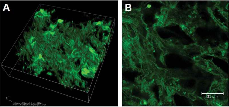
Confocal acquisitions of an eosin-stained NI3 liver decellularised matrix. A) 3D rendering of a 62 x 62 x 16 μm volume and B) detail of a central slice of the confocal scan showing a rich intra-lobular network with an average (equilibrium swollen) pore size of about 22 μm.