Abstract
Background: Injuries to the scapholunate can have severe long-term effects on the wrist. Early detection of these injuries can help identify pathology. The purpose of this study was to evaluate the motions of the scapholunate joint in normal wrists in a clenched fist and through radial and ulnar deviation using novel dynamic computed tomography (CT) imaging. Methods: Fifteen participants below 40 years of age consented to have their wrist scanned. Eight participants were randomized to have the right wrist scanned and 7 the left wrist. Volunteers were positioned at the back of the gantry with the wrist placed on the table, palmar side down. Participants began with the hand in a relaxed fist position and then proceeded through an established range of motion protocol. Dynamic CT imaging was captured throughout the range of motion. Results: The movement in the healthy scapholunate joint through a clenched fist and radial and ulnar deviation is minimal. The averages were 1.19, 1.01, and 0.95 mm, representing the middle, dorsal, and volar measurements, respectively. Conclusions: This novel dynamic CT scan of the wrist is a user-friendly way of measuring of the scapholunate distance, which is minimal in the normal wrist below 40 years of age.
Keywords: scapholunate, computed tomography, 4D CT, dynamic, clenched fist
Introduction
The scapholunate joint has been arguably the most studied aspect of the human wrist. The sequelae from injuries to the ligament can be debilitating and severely limit function and quality of life.13,20,24,25 The potential for deformity and damaging effects to other carpal joints makes recognition of these injuries very important. The human carpus has a tremendous amount of complexity stemming from the interactions of the bones and ligaments, and their behaviors through the various ranges of motion.3,19 Advances in imaging from specific radiographs14,18 to computed tomography (CT) and magnetic resonance imaging (MRI) have allowed better understanding of wrist kinematics and potentially improve diagnostic accuracy of wrist ligament pathology.1,12,15,21-23 Much of the dynamic CT studies of the scapholunate joint have centered around the movement described as dart throwers motion.6,7 However, dart throwers motion is predominantly a radial-ulnar deviation motion and does not assess stress of a clenched fist. MRI also provides excellent information and characterization of the ligamentous structures about the carpus, but lacks analysis of wrist kinematics.2
The clenched fist view has previously been studied in scapholunate dissociation, and our goal was to assess the normal joint behavior in this position using dynamic CT imaging.14,17,18 The advancements in CT scanning and software to utilize dynamic CT (4D CT) analysis have created a unique modality to examine the wrist.4 Our goal was to establish a protocol for measurements of the normal joint, on a novel dynamic CT wrist scan. The purpose of this study was to evaluate the motions of the scapholunate joint in normal wrists with a clenched fist view, as well as through radial and ulnar deviation. Understanding the appearance of a normal wrist scan would be crucial to interpreting the imaging of an injured wrist using this new test. The radiation exposure to participants for this exam was low when compared with the normal background radiation any individual experiences. This has been documented in previous dynamic studies of the scapholunate joint, adding improved benefits to patient safety.6,7
Materials and Methods
Research ethics approval was obtained from our institution prior to commencement of the study. Informed written consent was obtained from all individual participants included in this study. Fifteen volunteers consented to have their wrist scanned. The wrist to be scanned was selected with the flip of a coin. Eight participants were randomized to have the right wrist scanned and 7 the left wrist. All participants denied any previous significant trauma to the wrist to be scanned. No participants had previous wrist surgery. All participants were questioned regarding symptoms of instability (eg, pain, clicking, popping) or a history of cast immobilization and were excluded if any of these were present. All 15 scans were used in the analysis.
The images were acquired using 320 Multidetector CT Scanner (Aquilion ONE ViSION Edition, software version 7.0; Toshiba Medical Systems Inc, Toshigi-ken, Japan). The 4D Orthopaedic Analysis application was used via the SUREXtension software (Toshiba Medical Systems Inc) to further examine and analyze the data collected from these dynamic CT scans. Vitrea Fx Workstation (Vital Images, Inc, Minnetonka, Minnesota) was also used for analysis of the studies.
A protocol was designed to ensure adequate time (10 seconds), yet limited exposure, for volunteers completing the scan. The area to be scanned was set at 8 cm, to include the distal radioulnar joint and entire carpus. The protocol was developed on the scanner computer to ensure settings were the same for each participant. Individualized demonstrations were given by the study investigator to each participant to ensure understanding of the necessary movements to be completed and the appropriate speed and position of the wrist throughout. The investigator was present for all participants to ensure the process was understood. All scans were completed without complications.
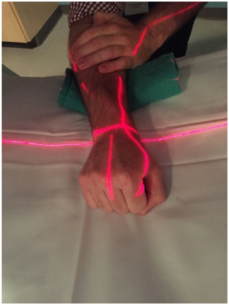
Volunteers were provided with protective lead gowns and thyroid shields and positioned at the back of the gantry. The selected wrist was then placed on the table, palmar side down. A folded towel was placed to elevate the radiocarpal joint to the level of the image acquisition sector and to avoid wrist extension with clenching of the fist.11 The elbow and forearm were rested on the table. The contralateral hand was used to steady the arm proximal to the wrist to prevent pronation, supination, or position changes through the elbow joint (Figure 1). Participants began with the hand in a relaxed fist position and then proceeded to clench the hand in a full fist and relax. Once relaxed again, the wrist was maximally ulnar deviated and then maximally radially deviated. This was all done in a fluid motion within 10 seconds (which was preset as the maximum detection time). All participants were able to complete the range of motion in the allotted time and some were able to stop the exam prior to the set timer elapsing (Supplemental Videos 1 and 2).
Figure 1.

Patient positioning in computed tomography scanner for study.
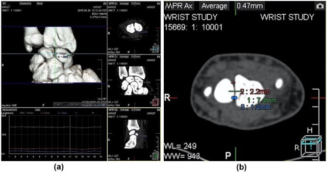
The points used for our measurements were carefully selected using all planes from the dynamic CT. The markers were placed within the scaphoid and lunate bones on the CT images to ensure they were within the bone in all planes. The cortex points were selected dorsal and volar of the estimated midpoint on the axial images (Figure 2).
Figure 2.
(a) 4D Orthopedic Analysis application (Toshiba Medical Systems Inc) analysis of individual study volunteer depicting three-dimensional, axial, coronal, and sagittal dynamic computed tomography with graphed measurements. Some measured lines are present on depicted slices and (b) axial cut depicting location of markers and measurement lines.
Results
As is evident in the results, the movement in the healthy scapholunate joint is minimal. The averages (and standard deviations) were 1.19 ± 0.84 mm, 1.01 ± 0.38 mm, and 0.95 ± 0.69 mm, representing the middle, dorsal, and volar measurements, respectively (Table 1).
Table 1.
Maximum Distances Between Marked Points on the Scaphoid and Lunate Measured on the Computed Tomographic Scans Using the 4D Orthopedic Analysis Application via SUREXtension Software (Toshiba Medical Systems Inc).
| Study | Green/middle (mm)a | Red/dorsal (mm)b | Blue/volar (mm)c |
|---|---|---|---|
| 1 | 0.4 | 0.4 | 0.5 |
| 2 | 1.1 | 1.2 | 0.9 |
| 3 | 1.1 | 1.2 | 0.5 |
| 4 | 1.0 | 1.0 | 1.0 |
| 5 | 0.9 | 0.8 | 0.9 |
| 6 | 1.5 | 1.2 | 0.8 |
| 7 | 0.6 | 0.7 | 0.7 |
| 8 | 0.9 | 1.1 | 0.8 |
| 9 | 1.7 | 1.1 | 1.1 |
| 10 | 3.8 | 2.0 | 3.2 |
| 11 | 0.8 | 0.8 | 0.4 |
| 12 | 1.1 | 0.8 | 1.0 |
| 13 | 0.5 | 0.7 | 0.3 |
| 14 | 1.9 | 1.4 | 1.5 |
| 15 | 0.5 | 0.7 | 0.6 |
| Average | 1.19 ± 0.84 | 1.01 ± 0.38 | 0.95 ± 0.69 |
Green. Measured between a point determined to fall within each of the scaphoid and lunate bones in all planes. This was made at the midpoint of the scapholunate joint in each of the sagittal, coronal, and axial planes.
Red. A line made dorsal to middle/green line on the axial images. This line was connecting points on the cortex of the scaphoid and lunate.
Blue. A line made volar to middle/green line on the axial images. This line was connecting points on the cortex of the scaphoid and lunate.
Discussion
The scapholunate joint has been well studied as a source of pathology in the wrist. Injuries to this ligament have been shown to lead to degenerative changes throughout the carpus and wrist.20,24 Instability through the joint can progress to dorsal intercalated segmental instability deformity that can further advance to a scapholunate advanced collapse (SLAC) wrist. The malposition of the scaphoid (flexed) and lunate (extended) leads to the progressive degenerative changes of the SLAC wrist described and classified by Watson and Ballet.24 Early diagnosis of these injuries has continued to challenge physicians, with the thought that earlier recognition and treatment could yield improved outcomes.
Concluding a diagnosis of scapholunate injuries can be a challenge. Plain radiographs pose a challenge in identifying injuries early, as they can be partial or complete. Partial injuries can take weeks to months to be present on plain film, thus severity and timing are factors in radiographic diagnosis. MRI certainly provides a better anatomic evaluation of the wrist ligaments, but lacks functional assessment.2 This study shows the setup and time to complete the dynamic CT study is minimal. Diagnostic arthroscopy of the wrist does provide a means of assessing kinematic behavior of the carpal bones, but this invasive measure could possibly be avoided with assessment via dynamic CT.5 There are 2 established arthroscopic classifications for the stages of scapholunate dissociation, the European Wrist Arthroscopy Society (EWAS) and Geissler classifications, for which plain radiographs have poor correlation to diagnosis of dynamic instability.10,16 Utilization of this dynamic CT could provide a noninvasive means of identifying the early stages of each classification system, without subjecting patients to arthroscopy.
Dynamic imaging is an advanced imaging modality readily available for diagnosis. Static imaging for the wrist has improved steadily over time. The addition of motion analysis allows us to visualize the relationship of bones as they go through functional range of motion. This novel approach of looking at the normal motion of the scapholunate joint, through clenching of the fist and radial and ulnar deviation, will help better understand normal kinematics. The next step is to compare these normal scans to wrists with known pathology who undergo dynamic CT scans as part of their surgical planning. Understanding normal imaging anatomy is imperative, prior to utilizing this dynamic CT for defining pathology.
The clenched fist view or posterior-anterior (PA) stress view has been used to visualize the dynamic loading of the scapholunate joint. The longitudinal force of the contracting forearm muscle drives the capitate proximally and widens the scapholunate interval if the ligament is disrupted. The PA film is captured with the patient forming a tight fist or while grasping an object, such as a pencil. Widening of the scapholunate >3 mm on the clenched fist view has been recognized as an indication of possible disruption of the scapholunate ligament.14,18
In our sample of normal wrists, the clenching of the fist and radial and ulnar deviation show minimal change in the scaphoid and lunate position relative to one another (<2 mm). The normal scapholunate ligament maintains the position of the 2 bones throughout a full range in the coronal plane, as well as with clenching the fist. We are able to show that dynamic CT is a user-friendly and timely means of imaging the motions of the scapholunate joint. One participant (Study 10) had measurements that were suggestive of increased scapholunate distance, which could be in keeping with an unknown injury or anatomic variation.
This novel technique requires proper setup and positioning of the patient’s wrist in the CT scanner, as well as availability of the necessary software to produce quality imaging. Elevating the wrist slightly off the table ensures it is captured by the imager throughout movement, as well as stabilizing the remaining forearm to capture true wrist motion. The scan field can be adjusted based on patient size and the specific area to be scanned, but should be limited to reduce radiation exposure risks.
Limitations of a dynamic/4D CT study include the fact that the images are an approximation based on tomographic image data.26 The landmarks used in the measurements were applied manually, and measurement error cannot be excluded. Our sample was small, and a larger scale study would be required to fully characterize the normal joint motion. The assumption of participant’s wrists being normal was based on their self-reported lack of pain or history of injury and certainly does not rule out the fact there could be underlying abnormalities in our study group. Further studies will need to be performed with documented or suspected scapholunate instability patients undergoing dynamic imaging, to assess if these measurements are indeed accurate ways of characterizing the scapholunate joint stability.
Diagnosing scapholunate instability can be challenging in radiographic evaluations. Our study shows dynamic CT scans of the wrist are a timely, minimally invasive technique to accurately image the scapholunate joint. Early recognition of scapholunate injuries has the potential to lead to early diagnosis and limit long-term functional disability. Being able to achieve a diagnosis early can lead to potential less invasive treatments for acute injuries, as healing potential of the scapholunate ligament declines with time and eliminates reduction/pinning or primary repair option.9 Even with established diagnosis, surgical repair or reconstruction techniques are challenging, and many different procedures have been described for scapholunate instability.8,9 Dynamic CT assessments of musculoskeletal patients is a field that continues to evolve, with the ultimate goal of improved patient care with limited risk.
Footnotes
Supplemental material is available in the online version of the article.
Ethical Approval: Approved by Heath Research Ethics Authority of Newfoundland and Labrador—HREB# 2015.040.
Statement of Human and Animal Rights: All procedures followed were in accordance with the ethical standards of the responsible committee on human experimentation (institutional and national) and with the Helsinki Declaration of 1975, as revised in 2008.
Statement of Informed Consent: Informed consent was obtained from all individual participants included in this study.
Declaration of Conflicting Interests: The author(s) declared no potential conflicts of interest with respect to the research, authorship, and/or publication of this article.
Funding: The author(s) received no financial support for the research, authorship, and/or publication of this article.
References
- 1. Alta TD, Bell SN, Troupis JM, et al. The new 4-dimensional computed tomographic scanner allows dynamic visualization and measurement of normal acromioclavicular joint motion in an unloaded and loaded condition. J Comput Assist Tomogr. 2012;26(6):749-754. [DOI] [PubMed] [Google Scholar]
- 2. Bateni CP, Bartolotta RJ, Richardson ML, et al. Imaging key wrist ligaments: what the surgeon needs the radiologist to know. AJR Am J Roentgenol. 2013;200(5):1089-1095. [DOI] [PubMed] [Google Scholar]
- 3. Berger RA. The gross and histologic anatomy of the scapholunate interosseous ligament. J Hand Surg Am. 1996;21(2):170-178. [DOI] [PubMed] [Google Scholar]
- 4. Blum A. Functional musculoskeletal CT: a century of innovation—RSNA 2014 TOSHIBA CT private seminar. https://www.youtube.com/watch?v=9w86X-fj1dg. Published December 16, 2014. Accessed September 13, 2016.
- 5. Cooney WP, Dobyns JH, Linscheid RL. Arthroscopy of the wrist: anatomy and classification of carpal instability. Arthroscopy. 1990;6(2):133-140. [DOI] [PubMed] [Google Scholar]
- 6. Edirisinghe Y, Troupis JM, Patel M, et al. Dynamic motion analysis of dart throwers motion visualized through computerized tomography and calculation of the axis of rotation. J Hand Surg Eur Vol. 2014;39(4):364-372. [DOI] [PubMed] [Google Scholar]
- 7. Garcia-Elias M, Alomar Serrallach X, Monill Serra J. Dart-throwing motion in patients with scapholunate instability: a dynamic four-dimensional computed tomography study. J Hand Surg Eur Vol. 2014;39(4):346-352. [DOI] [PubMed] [Google Scholar]
- 8. Garcia-Elias M, Lluch AL, Stanley JK. Three-ligament tenodesis for the treatment of scapholunate dissociation: indications and surgical technique. J Hand Surg Am. 2006;31(1):125-134. [DOI] [PubMed] [Google Scholar]
- 9. Geissler WB. Arthroscopic management of scapholunate instability. J Wrist Surg. 2013;2(2):129-135. [DOI] [PMC free article] [PubMed] [Google Scholar]
- 10. Geissler WB, Freeland AE, Savoie FH, et al. Intracarpal soft-tissue lesions associated with an intra-articular fracture of the distal end of the radius. J Bone Joint Surg Am. 1996;78(3):357-365. [DOI] [PubMed] [Google Scholar]
- 11. Gilula L. Commentary: the clenched pencil view. J Hand Surg Am. 2003;28(3):419-420. [DOI] [PubMed] [Google Scholar]
- 12. Halpenny D, Courtney K, Torreggiani WC. Dynamic four-dimensional 320 section CT and carpal bone injury—a description of a novel technique to diagnose scapholunate instability. Clin Radiol. 2012;67(2):185-187. [DOI] [PubMed] [Google Scholar]
- 13. Kuo CE, Wolfe SW. Scapholunate instability: current concepts in diagnosis and management. J Hand Surg Am. 2008;33(6):998-1013. [DOI] [PubMed] [Google Scholar]
- 14. Lawand A, Foulkes GD. The “clenched pencil” view: a modified clenched fist scapholunate stress view. J Hand Surg Am. 2003;28(3):414-418. [DOI] [PubMed] [Google Scholar]
- 15. Leng S, Zhao K, Qu M, et al. Dynamic CT technique for assessment of wrist joint instabilities. Med Phys. 2011;38(suppl 1):S50. [DOI] [PMC free article] [PubMed] [Google Scholar]
- 16. Messina JC, Van Overstraeten L, Luchetti R, et al. The EWAS classification of scapholunate tears: an anatomical arthroscopic study. J Wrist Surg. 2013;2(2):105-109. [DOI] [PMC free article] [PubMed] [Google Scholar]
- 17. Moneim MS. The tangential posteroanterior radiograph to demonstrate scapholunate dissociation. J Bone Joint Surg Am. 1981;63(8):1324-1326. [PubMed] [Google Scholar]
- 18. Patel RM, Kalainov DM, Chilelli BJ, et al. Comparisons of three radiographic views in assessing for scapholunate instability. HAND. 2015;10(2):233-238. [DOI] [PMC free article] [PubMed] [Google Scholar]
- 19. Rajan PV, Day CS. Scapholunate interosseous ligament anatomy and biomechanics. J Hand Surg Am. 2015;40(8):1692-1702. [DOI] [PubMed] [Google Scholar]
- 20. Rajan PV, Day CS. Scapholunate ligament insufficiency. J Hand Surg Am. 2015;40(3):583-585. [DOI] [PubMed] [Google Scholar]
- 21. Ramamurthy NK, Chojnowski AJ, Toms AP. Imaging in carpal instability. J Hand Surg Eur Vol. 2016;41(1):22-34. [DOI] [PubMed] [Google Scholar]
- 22. Shores JT, Demehri S, Chhabra A. Kinematic “4 dimensional” CT imaging in the assessment of wrist biomechanics before and after surgical repair. Eplasty. 2013;13:e9. [PMC free article] [PubMed] [Google Scholar]
- 23. Tay SC, Primak AN, Fletcher JG, et al. Four-dimensional computed tomographic imaging in the wrist: proof of feasibility in a cadaveric model. Skeletal Radiol. 2007;36(12):1163-1169. [DOI] [PubMed] [Google Scholar]
- 24. Watson HK, Ballet FL. The SLAC wrist: scapholunate advanced collapse pattern of degenerative arthritis. J Hand Surg. 1984;9(3):358-365. [DOI] [PubMed] [Google Scholar]
- 25. White NJ, Rollick NC. Injuries of the scapholunate interosseous ligament: an update. J Am Acad Orthop Surg. 2015;23(11):691-703. [DOI] [PubMed] [Google Scholar]
- 26. Zhao K, Breighner R, Holmes D, III, et al. A technique for quantifying wrist motion using four-dimensional computed tomography: approach and validation. J Biomech Eng. 2015;137(7):074501. [DOI] [PMC free article] [PubMed] [Google Scholar]


