Abstract
Background
The nonsurgical treatment of chest wall deformity by a vacuum bell or external brace is gradual, with correction taking place over months. Monitoring the progress of nonsurgical treatment of chest wall deformity has relied on the ancient methods of measuring the depth of the excavatum and the protrusion of the carinatum. Patients, who are often adolescent, may become discouraged and abandon treatment.
Methods
Optical scanning was utilized before and after the intervention to assess the effectiveness of treatment. The device measured the change in chest shape at each visit. In this pilot study, patients were included if they were willing to undergo scanning before and after treatment. Both surgical and nonsurgical treatment results were assessed.
Results
Scanning was successful in 7 patients. Optical scanning allowed a visually clear, precise assessment of treatment, whether by operation, vacuum bell (for pectus excavatum), or external compression brace (for pectus carinatum). Millimeter-scale differences were identified and presented graphically to patients and families.
Conclusion
Optical scanning with the digital subtraction of images obtained months apart allows a comparison of chest shape before and after treatment. For nonsurgical, gradual methods, this allows the patient to more easily appreciate progress. We speculate that this will increase adherence to these methods in adolescent patients.
Keywords: Imaging, Funnel chest, Pectus carinatum
Introduction
Recently, vacuum bell therapy for pectus excavatum and external brace treatment for pectus carinatum have gained acceptance as nonsurgical treatments of chest wall deformity. It has proven difficult to monitor the progress of treatment. External measurements of the chest can be made using ruler or caliper devices, but these give a limited picture of the entire chest shape. Computed tomography scanning, which gives a good 3-dimensional (3D) picture with reconstruction, subjects the patient to ionizing radiation. Magnetic resonance imaging is expensive and time-consuming. We sought a noninvasive way to follow the progress of treatment and to provide positive reinforcement to adolescent patients who grow tired of the treatment, which often takes more than a year of daily application.
Methods
An optical scanner was developed from a Kinect device by the engineering department at Old Dominion University (Fig. 1). The equipment was verified to measure distances from the probe accurately, and its ability to produce a 3D image was confirmed [1,2]. Measurements of accuracy and precision were made, and were found to be more than sufficient for use in a biological system [3–7]. The scanner was used on both male and female patients without breast development, obtaining images in the supine position, with a consistent position relative to the scanner. A total of 7 adolescent male patients, aged 9 to 18 years, were studied. The scanner utilized visible light, not ionizing radiation. It was used in the examination room of our clinic.
Fig. 1.
ODU scanner, showing a laptop containing our proprietary software and markings on the exam table to allow consistent patient placement in the supine position.
Patients were scanned before, during, and after treatment. Photographs were taken at the beginning and end of treatment. Measurements were taken with a dowel and ruler for the excavatum patients. A color map, using the colors that make up visible light, was used to indicate where the chest had changed in shape. This image, which demonstrated treatment progress, was then shown to patients and parents to encourage adherence to the treatment regimen. Formal patient satisfaction surveys were not utilized.
This study was approved by the Institutional Review Board of the Eastern Virginia Medical School (IRB approval no., #14-10-EX-0214-CHKD).
Results
The device was utilized successfully in all patients. A male patient who underwent the Nuss procedure for the surgical treatment of pectus excavatum was studied. This patient, who was 18 years old at the time of the operation, had a Haller index of 5.7 and a correction index of 34%. He showed a clinically normal chest. In scans taken 1 week apart (before and after the operation), the chest wall was seen to be elevated by almost 2 cm (Fig. 2).
Fig. 2.
Digital subtraction image of the chest contour before and after surgical correction in an 18-year-old male patient, showing elevation of the deepest part of the depression by 18.7 mm, and the surrounding areas by correspondingly smaller amounts.
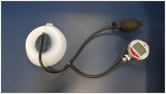
Vacuum bell treatment: A vacuum bell was applied twice daily by the patient or the patient’s parents, starting at 20 minutes each time and increasing to 2 hours twice daily (Figs. 3–7).
Fig. 3.
Vacuum bell (designed and manufactured by Eckhart Klobe in Germany).
Fig. 4.
Digital subtraction of the chest contour before and after vacuum bell treatment of pectus excavatum over a 13-month period in a 15-year-old male. The deepest area was elevated by 11 mm; the surrounding areas were also elevated, so that the chest approached a normal shape.
Fig. 5.
Digital subtraction image before and after 11 months of treatment of a 9-year-old male patient, showing elevation of a diffuse area of the central chest (in blue), and depression (normalization) of an adjacent area (in red and orange).
Fig. 6.
Digital subtraction image before and after 9 months of vacuum bell treatment in a 13-year-old male, showing elevation of a diffuse area of the deepest portion (in blue), with flattening of adjacent areas in the left chest wall (in orange and yellow).
Fig. 7.
Digital subtraction of the chest contour over a 12-month period, during which a 15-year-old male patient did not wear the vacuum bell as directed. The chest is objectively worse: the deepest part is about 10 mm deeper (in orange/red). Such an image facilitates a decision to change treatment.
Pectus carinatum is routinely treated with an external brace (Figs. 8–10).
Fig. 8.
External brace used for the treatment of pectus carinatum (FMF; Pampamed Inc., Buenos Aires, Argentina).
Fig. 9.
Digital subtraction of the chest contour before and after brace treatment for pectus carinatum over 9 months in a 14-year-old. This image shows flattening of the protrusion (in orange/red), which was resolved clinically.
Fig. 10.
Digital subtraction of the chest contour before and after 1 month of external brace treatment of pectus carinatum in a 12-year-old male, showing good progress in treatment of the protrusion (in orange and red).
Discussion
This technique produced images that were well-received by patients and their families. The positive feedback of an objective demonstration of improvement is especially valuable for the vacuum bell and brace treatments, which change the chest shape slowly. A daily exam in the mirror performed by the patient may not make the same impression on young patients, who are impatient for results.
We scanned the patients in the supine position, which is a reproducible position. Changes in the anterior chest due to spine flexion or rotation were minimized. Breast protrusion in adolescent patients with developing breast tissue is a technical challenge that remains unresolved.
In conclusion, optical scanning offers a low-risk, practical, immediate, inexpensive, and well-accepted way to follow nonsurgical treatment of chest wall deformity.
Acknowledgments
The authors gratefully acknowledge the technical assistance of Mrs. Kristina Laws and Mr. Antarius Daniel, and the manuscript preparation assistance of Mrs. Trisha Arnel.
Footnotes
Conflict of interest
No potential conflict of interest relevant to this article was reported. Drs. Kelly and Obermeyer are consultants for Zimmer-Biomet Inc., USA.
References
- 1.Obeid MF, Obermeyer R, Kidane N, et al. Investigating the fidelity of an improvement-assessment tool after one vacuum bell treatment session. Proceedings of the Summer Simulation Multicenter Conference; 2016 Jul 24–26; Montreal, Canada. Vista (CA): The Society for Modeling and Simulation International; 2016. [Google Scholar]
- 2.Rechowicz KJ, Kelly R, Goretsky M, et al. In: Herold KE, Vossoughi J, Bentley WE, editors. Development of an average chest shape for objective evaluation of the aesthetic outcome in the Nuss procedure planning process; Proceedings of the 26th Southern Biomedical Engineering Conference SBEC 2010; 2010 Apr 30–May 2; College Park, USA. Berlin: Springer; 2010. pp. 528–31. [Google Scholar]
- 3.Rechowicz KJ, Kelly R, Goretsky M, et al. A design for simulating and validating the Nuss procedure for the minimally invasive correction of pectus excavatum. Stud Health Technol Inform. 2011;163:473–5. [PubMed] [Google Scholar]
- 4.Obeid MF, Obermeyer RJ, Kidane N, et al. Investigating the fidelity of an improvement-assessment tool after one vacuum bell treatment session. Proceedings of the Summer Computer Simulation Conference; 2016 Jul 24–27; Montreal, Canada. San Diego (CA): The Society for Computer Simulation International; 2016. [Google Scholar]
- 5.McKenzie FD, Chemlal S, Hubbard T, et al. Engineering collaborations in medical modeling and simulation. J Med Surg Res. 2014;1:2–7. [Google Scholar]
- 6.Chemlal S, Rechowicz KJ, Obeid MF, Kelly RE, McKenzie FD. Developing clinically relevant aspects of the nuss procedure surgical simulator. Stud Health Technol Inform. 2014;196:51–5. [PubMed] [Google Scholar]
- 7.Zeng Q, Kidane N, Obeid MF, et al. Utilizing pre- and postoperative CT to validate an instrument for quantifying pectus excavatum severity. In: Zhang L, Song X, Wu Y, editors. Theory, methodology, tools and applications for modeling and simulation of complex systems; Proceedings of the 16th Asia Simulation Conference and SCS Autumn Simulation Multi-Conference, AsiaSim/SCS AutumnSim 2016; 2016 Oct 8–11. Beijing, China; Singapore: Springer; 2016. pp. 451–6. [DOI] [Google Scholar]


