

Abstract
A man in his 50s suffered an impalement on a crowbar after falling from the roof of a domestic shed. A helicopter-based prehospital emergency medical service team was called to assist in the patient’s care. The crowbar had entered from the left-upper quadrant and was tenting the skin of the right iliac fossa. Analgesia and prehospital sedation were provided to facilitate extrication. A series of improvisations were carried out to support the logistics of transferring the patient using an air ambulance to the regional major trauma centre with the crowbar in situ. The patient was taken to the operating theatre without any imaging and a section of perforated bowel was removed. He made a full recovery and was discharged home 9 days postincident.
Keywords: prehospital; trauma; accidents, injuries; sedation
Background
Prehospital emergency medicine (PHEM) offers unique and challenging perspectives to orthodox emergency medicine. Impalements are extremely rare and often require a multidisciplinary team approach in the prehospital setting. Most cases of impalements are fatal at scene. Patients surviving the initial trauma and arriving at the hospital alive, usually make good recovery with multidisciplinary team management underpinning the importance of meticulous prehospital care in transporting patients with impalements safely. This case highlights the role of PHEM clinicians as experts in clinical knowledge, decision making, leadership, team working skills and improvisations.
Case presentation
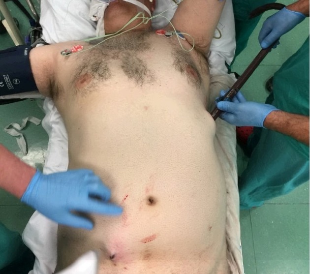
Magpas air ambulance medical team, comprising a critical care paramedic, a consultant paramedic and a senior emergency medicine physician, received an emergency dispatch call to a 51-year-old man, who had fallen from a height of 3 metres. He was repairing a tin roof with a crowbar when roof underneath gave way causing him to fall 10 feet to the ground below. The patient had fallen with his arm outstretched with the crowbar remaining in his hand during the fall. On impact with the ground the patient fell onto the crowbar which was then subsequently pushed into the ground. The patient was impaled by the crowbar through his left-upper abdomen tenting the skin of the right iliac fossa (figures 1–4). The patient was fixed to the ground by the crowbar. On air ambulance arrival, paramedic and fire and rescue services were present on scene. The patient had a wide-bore cannula and intravenous paracetamol was being infused.
Figure 1.

The patient was fixed to the ground with the impaled crowbar (arrow).
Figure 2.

The crowbar penetrated the ground by about 30cm.
Figure 3.

The lower end of the crowbar can be seen partially penetrating and tenting the skin over the right iliac fossa.
Figure 4.

Another view showing top-curved part of the crowbar buried in the ground rendering the patient immobile.
Primary survey revealed the following:
Airway: Patent and clear. C-spine had been manually immobilised.
Breathing: Chest had good air entry bilaterally with no signs of surgical emphysema on palpation. Trachea was central. Chest movements were symmetrical.
Circulation: Limited examination was possible due to impaled objected in situ and the fixed position of the patient. Abdomen had generalised tenderness.
Disability: Glasgow Coma Scale was 15/15. Pupils were bilaterally equal and reactive to light.
Exposure: No other injuries identified on exposure.
Observations were:
Pulse: 72/min.
Blood pressure: 142/76 mm Hg.
Saturations: 100% on non-breathing mask.
Respiratory rate: 32/min.
Nasal end tidal CO2: 3.5 kPa.
The patient had a history of ischaemic heart disease and was taking aspirin and bisoprolol.
Investigations
No investigations were carried out in the prehospital phase.
In the hospital, any prospect of imaging the patient through a CT scanner was ruled out due to the length and position of the crowbar impalement and the patient was transferred to the theatre from the emergency department (figure 5).
Figure 5.
Patient on the theatre table with crowbar in situ.
Initial blood results were as follows:
Haemoglobin: 15.1 g/dL.
White cell count: 13.13×10⁹/L.
Sodium: 133.7 mmol/L.
Potassium: 4.5 mmol/L.
Glucose: 6.7 mmol/L.
Lactate: 1.1.
Renal and liver functions were normal.
Laparotomy demonstrated that the metal bar had entered the abdominal cavity from the left flank in the mid-axillary line, skirting over the costal cartilage. It had traversed the anterior portion of the left abdomen, exiting the abdominal cavity through the medial aspect of rectus below the umbilicus. The crowbar had perforated the small bowel in two places within 30 cm (mid ileum) as well as the small bowel mesentery. No other visceral or vascular damage had occurred.
Whole body trauma CT scan was performed next day which did not identify any further injuries.
Treatment
Initial assessment revealed that the patient had severe abdominal pain and soon after the primary survey, fentanyl 1 μg/kg intravenously was administered. Other treatment given in prehospital phase included 250 ml of normal saline, 1 g tranexamic acid, antibiotics and ketamine for sedation.
Definitive treatment for managing impalements require vigilant removal of the object in the operation theatre. With the crowbar entering the left-upper abdomen, the potential injuries suspected were haemothorax, pneumothorax, diaphragmatic rupture, cardiac injury, abdominal viscera and major vessels. The helicopter emergency medical service (HEMS) team had discussed three possible clinical scenarios and strategies to transfer the patient to the hospital.
Scenario 1: If the patient remains in current stable haemodynamic state, the emphasis would be on meticulous, safe, though swift, extrication. The patient would be airlifted to a regional major trauma centre (MTC).
Scenario 2: Patient becomes hypotensive or develops signs of shock during any stage of extrication. In such a situation, land transport would be used to transfer the patient to the nearest trauma unit identified 21 miles away. Fluid strategy in suspected haemorrhagic shock would include 250 mL boluses of intravenous normal saline to maintain carotid pulse. The logistic of positioning a patient inside the ambulance was considered. The standard position of an ambulance trolley clamped to the left, inside the cabin, provides no room on the left side (figure 6). With crowbar penetrating from the left, a reverse configuration of placing the patient’s head towards the foot-end of the trolley would have allowed space for the crowbar. Keeping rush-hour traffic in view and using paramedics knowledge of local routes, travel by land to the nearest trauma unit appeared quicker with the added advantage of greater access to patient if clinical condition was to deteriorate further.
Figure 6.

The trolley, when clamped inside the ambulance, does not provide any space on the patient’s left side. The HEMS team had considered the option of placing the patient’s head towards the foot-end of the trolley to accommodate the crowbar.
Scenario 3: Patient loses cardiac output. in this situation, HEMS team had agreed to follow penetrating injury traumatic cardiac arrest (TCA) protocol by securing an airway with endotracheal intubation, performing bilateral thoracostomies, proceeding to clamshell thoracotomy to identify and relieve any cardiac tamponade.
Given the stable haemodynamics of our patient (scenario 1), the patient was taken by an air ambulance to the regional MTC.
As a three-member HEMS team, tasks were divided among team to execute the extrication plan. One team member stayed with the patient constantly assessing clinical deterioration and requirements for analgesia. Second took charge of communications liaising with control desk, receiving major trauma hospitals, pilots and the on call duty advice doctor(DAD). The third member coordinated the extrication with fire and rescue and ambulance services. At frequent intervals, team leaders from all services had brief meetings to share concerns ensuring safe progress.
Anticipating that patient would experience severe pain during extrication due to the impaled metal object, he was sedated using 60 mg of intravenous ketamine (0.6 mg/kg of estimated body weight). A total of 250 mcg of intravenous fentanyl was administered by HEMS team in 50 mcg small boluses to provide adequate analgesia through the extrication and transfer phase to hospital.
Figure 7 shows the position of the patient (a) on scene: fence (b) was taken down by the fire and rescue services and helicopter was relocated to the small car park (c).
Figure 7.

Extrication path was created for the patient (A) by taking down the wooden fence and metal railing, (B) into the car park, (C) where the helicopter had landed.
To tackle the logistics, a series of innovative steps described below were taken.
Alley leading to the outside of the house was too narrow to allow extrication of a patient with crowbar. A wood and metal fence to one side of the house that opened into a small car park was taken down by the fire and rescue service to create extrication route (figure 7).
To safely extricate the patient with the crowbar in situ, pilot repositioned the aircraft from initial landing site to the car park which required clearing to create landing space.
Garden tools available in the house were used to facilitate removing safely the part of the crowbar buried into the ground.
For extrication, patient was sedated using ketamine. Log roll was not possible due to crowbar being in situ. To place a scoop stretcher, patient was lifted off the ground vertically while maintaining C-spine immobilisation.
While working closely with fire and rescue service, various options were considered to attempt cutting the crowbar short for the ease of transport.1 2 A pedal cutter (figure 8) was chosen as the one with the least concern for vibration but several attempts to cut the crowbar were unsuccessful. Other cutting tools available were considered higher risk to cause vibration or twisting of the crowbar.
Considering the patient’s stable haemodynamics, rush-hour traffic and the single carriageway to major trauma centre, patient was airlifted instead of using land transport. Cabin size was considered and patient was accommodated in the aircraft with slight reconfiguration of team members seating positions.
Wedges and step blocks (figure 9) borrowed from fire services were used to stabilise the crowbar inside the air ambulance.
Figure 8.

An attempt to shorten the crowbar to facilitate transport failed with pedal cutter.
Figure 9.

Wedge blocks were borrowed from fire and rescue services to stabilise the crowbar during transfer.
Outcome and follow-up
The patient made a full recovery and was discharged home 9 days later.
He visited the MAGPAS air ambulance base to meet the team a few weeks following discharge expressing thanks to all the staff involved in his care. He shared with the team that he has kept the crowbar with him as a reminder of this incident (figure 10).
Fig 10:

The 92 cm in length crowbar has been nickel plated by the patient.
Discussion
With ever-improving safety standards in the western world, impalement injuries are rare3 but when they occur, they provide huge clinical and logistical challenges in the prehospital setting. A few dozen cases have been reported in literature each depicting unique set of challenges faced by the emergency care providers though adherence to basic trauma principles is still required. Instant fatality4 5 is high for most patients but those who survive the initial trauma and reach hospital alive have more favourable outcome6 with minimal morbidity, highlighting the importance of meticulous prehospital care.7–9
For a haemodynamically stable patient with impalement, it is vital for first responders to follow fundamentals of trauma life-support guidelines and take utmost care to limit the movements of an impaled object. It is important that during an initial medical assessment, an obvious, impaled object does not cause distraction and priority should remain to perform a primary survey to address life-threatening injuries. The object must be left in situ, though can be shortened in length if possible. Staff arriving first must ensure scene safety. Impalement, as in this case, occurring at construction site may include hazards like unstable structure, falling debris and exposed electric wires. Specific to TCA secondary to impalement, the paramedics are invaluable in delivering life-saving interventions while awaiting HEMS arrival, for example, controlling catastrophic haemorrhage, maintaining an airway and decompressing a tension pneumothorax. Fire and Rescue team carries cutting gear that plays a pivotal role in creating an open route for difficult extrications.
Most deaths after impalements occurs due to catastrophic haemorrhage.10 Impaled object may tamponade11 an injured vessel and minimal handling and movement of the object should be ensured during the transfer.12–15 Each case of impalement is unique in many ways. The location of incident, type and size of object, penetrating position and uncertainty about organs injured, all demands improvisation, dynamic planning and multidisciplinary execution. Impaled object should be left in situ in prehospital phase and be only removed in the controlled environment of operating theatre4 13
Our case report focuses on the prehospital aspects of impalement injuries and is unique as we did not find a single published case report with a thorough PHEM perspective where a patient with an impaled object in situ was airlifted to a MTC using an air ambulance. In this challenging clinical scenario, HEMS team brought tempo to the scene by making crucial decisions and by leading the extrication plan. HEMS team considered the strategies in stable and unstable patient and had the skills to perform life-saving procedures had they been required. Along with providing enhanced analgesia using fentanyl and sedation with ketamine, we made essential communications to liaise with the regional trauma desk and receiving MTC emergency department. Emphasis was given by HEMS staff on clinical management of impalement with minimal handling of impaled object throughout the extrication process with all team members. HEMS team brought experience in managing multidisciplinary teams by providing leadership at scene and maintained an overall situational awareness by frequently meeting with incident commander from both ambulance and fire and rescue service.
Patient’s perspective.
While working on the roof, I remember holding the crowbar in my left hand. As I was removing nails, the whole metal sheet started to slide. I started to back-peddle as fast as I could in an attempt to stay on the moving roof, but I couldn’t stop myself from falling.
I saw the crowbar sticking up through my chest. It was hard to breathe, I was fixed to the ground. I knew I was in serious danger, but I remained calm. I was thinking about my dog in case I never made it home again. My memories of the incident break up into weird segments like a slow-motion film after that. I think I was slipping in and out of consciousness.
I just could not believe how lucky I was to be alive. I had tears in my eyes and I felt so much gratitude for the emergency services who saved my life. After the accident I thought, I’ve got through this, I can get through anything and I don’t let the small stuff bother me any more. The air ambulance service has been great. I went to the airbase to meet the team. It was so good to talk to them and it was like coming full circle after the fall. My injuries have healed. Thanks to the incredible care I received. I’m in a new job that I love and life is good. I’ll always be grateful to the incredible team who came to my rescue that day. And I’ve kept the crowbar by the stairs in my house as a reminder of how lucky I am to still be here (figure 10).
Learning points.
Clarity of roles and tasks are essential to ensure efficient performance of the pre hospital emergency medical service team.
Improvisation is the key in rare cases.
Frequent huddles with key leaders of emergency services ensures shared mental model and robust execution.
In unfamiliar situations, be extra vigilant for human factors to avoid error.
Impaled object should be left in situ and should be only removed in operating theatre. Minimal handling and stabilisation of impalement are key to safe transfer to hospital.
Enhanced analgesia and sedation can assist in entrapments and difficult extrications.
Acknowledgments
Andrew Smith Critical Care Paramedic & Defence Specialist Adviser Paramedic Magpas Air Ambulance.
Footnotes
Contributors: SJ: doctor in the HEMS team that treated the patient, followed up patient after discharge to obtain consent, attended the meeting with patient and discussed the post discharge journey, intention to write the case report and gained the consent, also obtained patient’s perspective for the case report, literature search and review of previously published articles and case reports about impalements. Both authors discussed and planned the contents of the case report. Wrote the initial draft of the case report and finalised it after the initial draft was reviewed by coauthor DC. DC: consultant paramedic in the HEMS team which treated the patient, attended the meeting with patient and explained to the patient the educational benefits of publishing the case which assisted in obtaining the consent, took all the pictures of the incident on the day and have provided them for the publication, assisted in writing up the case report and made contributions to all the sections particularly abstract, background, presentation and treatment.
Funding: The authors have not declared a specific grant for this research from any funding agency in the public, commercial or not-for-profit sectors.
Competing interests: None declared.
Patient consent: Obtained.
Provenance and peer review: Not commissioned; externally peer reviewed.
References
- 1. Salomone JP. More than skin deep: use caution when treating impalement injuries. JEMS 2011;36:40–3. 10.1016/S0197-2510(11)70146-2 [DOI] [PubMed] [Google Scholar]
- 2. Angelopoulos S, Mantzoros I, Kyziridis D, et al. A rare case of a transabdominal impalement after a fall from a ladder. Int J Surg Case Rep 2016;22:40–3. 10.1016/j.ijscr.2016.03.011 [DOI] [PMC free article] [PubMed] [Google Scholar]
- 3. Kolahdouzan M, Rezaee MT, Shahabi S. Impalement thoracoabdominal trauma secondary to falling on metallic (iron) bars: an extremely rare and unique case. Arch Trauma Res 2016;5 10.5812/atr.18330 [DOI] [PMC free article] [PubMed] [Google Scholar]
- 4. Singhal M, Kumar MV, Prakash P, et al. Rare case of impalement of two occupants of a vehicle by the same object: insights into the management of complex thoracic impalements. Chin J Traumatol 2012;15:50–3. [PubMed] [Google Scholar]
- 5. Hansen CJ, Bernadas C, West MA, et al. Abdominal vena caval injuries: outcomes remain dismal. Surgery 2000;128:572–8. 10.1067/msy.2000.108054 [DOI] [PubMed] [Google Scholar]
- 6. Edwin F, Tettey M, Aniteye L, et al. Impalement injuries of the chest. Ghana Med J 2010;43 10.4314/gmj.v43i2.55320 [DOI] [PMC free article] [PubMed] [Google Scholar]
- 7. Malla G, Basnet B, Vohra R, et al. Thoraco- abdominal impalement injury: a case report. BMC Emerg Med 2014;14:7 10.1186/1471-227X-14-7 [DOI] [PMC free article] [PubMed] [Google Scholar]
- 8. Ayandipo OO, Irabor D, Afuwape O, et al. Multiple thoracoabdominal impalement injuries. Prehosp Disaster Med 2012;27:88–9. 10.1017/S1049023X11006820 [DOI] [PubMed] [Google Scholar]
- 9. Kim KT, Seo PW. A case of severe thoracoabdominal impalement by a steel bar. Korean J Thorac Cardiovasc Surg 2016;49:481–4. 10.5090/kjtcs.2016.49.6.481 [DOI] [PMC free article] [PubMed] [Google Scholar]
- 10. Offner P. Penetrating abdominal trauma treatment & management: approach considerations, prehospital care, surgical therapy. Emedicine.medscape.com 2018. https://emedicine.medscape.com/article/2036859-treatment#d9. [Google Scholar]
- 11. Kelly IP, Attwood SE, Quilan W, et al. The management of impalement injury. Injury 1995;26:191–3. 10.1016/0020-1383(94)00015-N [DOI] [PubMed] [Google Scholar]
- 12. Sawhney C, D’souza N, Mishra B, et al. Management of a massive thoracoabdominal impalement: a case report. Scand J Trauma Resusc Emerg Med 2009;17:50 10.1186/1757-7241-17-50 [DOI] [PMC free article] [PubMed] [Google Scholar]
- 13. Darbari A, Tandon S, Singh AK. Thoracic impalement injuries. Indian Journal of Thoracic and Cardiovascular Surgery 2005;21:229–31. 10.1007/s12055-005-0057-8 [DOI] [Google Scholar]
- 14. Lunca S, Morosanu C, Alexa O, et al. Severe thoracic impalement injury: Survival in a case with delayed surgical definitive care. Ulus Travma Acil Cerrahi Derg 2015;21:152-6 10.5505/tjtes.2015.38354 [DOI] [PubMed] [Google Scholar]
- 15. Lee SY, Lee JM, Choi SJ. Abdominal impalement injury caused by scaffolding pipe following a traffic accident - a case report. Journal of Trauma and Injury 2016;29:33–6. 10.20408/jti.2016.29.1.33 [DOI] [Google Scholar]