Abstract
We have isolated an endophytic fungus with heat stress alleviation potential from wild plant Euphorbia indica L. The phylogenetic analysis and 18S rDNA sequence homology revealed that the designated isolate was Aspergillus japonicus EuR-26. Analysis of A. japonicus culture filtrate displayed higher concentrations of salicylic acid (SA), indoleacetic acid (IAA), flavonoids, and phenolics. Furthermore, A. japonicus association with soybean and sunflower had improved plant biomass and other growth features under high temperature stress (40°C) in comparison to endophyte-free plants. In fact, endophytic association mitigated heat stress by negotiating the activities of abscisic acid, catalase, and ascorbic acid oxidase in both soybean and sunflower. The nutritional quality (phenolic, flavonoids, soluble sugars, proteins, and lipids) of the A. japonicus-associated seedlings has also improved under heat stress in comparison to endophyte-free plants. From the results, it is concluded that A. japonicus can modulate host plants growth under heat stress and can be used as thermal stress alleviator in arid and semiarid regions of the globe (where mean summer temperature exceeds 40°C) to sustain agriculture.
1. Introduction
Temperature stress is becoming more persistent, especially in arid, semiarid, and tropical zones of the globe, causing great extortions to food crops. Rising in the total mean global temperature also results in saline conditions through moisture evaporation from the soil. Plants are vulnerable to various stresses because of their sessile nature. In such abnormal state, reactive oxygen species (ROS) are generated in high concentrations, which cause apoptosis or premature cell death when exposed for a longer time. Different ROS species, including singlet oxygen (1O2), superoxide (O2−1), hydroxyl radical (OH) and hydrogen peroxide (H2O2) containing free electrons, are leaked in chloroplast and mitochondria from electron transport chain [1]. ROS are trapped by biological membranes where they are detoxified by local antioxidants except H2O2 [2]. But all plants contain a specific enzymatic antioxidant system in the form of superoxide dismutase (SOD), catalase (CAT), ascorbic acid oxidase (AAO), and peroxidase (POD) which neutralize ROS before they come into action to damage the cell components [3]. CAT and AAO specifically target ROS by lowering their energy or disturbing their oxidizing chain reactions [4]. CAT is known to have multigene family responsible for encoding vital plant proteins. These vital proteins have significance in localization, regulation, and expression of stress receptive genes in cells facing different environmental stresses [5]. Similarly, AAO helps in controlling the level of membrane bound antioxidant α-tocopherol as well as reorganization of cell wall during different environmental stresses [6]. Moreover, plants also have the potential to modulate biochemical and physiological modifications depending on variable capacity to identify stimuli and conduct signals [7].
Phytohormones, like jasmonic acid (JA), abscisic acid (ABA), and salicylic acid (SA), serve as a signaling compound during stress [8]. Different phytohormones respond differently during biotic and abiotic stresses; for example, ABA regulates stomatal closure, plant growth, and development under stressful conditions [9]. SA controls plant growth, development, flower induction, stomatal response, ethylene synthesis, and respiration [10]. Similarly, JA is known to have a role in the biosynthesis of defensive secondary metabolites and proteins [11]. JA also helps in modulation of many physiological phenomena like resistance to insects and pathogen, pollen development, senescence, and root growth [12]. Endophytic fungi that reside inside plant tissues without causing any visible symptoms are one of the best bio agents that help in restoration of plant growth under stress [13]. They also have a role in enhancement of host growth, increasing of nutrient absorption, reduction in disease severity, and improving of host resistance against environmental stresses [13]. Vital secondary metabolites like hormones (GAs, IAA, SA, and JA), flavonoids, phenolics, and proline are also secreted by endophytic fungi in their culture filtrate and probably in their host tissues [14]. Plants lacking in endophytic fungi are more susceptible to drought, salinity, heat, and pathogenic stresses hence overall yield is highly reduced in such abnormal situations as compared to endophyte-hosted plants [15]. The present study was an attempt to explore such endophytic fungi that have the potential to improve host plant growth and yield as well as enhance their immunity under thermal stress.
2. Materials and Methods
2.1. Plant Sample Collection and Endophyte Isolation
A wild plant Euphorbia indica L. was collected from the desert area of District Noshera Khyber Pakhtunkhwa, Pakistan, and processed for the isolation of endophytic fungi using Khan et al. [16] procedure. Fungal isolates were purified on potato dextrose agar (PDA) medium and kept in refrigerator at 4°C. For longer storage, PDA slants were made [17]. For the collection and analysis of fungal secondary metabolites, endophyte isolates were grown in 50 ml of Czapek medium for 7 days in a shaking incubator set at 120 rpm at 28°C.
2.2. Screening Bioassay of fungal Filtrate on Rice Seedlings
Screening bioassay of fungal filtrate was carried out on rice seedlings by applying 100μl of fungal filtrate on the tip of rice seedlings (Fakhr-e-Malakand variety provided by the Agricultural Biotechnology Institute, National Agricultural Research Center, Islamabad, Pakistan) at the two leaves stage grown in 0.8% water-agar medium in growth chamber (day/night cycle: 14 h, 28°C ± 0.3; 10 h, 25°C ± 0.3; relative humidity 70%) for 7 days. Root-shoot length and fresh and dry weights were analyzed after one week of filtrate treatment and compared with Czapek and distilled water treated plants.
2.3. Fungal Identification
For molecular identification of endophytic fungi, Khan et al. [16] method was applied. Internal transcribed region of 18S rRNA was amplified by using primers ITS1 (5′-TCC GTA GGT GAA CCT GCG G-3′) and ITS4 (5′-TCC TCC GCT TAT TGA TAT GC-3′). The sequence obtained, was then imperiled to BLASTn1 for sequence homology approximation. Neighbor joining (NJ) tree was applied for the phylogenetic analysis using the MEGA 7 software.
2.4. Inoculation of Aspergillus japonicus EuR-26 to Soybean and Sunflower
Endophytic fungus A. japonicus was grown in 250 ml conical flask containing 50 ml Czapek broth and kept in shaking incubator set at 120 rpm for 7 days at 28°C. Pellet and supernatant was separated using filter paper. One milligram of fungal biomass per 100 g of sterilized sand was inoculated to pots containing 9 seeds of soybean and sunflower each and shifted to growth chambers set at 25°C and 40°C for two weeks while supernatant was used for the determination of secondary metabolites. Half strength Hoagland solution (10 ml) was applied to the pots at 48 h intervals. Growth parameters were evaluated after 2 weeks of incubation in growth chamber [18]. The experiments were performed in triplicate (each replicate consisted of 9 seedlings).
2.5. IAA and SA Analysis in the Culture Filtrate of A. japonicus
For the determination of IAA in the filtrate of A. japonicus Benizri et al. [19] protocol was used. Two ml Salikowski reagent was mixed well with 1 ml of pure fungal filtrate and then kept in the dark for half an hour at 25°C. OD was taken at 540 nm with the help of PerkinElmer Lambda 25 spectrophotometer. Different concentrations (10, 20, 30, 40, 60, 80, and 100 μg/ml) of the IAA (Sigma Aldrich) were used to construct the standard curve.
For the determination of SA the method of Warrier et al. [20] was followed. Exactly, 2.99 ml freshly prepared FeCl3 (0.1%) solution was mixed with 100 μl of fungal filtrate. After the appearance of violet color, OD was observed at 540 nm. A standard curve was plotted by taking different concentrations (100, 200, 300, 400و and 500 μg/ml) of pure SA (Sigma Aldrich).
2.6. ABA Analysis in Sunflower and Soybean
For the analysis of ABA contents in soybean and sunflower the protocol of Yoon et al. [21] was followed. Fresh leaves of soybean and sunflower (0.5 g each) were ground in liquid nitrogen. A mixture of isopropanol (1.5 ml) and glacial acetic acid (28.5 ml) was added to the grounded leaves. The mixture was filtered and dehydrated by means of a rotary evaporator. Diazomethane was then added to the mixture and analyzed by GC–MS SIM (6890N network GC system equipped with 5973 network mass selective detector; Agilent Technologies, Palo Alto, CA, USA). The Lab-Base (Thermo Quset, Manchester, UK) data system software was used to observe responses to ions with m/z values of 162 and 190 for Me-ABA and 166 and 194 for Me-[2H6]-ABA. ABA ([2H6]-ABA) from Sigma Aldrich was used as internal standard.
2.7. Analysis of Antioxidants in Soybean and Sunflower Seedlings
Luck [22] protocol was followed in the determination of CAT concentration in soybean and sunflower seedlings. Fresh leaves (2 g) of soybean and sunflower were crushed in phosphate buffer (10 ml) and centrifuged for five minutes at 10,000 rpm. H2O2-phosphate buffer (3 ml) was then added to the supernatant (40 μl) and the OD was observed at 240 nm. H2O2-free phosphate buffer solution was used as blank. The enzyme unit was calculated as the amount of enzyme required to decrease the absorbance at 240 nm by 0.05. Oberbacher and Vines [23] protocol was used to measure the concentration of AAO in soybean and sunflower seedlings. Fresh leaves (0.1 g) of soybean and sunflower were ground in phosphate buffer (2 ml) and centrifuged for five minutes at 3000 rpm. About 3 ml of the substrate solution (8.8 mg of ascorbic acid in 300 ml phosphate buffer, pH 5.6) was mixed with 100 μl of supernatant and the OD was noted after every 30 seconds till 5 minutes at 265 nm.
2.8. Analysis of Phenolics, Proline and Flavonoids in the Culture Filtrate of A. japonicus, Sunflower, and Soybean
Cai et al. [24] procedure was employed for the analysis of phenolics in soybean, sunflower, and fungal filtrate. Different concentrations (100, 200, 300, 500, 600, 700, and 900 mg/ml) of gallic acid (Sigma Aldrich) were used to construct a standard curve. For the determination of proline, the method of Bates et al. [25] was followed, but after some modifications. A standard curve was plotted against different concentrations (2, 4, 6, 8, and 10 μg/ml) of standard proline (Sigma Aldrich) and the OD was taken at 520 nm. For the analysis of total flavonoids, the method of Mervat et al. [26] was used. A standard curve was constructed by using different concentrations of pure quercetin (15, 30, 60, 120, 240, and 480 μg/ml, Sigma Aldrich) and the OD were taken at 415 nm.
2.9. Analysis of Total Proteins, Lipids, and Soluble Sugars
Lowry et al. [27] method was employed for the analysis of total proteins in the seedlings of soybean and sunflower. A standard curve was drawn using different concentrations (20, 40, 60, 80, and 100 μg/ml) of BSA (Sigma Aldrich) and the OD was observed at 650 nm. For the determination of total lipids, we used the method of Van Hand et al. [28] with some modifications. A standard curve was constructed using different concentrations of canola oil (10, 40, 70, 100, 130, and 160 μg/ml) and the OD was noticed at 490 nm. For the analysis of total soluble sugars in the soybean and sunflower seedlings, the method of Mohammadkhani and Heidari [29] was used. A standard curve was constructed by using different concentrations (20, 40, 60, 80, and 100 μg/ml) of glucose (Sigma Aldrich) and the OD was measured at 485 nm.
2.10. Statistical Analysis
All the experiments were performed in triplicate. ANOVA (one-way analysis of variance) was used for the analysis of data and means were compared by Tukey HSD test at p < 0.05, using SPSS-20 (SPSS Inch., Chicago, IL, USA) for windows.
3. Results
3.1. Endophytes Isolation and Their Preliminary Screening on Rice Seedlings
We isolated 14 different endophytic fungi (Figure S1) from the wild plant of Euphorbia indica L. and initially screened on rice seedlings for their growth promoting or inhibiting activities (Figure S2). Endophytes were first isolated on Hagem minimal medium and then purified on PDA on the basis of morphological differences. After screening on rice seedlings at the two leaves stage, growth parameters were recorded after 1 week of filtrate treatment (Table 1). EuR-26 isolate was found to be growth promoter and chosen for molecular identification.
Table 1.
Effect of A. japonicus filtrate on the growth of rice seedlings.
| Growth attributes | Control (DW) | Control (Czk) | A. japonicus |
|---|---|---|---|
| Shoot Length (cm) | 12.6 ± 0.25 a | 13.8 ± 0.34 ab | 15.67 ± 0.52 b |
| Root Length (cm) | 6.3 ± 0.38 a | 6.6 ± 0.18 ab | 8.1 ± 0.35 b |
| Dry Weight Shoot (g) | 0.0034 ± 0.001 a | 0.0049 ± 0.003 a | 0.0086 ± 0.004 b |
| Dry Weight Root (g) | 0.0147 ± 0.001 a | 0.0155 ± 0.004 a | 0.0182 ± 0.004 b |
| Chlorophyll (SPAD) | 20.73 ± 1.5 a | 22.7 ± 0.56 b | 25.9 ± 0.18 c |
DW = distilled water, Czk = Czapek broth medium. Data are means of 3 replicates (each replicate consisted of 9 seedlings) with standard error. For each set of treatment, the different letter indicates significant differences at p (< 0.05) as estimated by Tukey HSD test.
3.2. Molecular Identification of Fungal Isolate EuR-26
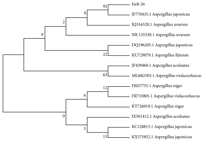
Fresh mycelium was used for the extraction of genomic DNA while, applying ITS1 (5′-TCC GTA GGT GAA CCT GCG G-3′) and ITS4 (5′-TCC TCC GCT TAT TGA TAT GC-3′). Sequence of ITS region of related fungi was compared with the nucleotide sequence of our fungal isolate EuR-26 with the help of the BLAST search program. Sequence of 18S rDNA exhibited maximum similarity (92%) with A. Japonicas. The phylogenetic consensus tree was built from 14 (13 reference and 1 clone) by neighbor joining (NJ) method using MEGA 7 software (Figure 1). Our isolate EuR-26 made a clad with A. japonicus supported by 92% bootstrap value in the consensus tree. Combining results of phylogenetic analysis and sequence homology suggested that the strain EuR-26 was A. japonicus.
Figure 1.
Phylogenetic consensus tree using neighbor joining (NJ) method for the identification of isolate EuR-26, using 14 taxa, 13 references, and 1 clone. The isolate was identified as Aspergillus japonicus as having 92% bootstrap value.
3.3. Analysis of Secondary Metabolites in the Culture Filtrates of A. japonicus
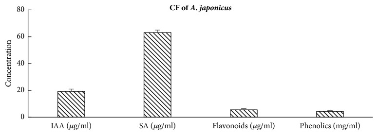
Culture filtrate of A. japonicus was analyzed for the presence of important secondary metabolites including IAA, SA, flavonoids, and phenolics. Different concentrations of secondary metabolites were IAA (19.19 μg/ml), SA (63.11μg/ml), flavonoids (5.56 μg/ml), and phenolic (4.4 mg/ml) (Figure 2).
Figure 2.
Analysis of secondary metabolites secreted by A. japonicus (EuR-26) in their culture filtrate (CF) grown for 7 days in Czapek medium in shaking incubator set at 120 rpm at 28°C. The bars labeled with different letters are significantly different at p < 0.05 as estimated by Tukey HSD test. The bars represent SE of a triplicate data (each replicate consisted of 9 seedlings).
3.4. Growth Features of A. japonicus-Associated Soybean and Sunflower
Both A. japonicus-associated soybean and sunflower at 25°C and 40°C showed significant increase in various growth parameters, including chlorophyll content, shoot length, shoot fresh, and dry weights as compared to endophyte-free plants. At room temperature (25°C) soybean (Table 2) and sunflower (Table 3) seedlings have more chlorophyll, root-shoot length, and dry weight than under heat stress condition (40°C).
Table 2.
Effect of A. japonicus on the growth features of soybean.
| Growth attributes | 25°C | 40°C | ||
|---|---|---|---|---|
| Control | A. japonicus | Control | A. japonicus | |
| Chlorophyll (SPAD) | 32.6 ± 0.21 a | 37.4 ± 0.4 d | 29 ± 2.46 a | 33.2 ± 0.07 c |
| Shoot Length (cm) | 38.7 ± 1.76 b | 48.3 ± 1.2 e | 26 ± 0.93 a | 35.5 ± 1.26 bc |
| Root Length (cm) | 16.3 ± 3.9 abc | 13.3 ± 2.33 ab | 10 ± 0.6 ab | 12 ± 2.6 abcd |
| Dry Weight Shoot (g) | 0.14 ± 0.01 a | 0.147 ± 0.01 ab | 0.08 ± 0.0003 ab | 0.09 ± 0.0001 ab |
| Dry Weight Root (g) | 0.08 ± 0.002 ab | 0.10 ± 0.01 bc | 0.01 ± 0.0001 ab | 0.062 ± 0.001 e |
Data are means of three replicates (each replicate consisted of 9 seedlings) with standard errors. For each set of treatment, the different letter indicates significant differences at p (< 0.05) as estimated by Tukey HSD test.
Table 3.
Effect of A. Japonicas on the growth features of sunflower.
| Growth attributes | 25°C | 40°C | ||
|---|---|---|---|---|
| Control | A. japonicus | Control | A. japonicus | |
| Chlorophyll (SPAD) | 39 ± 1.29 a | 43.3 ± 0.6 ab | 38.4 ± 1.95 a | 39.9 ± 3.12 a |
| Shoot Length (cm) | 23.7 ± 0.88 a | 25.2 ± 0.43 ab | 20.5 ± 0.45 a | 21 ± 1.53 ab |
| Root Length (cm) | 9 ± 0.58 a | 11.6 ± 0.87 abcd | 6 ± 0.58 b | 6 ± 0.25 b |
| Dry Weight Shoot (g) | 0.085 ± 0.009 ab | 0.088 ± 0.007 ab | 0.04 ± 0.0006 b | 0.037 ± 0.0002 a |
| Dry Weight Root (g) | 0.024 ± 0.001 a | 0.028 ± 0.002 a | 0.014 ± 0.0003 b | 0.02 ± 0.0002 c |
Data are means of three replicates (each replicate consisted of 9 seedlings) with standard errors. For each set of treatment, the different letter indicates significant differences at p (< 0.05) as estimated by Tukey HSD test.
3.5. Modulation in A. japonicus-Associated Soybean and Sunflower's Endogenous Flavonoids, Phenols, and Proline
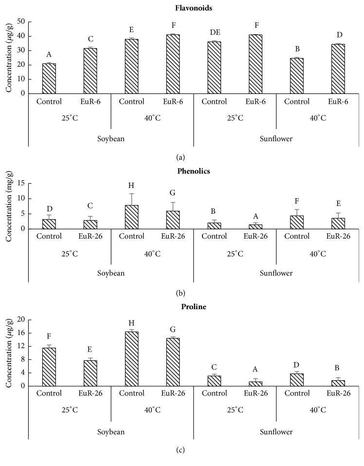
Important secondary metabolites like flavonoids, phenolics, and prolines were analyzed in soybean and sunflower seedlings inoculated with A. japonicus at 25°C and 40°C in growth chamber. An increase has been noted in A. japonicus-associated (41.13 μg/g) and control (37.96 μg/g) soybean seedlings at 40°C as compared at 25°C (31.53 μg/g in experimental and 20.91μg/g in control seedlings), while endophyte-aligned seedlings have more flavonoids content as compared to the endophyte-free seedlings. Similarly, sunflower seedlings aligned with A. japonicus have more flavonoids (41.04 μg/g at 25°C and 34.43 μg/g at 40°C) than control plants (36.18 μg/g at 25°C and 24.64 μg/g at 40°C) at both temperatures, while a decrease has been noted at thermal stress as compared to normal condition (Figure 3(a)). We also found a slight reduction in the phenolics and proline concentration in A. japonicus-aligned soybean and sunflower seedlings as compared to control plants. At 25°C endophyte-aligned soybean has 2.8 mg/g and endophyte-free seedlings have 3.13 mg/g of phenolics while, at 40°C, endophyte-aligned soybean has 5.89 mg/g and endophyte-free seedlings have 7.8 mg/g of phenolics. Sunflower seedlings inoculated with A. japonicus at 25°C have 1.38 mg/g and control plants have 2.01 mg/g of total phenolics while, at 40°C, endophyte-associated sunflower has 3.53 mg/g and endophyte-free seedlings have 4.34 mg/g of phenolics (Figure 3(b)). A significant reduction was found in the total proline concentration in soybean (7.71 μg/g at 25°C and 14.46 μg/g at 40°C) and sunflower (1.29 μg/g at 25°C and 1.68 μg/g at 40°C) seedlings aligned with A. japonicus as compared to control soybean (11.5 μg/g at 25°C and 16.4 μg/g at 40°C) and sunflower (3.09 μg/g at 25°C and 3.73 μg/g at 40°C) seedlings (Figure 3(c)).
Figure 3.
Effect of A. japonicus on the concentration of (a) flavonoid, (b) phenolics, and (c) proline in soybean and sunflower seedlings grown at 25°C and 40°C. For each set of treatment, the different letter indicates significant differences at p (< 0.05) as estimated by Tukey HSD test. The bars represent SE of a triplicate data (each replicate consisted of 9 seedlings).
3.6. Reduction in the Concentration of CAT and AAO
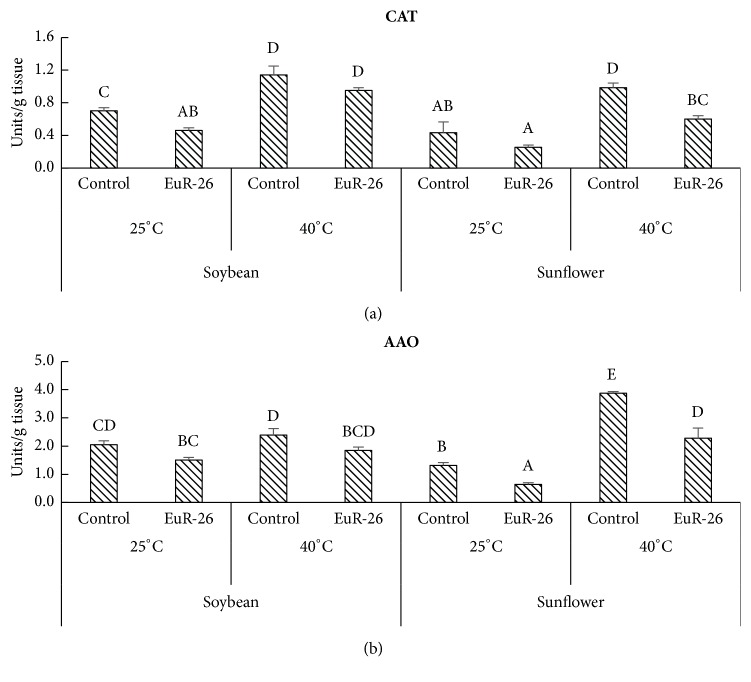
A significant reduction was detected in the concentration of CAT and AAO in soybean and sunflower seedlings associated with A. japonicus as compared to control plants. Endophyte-aligned soybean has 0.46 enzyme unit/g tissue and sunflower had 0.25 enzyme unit/g tissue CAT at 25°C as compared to endophyte-free soybean 0.7 enzyme unit/g tissue and sunflower 0.43 enzyme unit/g tissue. At 40°C, the soybean seedlings had 0.95 enzyme unit/g tissue and sunflower had 0.6 units of enzyme unit/g tissue as compared to control soybean 1.14 enzyme unit/g tissue and sunflower 0.98 enzyme unit/g tissue (Figure 4(a)). A similar decrease was found in the amount of AAO in soybean and sunflower aligned with A. japonicus at 25°C and 40°C as compared to endophyte-free soybean and sunflower (Figure 4(b)).
Figure 4.
Effect of A. japonicus on the concentration of (a) CAT and (b) AAO in soybean and sunflower seedlings grown at 25°C and 40°C. One enzyme unit was calculated as the amount of enzyme required to decrease the absorbance at 240 nm for CAT and 265 nm for AAO by 0.05 units. For each set of treatment, the different letter indicates significant differences at p (< 0.05) as estimated by Tukey HSD test. The bars represent SE of triplicate data (each replicate consisted of 9 seedlings).
3.7. Reduction in Endogenous ABA Content in Soybean and Sunflower
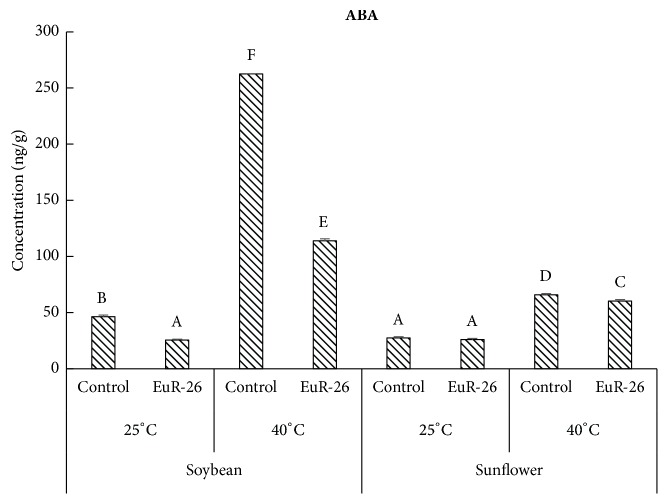
A significant reduction was found in the concentration of ABA in soybean and sunflower seedlings associated with A. japonicus as compared to control seedlings at 40°C. Endophyte-associated soybean has 25.56 ng/g and endophyte-free seedling had 46.42 ng/g at 25°C. At 40°C, the experimental soybean had 113.78 ng/g and control seedling has 262.46 ng/g of ABA concentrations. Similarly, Endophyte-associated sunflower has 26.05 ng/g and endophyte-free seedling has 27.31 ng/g at 25°C while, at 40°C, experimental sunflower has 60.32 ng/g and control seedling has 65.78 ng/g of ABA concentrations (Figure 5).
Figure 5.
GC MS quantification of endogenous ABA concentration in soybean and sunflower grown at 25°C and 40°C with and without A. japonicus. For each set of treatment, the different letter indicates significant differences at p (< 0.05) as estimated by Tukey HSD test. The bars represent SE of a triplicate data (each replicate consisted of 9 seedlings).
3.8. Enhancement in the Nutritive Value of Soybean and Sunflower
Nutritive values of soybean and sunflower were determined by quantifying total sugar, protein, and lipid contents. A. japonicus-associated soybean has 202.8 μg/g while control plants have 196.39 μg/g of soluble sugars at 25°C and, at 40°C, experimental soybean seedlings had 169.03 μg/g while control seedlings have 133.75 μg/g total sugar content. A similar increase was observed in the total sugar concentrations of sunflower seedlings inoculated with an endophytic fungus as compared to endophyte-free seedlings under heat stress (Figure 6(a)). A significant increase was detected in the total protein contents of soybean (302.83 μg/g at 25°C and at 240.8 μg/g 40°C) and sunflower (189.15 μg/g at 25°C and at 227.29 μg/g 40°C) aligned with A. japonicus as compared control seedlings of soybean (213.67 μg/g at 25°C and at 163.04 μg/g 40°C) and sunflower (204.13 μg/g at 25°C and at 125.84 μg/g 40°C) (Figure 6(b)). An enhancement in the total lipids was found in A. japonicus-inoculated soybean and sunflower as compared to A. japonicus-free soybean and sunflower at 25°C and 40°C (Figure 6(c)).
Figure 6.
Analysis of (a) soluble sugars, (b) proteins, and (c) lipids of soybean and sunflower seedlings grown at 25°C and 40°C with- and without-endophytic fungal strain A. japonicus. For each set of treatment, the different letter indicates significant differences at p (< 0.05) as estimated by Tukey HSD test. The bars represent SE of a triplicate data (each replicate consisted of 9 seedlings).
4. Discussion
Endophytic fungi are known to have symbiotic association with plants in natural ecosystem [30]. The endophytes are responsible for synthesizing a large number of vital bioactive secondary metabolites, including phenolics, flavonoids, and alkaloids, that have importance in the fields of medicine, food, and agriculture [31]. Endophytes have a defensive role for their host plant in providing resistance against abiotic and biotic stresses as well as improving overall growth and yield [32]. We isolated endophytic fungi from wild plant Euphorbia indica L. found in xeric condition and screened them for their growth promoting potential under thermal stress. Fungal filtrate was initially screened on rice seedlings (Fakhr-e-Malakand variety provided by the Agricultural Biotechnology Institute, National Agricultural Research Center, Islamabad, Pakistan) for their growth promoting or inhibiting activities. The rice was selected because their response is very easy and quick to growth hormones, like GAs and IAA, secreted by endophytic fungi [33]. Culture filtrate of A. japonicus significantly enhanced rice growth features and suggested that this endophyte has the potential for plant growth promotion, which was later on confirmed by detecting IAA in their culture filtrate. Plant growth promoting potential of endophytic fungi and bacteria was previously reported by Mei and Flinn [34]. Endophytic fungi improve plant growth and development by secreting bioactive secondary metabolites and sharing of mutualistic genes that can work better for host performance [32]. Prolonged exposure of plants to different stresses like drought, salinity, and heat results in growth inhibition, acceleration of apoptosis, and premature cell death [35]. Present study on the association of A. japonicus with roots of soybean and sunflower caused a significant increase in chlorophyll content, root-shoot length, and biomass of host plants. The increase in chlorophyll contents in endophyte-associated plants might be associated with increased photosynthetic rate. Sun et al. [36] reported similar observations that the endophyte Piriformospora indica successfully inhibited the drought initiated losses in chlorophyll contents by maintaining the photosynthetic rate. Similarly, plant endophytes have been noticed to promote plant growth and biomass under abiotic stress conditions. Penicillium resedanum, Curvularia protuberate, Alternaria sp., and Trichoderma harzianum are one of the best examples that have controlled the shoot and root lengths and biomass production of the host plants under abiotic stress [37–39]. The host plant growth promotion by the endophytes under abiotic stress can be attributed to the IAA and GA production [40–42]. This means that A. japonicus as an endophyte helped the host plants (soybean and sunflower) to grow normal under heat stress, i.e., 40°C. Also, it is possible that the IAA producing A. japonicus might also be helpful in alleviating biotic and abiotic stresses in host plant species. Our results strongly supported and confirmed the findings of Mei and Flinn [34], who reported that IAA producing fungal and bacterial endophytes can improve rice growth under drought, salinity, and high temperature stress. Besides the plant growth promotion, A. japonicas have also improved the nutritional quality of both soybean and sunflower under stress condition. The A. japonicas associated soybean and sunflower seedlings have higher amounts of soluble sugars, total proteins, and total lipids as compared to the nonassociated seedlings of both species, when grown under 40°C. Similar observations have been noted by Clifton et al. [43], who said that the alleviation in nutritional quality of soybean was due to fungi, M. brunneum. Certainly, the endophytes might help the host plants to absorb optimum amounts of nutrients from rhizosphere to fulfill its nutrient requirements even under stress conditions.
Signaling by phytohormones especially ABA is one of the important defense strategies by higher plants under abiotic stresses. ABA significantly increases during abiotic stresses, like high temperature in rice seedlings [44]. Heat stress cause upregulation of genes responsible for ABA biosynthesis, while it downregulates the genes involved in ABA catabolism [45]. We found low levels of ABA in A. japonicus-aligned soybean and sunflower exposed to thermal stress as compared to control plants, indicating that endophytic fungi have the capacity to lower the effect of heat stress for host plants.
Low concentration of ROS is required for better signaling, growth and development while their high concentration is injurious to plant tissues leading to premature cell death. Plants display a remarkable collection of enzymatic and nonenzymatic antioxidant defense systems that neutralize ROS toxicity in stressful conditions [46]. CAT is known to be involved directly while AAO as indirectly in ROS detoxification. High concentration of antioxidants, including CAT and AAO, determines stress severity and susceptibility in plants [47]. A significant decrease was found in the activities of CAT and AAO in A. japonicus-associated soybean and sunflower under thermal stress as compared to endophyte-free plants. CAT and AAO have a role in degrading excess of H2O2 formed in microbodies and mitochondria of plants under stress as well as regulate stress responses in plants. Our results confirmed the work of Waqas et al. [10], who observed that endophytes-hosted plants are more resistant to environmental stresses than endophyte-free plants.
Plants accumulate high content of proline in response to different environmental stresses as an osmolyte, a buffering agent for cellular redox and a scavenger of free radicals potential [48]. It also plays a role in alleviating cytoplasmic acidosis and keeping proper proportion of NADP+/NADPH necessary for harmonious plant-cellular-metabolism [49] and proteins [50]. After the harsh stressful condition, hoarded proline quickly disintegrates, releasing high concentration of powerful reducing agents, which speed up ATP synthesis and renovation of stress-induced damage [51]. Some abiotic stress responsive genes that have proline responsive elements, like PRE, ACTCAT in their promoter regions, are also expressed by proline accumulated under stress [52]. High concentration of proline in our controlled soybean and sunflower as compared to A. japonicus-associated plants confirmed that proline accumulation enhances under abiotic stresses including high temperature [53], while endophytic fungi help in reducing the stress severity for the host plant.
Phenolics are natural compounds synthesized in response of changing environmental conditions and essential for plants in defensive mechanisms [54]. Different phenolic compounds are accumulated by higher plants as defense tools against different biotic and abiotic stresses [55]. Accumulation of phenolics by plants during stressful condition is one of the common indicators of plants. Thus, our results that temperature stress enhances phenolics in soybean and sunflower strongly support the research work of Lattanzio et al. [56]. Moreover, decrease in the amount of phenolics in A. japonicus-associated soybean and sunflower suggests that endophytic fungi have a role in alleviating high temperature stress in host plants. On the contrary, the amounts of flavonoids increased in A. japonicus-associated soybean and sunflower seedlings in comparison to the A. japonicus-free seedlings. From the results, we can argue that A. japonicas can ameliorate the production of flavonoids in both species to improve its health protective effect on human beings. The results of the present study are in accordance with the observations of Yang et al. [57], who observed high total flavonoids in fungal inoculated wine grapes.
5. Conclusion
The current study revealed that endophyte-associated plants have promoted the growth of soybean and sunflower seedlings under normal as well as high temperature stress as compared to endophyte-free plants. Moreover, the interaction of endophytic fungi with economically vital crops can significantly improve their nutritive quality and quantity. Hence, the uses of endophytic fungi not only promote growth of the plants under normal condition, but also alleviate thermal stress in crop plants. Therefore, the application of A. japonicus to crop plants is suggested in the future for sustainable agriculture.
Acknowledgments
This research was supported by the Basic Science Research Program through the National Research Foundation of Korea (NRF) funded by the Ministry of Education (2017R1D1A1B04035601).
Contributor Information
Muhammad Hamayun, Email: hamayun@awkum.edu.pk.
Amjad Iqbal, Email: amjadiqbal147@yahoo.com.
In-Jung Lee, Email: ijlee@knu.ac.kr.
Data Availability
The authors confirm that the whole data is presented in the manuscript and supplementary files.
Ethical Approval
Our study does not involve any human, animal or endangered species.
Consent
No consent/approval at the national or international level or appropriate permissions and/or licenses for the study was required.
Conflicts of Interest
The authors declare that there are no conflicts of interest of financial or nonfinancial nature related to this manuscript.
Authors' Contributions
All the authors have equally contributed in this manuscript.
Supplementary Materials
Supplementary 1. Figure S1: colonies of endophytic fungi grown on Hagem minimal medium and purified on PDA media plates, isolated from Euphorbia indica L. Different fungal colonies (14) were isolated from the host plant, 9 from roots, and 5 from stem; i.e., EuR-1, EuR-2, EuR-3, EuR-4, EuR-5, EuR-6, EuR-8, EuR-23, EuR-26, EuS-1, EuS-2, EuS-3, EuS-5, and EuS-14 (EuR represents Euphorbia indica L. root while EuS represents Euphorbia indica L. stem). Supplementary 2. Figure S2: screening bioassay of fungal culture filtrates (100μl) isolated from Euphorbia indica L. on rice seedlings at 2 leaves stage grown in 0.8% water-agar medium for 2 weeks at 25°C. Reading taken after 1 week of culture filtrate application. 14 sets of pots (3 pots in each set) are shown. Each set has 3 treatments including Czapek control (right), distilled water control (left), and endophyte cultural filtrate (middle).
References
- 1.Ashraf M., Harris P. J. C. Potential biochemical indicators of salinity tolerance in plants. Journal of Plant Sciences. 2004;166(1):3–16. doi: 10.1016/j.plantsci.2003.10.024. [DOI] [Google Scholar]
- 2.Quan L. J., Zhang B., Shi W. W., Li H. Y. Hydrogen peroxide in plants: a versatile molecule of the reactive oxygen species network. Journal of Integrative Plant Biology. 2008;50(1):2–18. doi: 10.1111/j.1744-7909.2007.00599.x. [DOI] [PubMed] [Google Scholar]
- 3.Alici E. H., Arabaci G. Determination of SOD, POD, PPO and cat enzyme activities in Rumex obtusifolius L. Annual Research & Review in Biology. 2016;11(3):1–7. [Google Scholar]
- 4.Mhamdi A., Queval G., Chaouch S., Vanderauwera S., Van Breusegem F., Noctor G. Catalase function in plants: a focus on Arabidopsis mutants as stress-mimic models. Journal of Experimental Botany. 2010;61(15):4197–4220. doi: 10.1093/jxb/erq282. [DOI] [PubMed] [Google Scholar]
- 5.Su Y., Guo J., Ling H., et al. Isolation of a novel peroxisomal catalase gene from sugarcane, which is responsive to biotic and abiotic stresses. PLoS ONE. 2014;9(1) doi: 10.1371/journal.pone.0084426.e84426 [DOI] [PMC free article] [PubMed] [Google Scholar]
- 6.Conklin P. L. Recent advances in the role and biosynthesis of ascorbic acid in plants. Plant, Cell & Environment. 2001;24(4):383–394. doi: 10.1046/j.1365-3040.2001.00686.x. [DOI] [Google Scholar]
- 7.Shao H., Liang Z., Shao M. Changes of some anti-oxidative enzymes under soil water deficits among 10 wheat genotypes at tillering stage. Journal of the Science of Food and Agriculture. 2005;86 [Google Scholar]
- 8.Shinozaki K., Yamaguchi-Shinozaki K. Gene networks involved in drought stress response and tolerance. Journal of Experimental Botany. 2007;58(2):221–227. doi: 10.1093/jxb/erl164. [DOI] [PubMed] [Google Scholar]
- 9.Wasilewska A., Vlad F., Sirichandra C., et al. An update on abscisic acid signaling in plants and more ⋯. Molecular Plant. 2008;1(2):198–217. doi: 10.1093/mp/ssm022. [DOI] [PubMed] [Google Scholar]
- 10.Waqas M., Khan A. L., Kamran M., et al. Endophytic fungi produce gibberellins and indoleacetic acid and promotes host-plant growth during stress. Molecules. 2012;17(9):10754–10773. doi: 10.3390/molecules170910754. [DOI] [PMC free article] [PubMed] [Google Scholar]
- 11.Balbi V., Devoto A. Jasmonate signalling network in Arabidopsis thaliana: Crucial regulatory nodes and new physiological scenarios. New Phytologist. 2008;177(2):301–318. doi: 10.1111/j.1469-8137.2007.02292.x. [DOI] [PubMed] [Google Scholar]
- 12.Lorenzo O., Chico J. M., Sánchez-Serrano J. J., Solano R. JASMONATE-INSENSITIVE1 encodes a MYC transcription factor essential to discriminate between different jasmonate-regulated defense responses in arabidopsis. The Plant Cell. 2004;16(7):1938–1950. doi: 10.1105/tpc.022319. [DOI] [PMC free article] [PubMed] [Google Scholar]
- 13.Rodriguez R. J., Woodward C. J., Redman R. S. Fungal influence on plant tolerance to stress. Biocomplexity of Plant-Fungal Interactions. 2012:155–163. [Google Scholar]
- 14.Sun X., Guo L.-D., Hyde K. D. Community composition of endophytic fungi in Acer truncatum and their role in decomposition. Fungal Diversity. 2011;47:85–95. doi: 10.1007/s13225-010-0086-5. [DOI] [Google Scholar]
- 15.Saikkonen K., Saari S., Helander M. Defensive mutualism between plants and endophytic fungi? Fungal Diversity. 2010;41:101–113. doi: 10.1007/s13225-010-0023-7. [DOI] [Google Scholar]
- 16.Khan S. A., Hamayun M., Yoon H., et al. Plant growth promotion and Penicillium citrinum. BMC Microbiology. 2008;8 doi: 10.1186/1471-2180-8-231. [DOI] [PMC free article] [PubMed] [Google Scholar]
- 17.Khan S. A., Hamayun M., Kim H.-Y., et al. A new strain of Arthrinium phaeospermum isolated from Carex kobomugi Ohwi is capable of gibberellin production. Biotechnology Letters. 2009;31(2):283–287. doi: 10.1007/s10529-008-9862-7. [DOI] [PubMed] [Google Scholar]
- 18.Misra N., Dwivedi U. N. Genotypic difference in salinity tolerance of green gram cultivars. Journal of Plant Sciences. 2004;166(5):1135–1142. doi: 10.1016/j.plantsci.2003.11.028. [DOI] [Google Scholar]
- 19.Benizri E., Courtade A., Picard C., Guckert A. Role of maize root exudates in the production of auxins by Pseudomonas fluorescens M.3.1. Soil Biology & Biochemistry. 1998;30(10-11):1481–1484. doi: 10.1016/S0038-0717(98)00006-6. [DOI] [Google Scholar]
- 20.Warrier R., Paul M., Vineetha M. Estimation of salicylic acid in Eucalyptus leaves using spectrophotometric methods. Genetics and Plant Physiology. 2013;3(1-2):90–97. [Google Scholar]
- 21.Yoon J. Y., Hamayun M., Lee S.-K., Lee I.-J. Methyl jasmonate alleviated salinity stress in soybean. Journal of Crop Science and Biotechnology. 2009;12(2):63–68. doi: 10.1007/s12892-009-0060-5. [DOI] [Google Scholar]
- 22.Luck H. Methods in Enzymatic Analysis. New York, NY, USA: Academic Press; 1974. [Google Scholar]
- 23.Oberbacher M. F., Vines H. M. Spectrophotometric assay of ascorbic acid oxidase. Nature. 1963;197(4873):1203–1204. doi: 10.1038/1971203a0. [DOI] [PubMed] [Google Scholar]
- 24.Cai Y., Luo Q., Sun M., Corke H. Antioxidant activity and phenolic compounds of 112 traditional Chinese medicinal plants associated with anticancer. Life Sciences. 2004;74(17):2157–2184. doi: 10.1016/j.lfs.2003.09.047. [DOI] [PMC free article] [PubMed] [Google Scholar]
- 25.Bates L. S., Waldren R. P., Teare I. D. Rapid determination of free proline for water-stress studies. Plant and Soil. 1973;39(1):205–207. doi: 10.1007/BF00018060. [DOI] [Google Scholar]
- 26.El Far M. M. M., Taie H. A. A. Antioxidant activities, total anthocyanins, phenolics and flavonoids contents of some sweetpotato genotypes under stress of different concentrations of sucrose and sorbitol. Australian Journal of Basic and Applied Sciences. 2009;3(4):3609–3616. [Google Scholar]
- 27.Lowry O. H., Rosebrough N. J., Farr A. L., Randall R. J. Protein measurement with the Folin phenol reagent. The Journal of Biological Chemistry. 1951;193(1):265–275. [PubMed] [Google Scholar]
- 28.Van Handel E. Rapid determination of glycogen and sugars in mosquitoes. Journal of the American Mosquito Control Association. 1985;1(3):299–301. [PubMed] [Google Scholar]
- 29.Mohammadkhani N., Heidari R. Drought-induced accumulation of soluble sugars and proline in two maize varieties. World Applied Sciences Journal. 2008;3(3):448–453. [Google Scholar]
- 30.Zhao J., Zhou L., Wang J., et al. Endophytic fungi for producing bioactive compounds originally from their host plants. Current Research, Technology and Education Topics in Applied Microbiology and Microbial Biotechnology. 2010;1:567–576. [Google Scholar]
- 31.Liu C., Liu T., Yuan F., Gu Y. Isolating endophytic fungi from evergreen plants and determining their antifungal activities. African Journal of Microbiology Research. 2010;4(21):2243–2248. [Google Scholar]
- 32.Rodriguez R. J., White J. F., Jr., Arnold A. E., Redman R. S. Fungal endophytes: diversity and functional roles. New Phytologist. 2009;182(2):314–330. doi: 10.1111/j.1469-8137.2009.02773.x. [DOI] [PubMed] [Google Scholar]
- 33.Hamayun M., Hussain A., Khan S. A., et al. Kinetin modulates physio-hormonal attributes and isoflavone contents of soybean grown under salinity stress. Frontiers in Plant Science. 2015;6:p. 377. doi: 10.3389/fpls.2015.00377. [DOI] [PMC free article] [PubMed] [Google Scholar]
- 34.Mei C., Flinn B. S. The use of beneficial microbial endophytes for plant biomass and stress tolerance improvement. Recent Patents on Biotechnology. 2010;4(1):81–95. doi: 10.2174/187220810790069523. [DOI] [PubMed] [Google Scholar]
- 35.Iqbal M., Ashraf M. Gibberellic acid mediated induction of salt tolerance in wheat plants: Growth, ionic partitioning, photosynthesis, yield and hormonal homeostasis. Environmental and Experimental Botany. 2013;86:76–85. doi: 10.1016/j.envexpbot.2010.06.002. [DOI] [Google Scholar]
- 36.Sun C., Johnson J. M., Cai D., Sherameti I., Oelmüller R., Lou B. Piriformospora indica confers drought tolerance in Chinese cabbage leaves by stimulating antioxidant enzymes, the expression of drought-related genes and the plastid-localized CAS protein. Journal of Plant Physiology. 2010;167(12):1009–1017. doi: 10.1016/j.jplph.2010.02.013. [DOI] [PubMed] [Google Scholar]
- 37.Khan A. L., Waqas M., Lee I.-J. Resilience of Penicillium resedanum LK6 and exogenous gibberellin in improving Capsicum annuum growth under abiotic stresses. Journal of Plant Research. 2015;128(2):259–268. doi: 10.1007/s10265-014-0688-1. [DOI] [PubMed] [Google Scholar]
- 38.Redman R. S., Kim Y. O., Woodward C. J., et al. Increased fitness of rice plants to abiotic stress via habitat adapted symbiosis: a strategy for mitigating impacts of climate change. PLoS ONE. 2011;6(7) doi: 10.1371/journal.pone.0014823.e14823 [DOI] [PMC free article] [PubMed] [Google Scholar]
- 39.Azad K., Kaminskyj S. A fungal endophyte strategy for mitigating the effect of salt and drought stress on plant growth. Symbiosis. 2016;68(1–3):73–78. doi: 10.1007/s13199-015-0370-y. [DOI] [Google Scholar]
- 40.Hussain A., Shah S. T., Rahman H., Irshad M., Iqbal A. Effect of IAA on in vitro growth and colonization of Nostoc in plant roots. Frontiers in Plant Science. 2015;6 doi: 10.3389/fpls.2015.00046. [DOI] [PMC free article] [PubMed] [Google Scholar]
- 41.Hamayun M., Hussain A., Khan S. A., et al. Gibberellins producing endophytic fungus Porostereum spadiceum AGH786 rescues growth of salt affected soybean. Frontiers in Microbiology. 2017;8 doi: 10.3389/fmicb.2017.00686. [DOI] [PMC free article] [PubMed] [Google Scholar]
- 42.Bilal L., Asaf S., Hamayun M., et al. Plant growth promoting endophytic fungi Asprgillus fumigatus TS1 and Fusarium proliferatum BRL1 produce gibberellins and regulates plant endogenous hormones. Symbiosis. 2018:1–11. [Google Scholar]
- 43.Clifton E. H., Jaronski S. T., Coates B. S., Hodgson E. W., Gassmann A. J. Effects of endophytic entomopathogenic fungi on soybean aphid and identification of Metarhizium isolates from agricultural fields. PLoS ONE. 2018;13(3) doi: 10.1371/journal.pone.0194815.e0194815 [DOI] [PMC free article] [PubMed] [Google Scholar]
- 44.Raghavendra A. S., Gonugunta V. K., Christmann A., Grill E. ABA perception and signalling. Trends in Plant Science. 2010;15(7):395–401. doi: 10.1016/j.tplants.2010.04.006. [DOI] [PubMed] [Google Scholar]
- 45.Toh S., Imamura A., Watanabe A., et al. High temperature-induced abscisic acid biosynthesis and its role in the inhibition of gibberellin action in Arabidopsis seeds. Plant Physiology. 2008;146(3):1368–1385. doi: 10.1104/pp.107.113738. [DOI] [PMC free article] [PubMed] [Google Scholar]
- 46.Agrawal G. K., Tamogami S., Iwahashi H., Agrawal V. P., Rakwal R. Transient regulation of jasmonic acid-inducible rice MAP kinase gene (OsBWMK1) by diverse biotic and abiotic stresses. Plant Physiology and Biochemistry. 2003;41(4):355–361. doi: 10.1016/S0981-9428(03)00030-5. [DOI] [Google Scholar]
- 47.Waller F., Achatz B., Baltruschat H., et al. The endophytic fungus Piriformospora indica reprograms barley to salt-stress tolerance, disease resistance, and higher yield. Proceedings of the National Acadamy of Sciences of the United States of America. 2005;102(38):13386–13391. doi: 10.1073/pnas.0504423102. [DOI] [PMC free article] [PubMed] [Google Scholar]
- 48.Kavi Kishor P. B., Sangam S., Amrutha R. N., et al. Regulation of proline biosynthesis, degradation, uptake and transport in higher plants: Its implications in plant growth and abiotic stress tolerance. Current Science. 2005;88(3):424–438. [Google Scholar]
- 49.Hare P. D., Cress W. A. Metabolic implications of stress-induced proline accumulation in plants. Plant Growth Regulation. 1997;21(2):79–102. doi: 10.1023/A:1005703923347. [DOI] [Google Scholar]
- 50.Srinivas V., Balasubramanian D. Proline is a protein-compatible hydrotrope. Langmuir. 1995;11(7):2830–2833. doi: 10.1021/la00007a077. [DOI] [Google Scholar]
- 51.Hare P. D., Cress W. A., Van Staden J. Dissecting the roles of osmolyte accumulation during stress. Plant, Cell & Environment. 1998;21(6):535–553. doi: 10.1046/j.1365-3040.1998.00309.x. [DOI] [Google Scholar]
- 52.Chinnusamy V., Jagendorf A., Zhu J.-K. Understanding and improving salt tolerance in plants. Crop Science. 2005;45(2):437–448. doi: 10.2135/cropsci2005.0437. [DOI] [Google Scholar]
- 53.Claussen W. Proline as a measure of stress in tomato plants. Journal of Plant Sciences. 2005;168(1):241–248. doi: 10.1016/j.plantsci.2004.07.039. [DOI] [Google Scholar]
- 54.Caretto S., Linsalata V., Colella G., Mita G., Lattanzio V. Carbon fluxes between primary metabolism and phenolic pathway in plant tissues under stress. International Journal of Molecular Sciences. 2015;16(11):26378–26394. doi: 10.3390/ijms161125967. [DOI] [PMC free article] [PubMed] [Google Scholar]
- 55.Nakabayashi R., Saito K. Integrated metabolomics for abiotic stress responses in plants. Current Opinion in Plant Biology. 2015;24:10–16. doi: 10.1016/j.pbi.2015.01.003. [DOI] [PubMed] [Google Scholar]
- 56.Lattanzio V., Cardinali A., Ruta C., et al. Relationship of secondary metabolism to growth in oregano (Origanum vulgare L.) shoot cultures under nutritional stress. Environmental and Experimental Botany. 2009;65(1):54–62. doi: 10.1016/j.envexpbot.2008.09.002. [DOI] [Google Scholar]
- 57.Yang M., Ma M., Yuan M., et al. Fungal endophytes as a metabolic fine-tuning regulator for wine grape. PLoS ONE. 2016;11(9) doi: 10.1371/journal.pone.0163186.e0163186 [DOI] [PMC free article] [PubMed] [Google Scholar]
Associated Data
This section collects any data citations, data availability statements, or supplementary materials included in this article.
Supplementary Materials
Supplementary 1. Figure S1: colonies of endophytic fungi grown on Hagem minimal medium and purified on PDA media plates, isolated from Euphorbia indica L. Different fungal colonies (14) were isolated from the host plant, 9 from roots, and 5 from stem; i.e., EuR-1, EuR-2, EuR-3, EuR-4, EuR-5, EuR-6, EuR-8, EuR-23, EuR-26, EuS-1, EuS-2, EuS-3, EuS-5, and EuS-14 (EuR represents Euphorbia indica L. root while EuS represents Euphorbia indica L. stem). Supplementary 2. Figure S2: screening bioassay of fungal culture filtrates (100μl) isolated from Euphorbia indica L. on rice seedlings at 2 leaves stage grown in 0.8% water-agar medium for 2 weeks at 25°C. Reading taken after 1 week of culture filtrate application. 14 sets of pots (3 pots in each set) are shown. Each set has 3 treatments including Czapek control (right), distilled water control (left), and endophyte cultural filtrate (middle).
Data Availability Statement
The authors confirm that the whole data is presented in the manuscript and supplementary files.


